. 2019 Aug 6;4(4):e00285-19.
doi: 10.1128/mSystems.00285-19.

Sulfur Assimilation Alters Flagellar Function and Modulates the Gene Expression Landscape of Serratia marcescens

Affiliations

Sulfur Assimilation Alters Flagellar Function and Modulates the Gene Expression Landscape of Serratia marcescens

Mark T Anderson et al. mSystems. .

Abstract

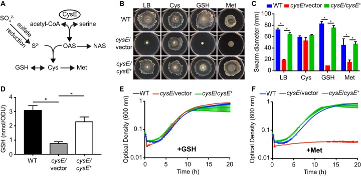
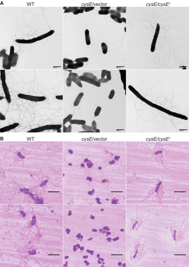
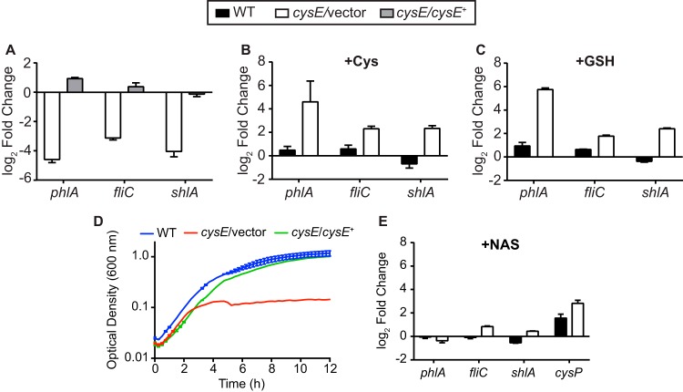
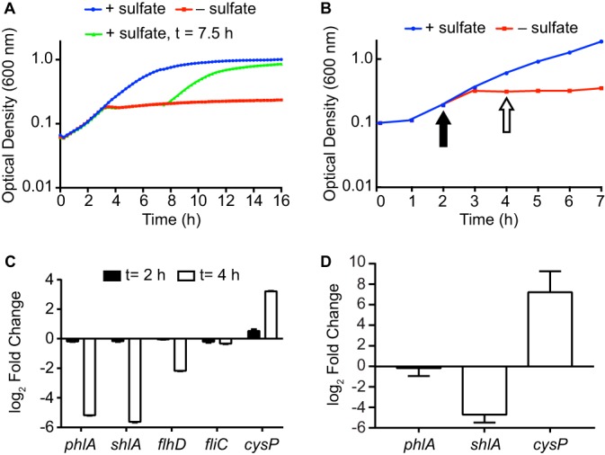
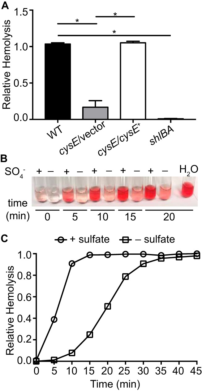
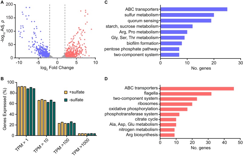
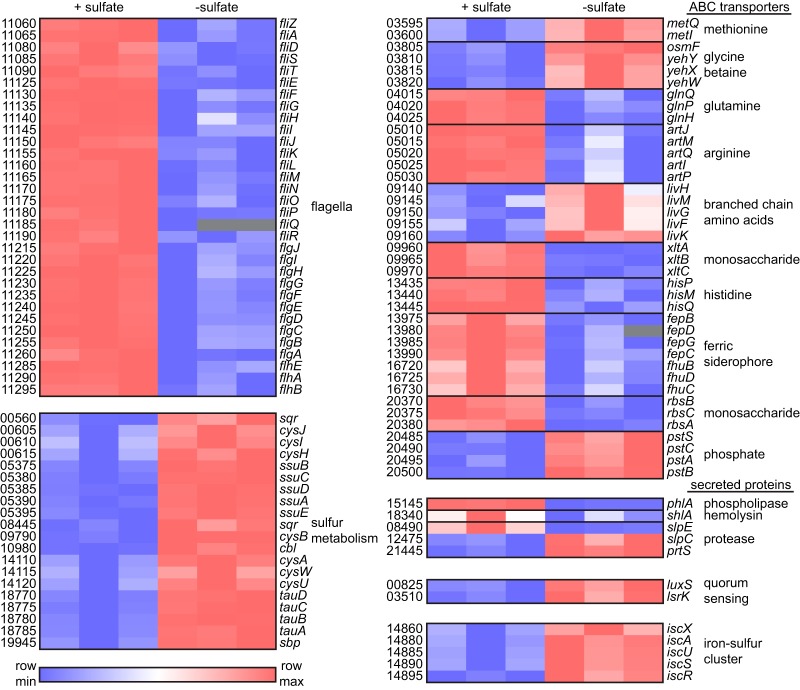
Sulfur is an essential nutrient that contributes to cellular redox homeostasis, transcriptional regulation, and translation initiation when incorporated into different biomolecules. Transport and reduction of extracellular sulfate followed by cysteine biosynthesis is a major pathway of bacterial sulfur assimilation. For the opportunistic pathogen Serratia marcescens, function of the cysteine biosynthesis pathway is required for extracellular phospholipase activity and flagellum-mediated surface motility, but little else is known about the influence of sulfur assimilation on the physiology of this organism. In this work, it was determined that an S. marcescens cysteine auxotroph fails to differentiate into hyperflagellated and elongated swarmer cells and that cysteine, but not other organic sulfur molecules, restores swarming motility to these bacteria. The S. marcescens cysteine auxotroph further exhibits reduced transcription of phospholipase, hemolysin, and flagellin genes, each of which is subject to transcriptional control by the flagellar regulatory system. Based on these data and the central role of cysteine in sulfur assimilation, it was reasoned that environmental sulfur availability may contribute to the regulation of these functions in S. marcescens Indeed, bacteria that are starved for sulfate exhibit substantially reduced transcription of the genes for hemolysin, phospholipase, and the FlhD flagellar master regulator. A global transcriptomic analysis further defined a large set of S. marcescens genes that are responsive to extracellular sulfate availability, including genes that encode membrane transport, nutrient utilization, and metabolism functions. Finally, sulfate availability was demonstrated to alter S. marcescens cytolytic activity, suggesting that sulfate assimilation may impact the virulence of this organism.IMPORTANCE Serratia marcescens is a versatile bacterial species that inhabits diverse environmental niches and is capable of pathogenic interactions with host organisms ranging from insects to humans. This report demonstrates for the first time the extensive impacts that environmental sulfate availability and cysteine biosynthesis have on the transcriptome of S. marcescens The finding that greater than 1,000 S. marcescens genes are differentially expressed depending on sulfate availability suggests that sulfur abundance is a crucial factor that controls the physiology of this organism. Furthermore, the high relative expression levels for the putative virulence factors flagella, phospholipase, and hemolysin in the presence of sulfate suggests that a sulfur-rich host environment could contribute to the transcription of these genes during infection.

Keywords: Serratia; cysteine; flagella; hemolysin; phospholipase; sulfur.


Figures

FIG 1
FIG 1
Cysteine limitation prevents swarming motility of S. marcescens. (A) Model of sulfate assimilation and cysteine biosynthesis derived from established pathways in E. coli. Intermediate steps in the glutathione (GSH), methionine, and sulfate reduction pathways are not depicted. (B) Representative images of swarming motility for S. marcescens strains on soft agar plates. Wild-type (WT), cysE mutant (cysE/vector), and the complemented mutant (cysE/cysE+) strains were spotted onto the surfaces of LB agar plates or LB agar supplemented with 1 mM cysteine, glutathione, or methionine and allowed to swarm for 16 h. (C) Quantitation of swarming motility. The swarm zone diameters were determined for triplicate cultures and the mean diameters (± standard deviations) are presented. *, P < 0.01 by t test. (D) Total intracellular glutathione from bacteria cultured in LB was measured for each strain and normalized to the culture optical density. Bars represent the means (± standard deviations) from triplicate cultures. *, P < 0.01 by t test. (E) Growth of S. marcescens strains in M9 medium supplemented with 1 mM glutathione. Growth was measured by optical density in 15-min intervals. Values represent the means (± standard deviations) from triplicate cultures. (F) Growth of S. marcescens strains in M9 medium supplemented with 1 mM methionine as described for panel E.
FIG 2
FIG 2
Cysteine-dependent swarm cell differentiation of S. marcescens. (A) Wild-type (WT), cysE mutant (cysE/vector), and complemented mutant (cysE/cysE+) strains were collected from the leading edges of actively growing swarm cultures and applied to grids for staining with 1% phosphotungstic acid. Bacterial cells were visualized by transmission electron microscopy, and representative images are shown. (B) Bacteria collected from swarm zones as described for panel A were stained for flagella on glass slides. Representative images of stained S. marcescens strains collected by light microscopy using a 100× lens objective are shown. Bars, 5 μm.
FIG 3
FIG 3
Products of sulfur metabolism contribute to expression of flagellar regulon genes. (A) Transcript levels were measured by qRT-PCR in both the cysE mutant (cysE/vector) and the complemented mutant (cysE/cysE+) relative to that in wild-type (WT) bacteria. All strains were cultured in M9 medium with Casamino Acids for this experiment. Transcript levels for the wild type and the cysE mutant strain were also determined for bacteria cultured in M9 medium containing Casamino Acids and supplemented with either 1 mM cysteine (B) or 1 mM glutathione (GSH) (C) relative to that in unsupplemented M9. (D) Wild-type, cysE mutant, and complemented mutant strains were cultured in M9 medium supplemented with 10 mM NAS. Growth was measured by optical density in 15-min intervals from triplicate cultures (means ± standard deviations). (E) Gene expression was determined for bacterial strains cultured in M9 medium with Casamino Acids and supplemented with 10 mM NAS relative to that in unsupplemented cultures. All qRT-PCR data are reported as the means (± standard deviations) from triplicate determinations.
FIG 4
FIG 4
Sulfate starvation inhibits phospholipase and hemolysin gene expression. (A) The kinetics of sulfate starvation were established by culturing the wild-type strain UMH9 in sulfate-replete M9 medium (+ sulfate) or sulfate-limited medium (− sulfate). Magnesium sulfate was added after growth arrest (7.5 h postinoculation) to sulfate-limited cultures to demonstrate that sulfate was the growth-limiting nutrient under the tested conditions. (B) Strain UMH9 was cultured in sulfate-replete or sulfate-limited medium as described for panel A and used for qRT-PCR gene expression analysis. Arrows indicate the points postinoculation at which aliquots from both cultures were removed for RNA isolation and cDNA synthesis. Growth curves represent the means (± standard deviations) optical densities from triplicate cultures measured in 15-min intervals. (C) Relative expression of the indicated genes was determined by qRT-PCR for cDNA generated from the experiment in panel B. Transcript levels from sulfate-starved bacteria relative to those in sulfate-replete bacteria are presented from 2 and 4 h after inoculation. (D) Gene expression in the S. marcescens fliP mutant for bacteria cultured under sulfate-limited conditions relative to that for sulfate-replete bacteria. All qRT-PCR data are reported as the means (± standard deviations) from triplicate determinations.
FIG 5
FIG 5
Sulfate starvation inhibits Serratia hemolysin activity. (A) Hemolytic activity of S. marcescens wild-type (WT), cysE mutant (cysE/vector), complemented cysE mutant (cysE/cysE+), and hemolysin null (shlBA) strains. Bacterial suspensions in PBS were normalized by optical density and mixed with 8% defibrinated sheep blood. Hemolytic activity was determined by measuring absorbance (405 nm) after 1 h from cleared suspensions relative to that of the positive control. Bars represent the means (± standard deviations) from triplicate assays. *, P < 0.001 by t test. (B) Representative image of a hemolytic activity assay for wild-type strain UMH9 cultured in sulfate-replete (+) and sulfate-limited (−) media. Bacterial suspensions were mixed with defibrinated sheep blood and assayed for hemolytic activity in 5-min intervals as described for panel A. (C) Quantitation of relative hemolysis over time for the wild-type strain cultured with sulfate (+ sulfate) and without sulfate (− sulfate). Results are representative of three experiments.
FIG 6
FIG 6
Sulfate availability alters the transcriptome of S. marcescens. Global gene expression of wild-type strain UMH9 was determined by RNA-seq for bacteria cultured in the presence of sulfate relative to that for sulfate-limited bacteria. (A) Fold change in expression was plotted for each mapped gene relative to the adjusted P value. A threshold of log2 fold change >2.0 or <−2.0 (adjusted P < 0.05) was used to identify significantly upregulated (red) and downregulated (blue) genes. (B) The proportions of total UMH9 genes having transcripts per million (TPM) values between 1 and 1,000 under the two tested conditions. (C and D) Protein-encoding genes were annotated by KEGG BlastKOALA to identify the top biological pathways affected by sulfate availability. Upregulated and downregulated pathways are colored as described for panel A.
FIG 7
FIG 7
Selected S. marcescens transcripts that are differentially regulated by sulfate availability. Relative expression of individual genes as determined by RNA-seq for triplicate cultures of UMH9 in sulfate-replete (+ sulfate) and sulfate-limited (− sulfate) media. Putative gene names were assigned based on homology to genes of known function in other bacterial species, and numbers designate open reading frames according to the NCBI UMH9 reference sequence annotation (e.g., BVG96_RS11060, fliZ). The heat map was generated using log2-transformed TPM values. Gray boxes indicate missing values.

Similar articles

Cited by

References

    1. Kredich NM. 2008. Biosynthesis of cysteine. EcoSal Plus 3:6.1.11. doi:10.1128/ecosalplus.3.6.1.11. - DOI - PubMed
    1. Kredich NM. 1992. The molecular basis for positive regulation of cys promoters in Salmonella Typhimurium and Escherichia coli. Mol Microbiol 6:2747–2753. doi:10.1111/j.1365-2958.1992.tb01453.x. - DOI - PubMed
    1. Kredich NM, Tomkins GM. 1966. The enzymic synthesis of l-cysteine in Escherichia coli and Salmonella Typhimurium. J Biol Chem 241:4955–4965. - PubMed
    1. Ostrowski J, Kredich NM. 1989. Molecular characterization of the cysJIH promoters of Salmonella Typhimurium and Escherichia coli: regulation by cysB protein and N-acetyl-l-serine. J Bacteriol 171:130–140. doi:10.1128/jb.171.1.130-140.1989. - DOI - PMC - PubMed
    1. Hryniewicz MM, Kredich NM. 1991. The cysP promoter of Salmonella Typhimurium: characterization of two binding sites for CysB protein, studies of in vivo transcription initiation, and demonstration of the anti-inducer effects of thiosulfate. J Bacteriol 173:5876–5886. doi:10.1128/jb.173.18.5876-5886.1991. - DOI - PMC - PubMed

LinkOut - more resources