. 2019 Aug 20;116(34):16717-16722.
doi: 10.1073/pnas.1908006116. Epub 2019 Aug 6.

Application of millisecond time-resolved solid state NMR to the kinetics and mechanism of melittin self-assembly

Affiliations

Application of millisecond time-resolved solid state NMR to the kinetics and mechanism of melittin self-assembly

Jaekyun Jeon et al. Proc Natl Acad Sci U S A. .

Abstract

Common experimental approaches for characterizing structural conversion processes such as protein folding and self-assembly do not report on all aspects of the evolution from an initial state to the final state. Here, we demonstrate an approach that is based on rapid mixing, freeze-trapping, and low-temperature solid-state NMR (ssNMR) with signal enhancements from dynamic nuclear polarization (DNP). Experiments on the folding and tetramerization of the 26-residue peptide melittin following a rapid pH jump show that multiple aspects of molecular structure can be followed with millisecond time resolution, including secondary structure at specific isotopically labeled sites, intramolecular and intermolecular contacts between specific pairs of labeled residues, and overall structural order. DNP-enhanced ssNMR data reveal that conversion of conformationally disordered melittin monomers at low pH to α-helical conformations at neutral pH occurs on nearly the same timescale as formation of antiparallel melittin dimers, about 6 to 9 ms for 0.3 mM melittin at 24 °C in aqueous solution containing 20% (vol/vol) glycerol and 75 mM sodium phosphate. Although stopped-flow fluorescence data suggest that melittin tetramers form quickly after dimerization, ssNMR spectra show that full structural order within melittin tetramers develops more slowly, in ∼60 ms. Time-resolved ssNMR is likely to find many applications to biomolecular structural conversion processes, including early stages of amyloid formation, viral capsid formation, and protein-protein recognition.

Keywords: melittin; protein folding; self-assembly; solid-state NMR.


Conflict of interest statement

The authors declare no conflict of interest.

Figures

Fig. 1.
Fig. 1.
(A) Conceptual representation of the conversion of 4 unfolded melittin monomers to a melittin tetramer (Protein Data Bank ID 2MLT) after a rapid pH jump. When melittin is 13C-labeled at all carbon sites of Gly3, Leu6, and Ile20, helix formation leads to intramolecular 13C–13C distances less than 4.5 Å between Gly3 and Leu6, while tetramer formation leads to intermolecular 13C–13C distances less than 4.0 Å between Leu6 and Ile20. (B and C) CD spectra of 300 μM melittin in 20% (vol/vol) glycerol with 75 mM NaHPO4 as a function of pH at 20 °C and as a function of temperature at pH 7.0.
Fig. 2.
Fig. 2.
(A) Apparatus for rapid mixing and freeze-trapping. A copper plate (1) is precooled by immersion in a liquid-nitrogen bucket (2), and then rotated continuously by a motor (3). Two solutions are pumped through a mixer (4) that sweeps across the rotating plate under the control of a stepper motor (5). A jet of mixed solution freezes on the rotating plate after traveling a variable distance df that determines the structural evolution time τe. After rotation stops, the plate is immersed in liquid nitrogen. The frozen solution is scraped from the plate and packed into a MAS rotor (6) for low-temperature ssNMR measurements. (B) DNP-enhanced, DQ-filtered 13C ssNMR spectra of frozen melittin solutions with the indicated values of τe. The dashed blue and red lines indicate 13CO and 13Cα chemical shifts for isotopically labeled residues at τe = ∞. Spectra were acquired at 30 K, 100.8-MHz 13C NMR frequency, and 7.00-kHz MAS frequency.
Fig. 3.
Fig. 3.
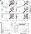
(A) DNP-enhanced 2D 13C ssNMR spectra of frozen melittin solutions with the indicated values of τe, obtained with a 13C–13C spin diffusion period τSD = 25 ms. The dashed lines indicate the 13Cα chemical shifts for Gly3, Leu6, and Ile20 in the fully folded tetrameric state at pH 7.0 (i.e., τe = ∞). Contour levels increase by successive factors of 1.3. (B) Dependence on τe of the coefficients cq of the 6 PC spectra, from singular value decomposition of the aliphatic regions of the 6 experimental 2D spectra. (C) Comparison of the dependences of the singular values sq on q for the experimental 2D spectra, for spectra reconstructed with only the first 2 PCs of the experimental spectra, and for spectra reconstructed with only the first 2 PCs of the experimental spectra plus additional random noise. A logarithmic scale is used in the Inset.
Fig. 4.
Fig. 4.
(A and B) DNP-enhanced 2D 13C ssNMR spectra of frozen melittin solutions with τSD = 25 ms and τSD = 1.0 s, respectively, and with τe = 4.6 ms. Signal amplitudes are displayed on continuous color scales. The numbered regions contain interresidue Gly3–Leu6 (1) and Leu6–Ile20 (3) cross-peak signals or intraresidue Leu6 (2) and Ile20 (4) cross-peak signals. (C) Dependences on τe of interresidue cross-peak volume ratios (V1/V2 for Gly3–Leu6; V3/V4 for Leu6–Ile20, where Vm is the volume within region m). Ratios from spectra with τSD = 25 ms are subtracted from those with τSD = 1 s to correct for overlap between intraresidue and interresidue cross-peaks. Error bars are derived from the rms noise in the 2D spectra. The color-coded dashed lines are least-squares fits to the form ABexp(τe/τbu), with indicated values of τbu. (D) Dependence on τe of the coefficient of the second PC for 2D spectra with τSD = 25 ms, from Fig. 3B. The dashed line is a least-squares fit to the form ABexp[(τe/τbu)β], with indicated values of τbu and β.

References

    1. Roder H., Maki K., Cheng H., Early events in protein folding explored by rapid mixing methods. Chem. Rev. 106, 1836–1861 (2006). - PMC - PubMed
    1. Eaton W. A., et al. , Fast kinetics and mechanisms in protein folding. Annu. Rev. Biophys. Biomol. Struct. 29, 327–359 (2000). - PMC - PubMed
    1. Gelman H., Gruebele M., Fast protein folding kinetics. Q. Rev. Biophys. 47, 95–142 (2014). - PMC - PubMed
    1. Royer C. A., Probing protein folding and conformational transitions with fluorescence. Chem. Rev. 106, 1769–1784 (2006). - PubMed
    1. Lerner E., et al. , Toward dynamic structural biology: Two decades of single-molecule Forster resonance energy transfer. Science 359, eaan1133 (2018). - PMC - PubMed

Publication types

LinkOut - more resources