

Brainstem and spinal cord MRI identifies altered sensorimotor pathways post-stroke
- PMID: 31388003
- PMCID: PMC6684621
- DOI: 10.1038/s41467-019-11244-3
Brainstem and spinal cord MRI identifies altered sensorimotor pathways post-stroke
Erratum in
-
Author Correction: Brainstem and spinal cord MRI identifies altered sensorimotor pathways post-stroke.Nat Commun. 2020 Jul 6;11(1):3433. doi: 10.1038/s41467-020-17024-8. Nat Commun. 2020. PMID: 32632101 Free PMC article.
Abstract
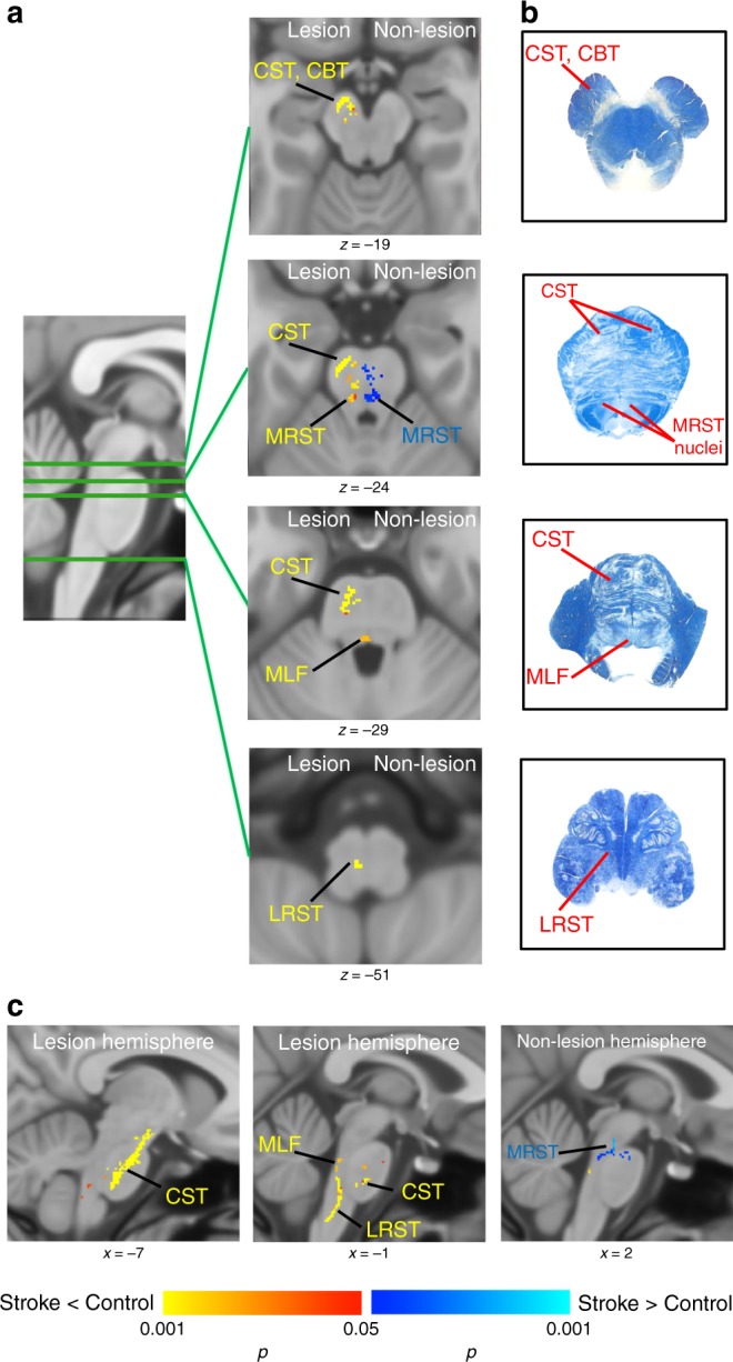
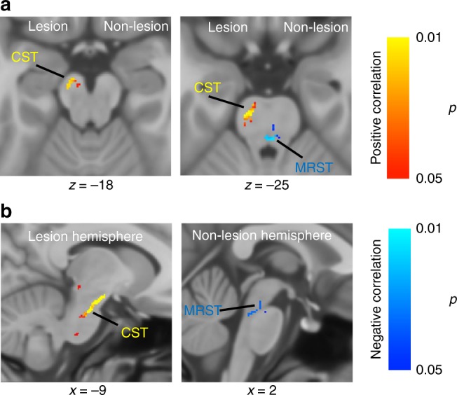
Damage to the corticospinal tract is widely studied following unilateral subcortical stroke, whereas less is known about changes to other sensorimotor pathways. This may be due to the fact that many studies investigated morphological changes in the brain, where the majority of descending and ascending brain pathways are overlapping, and did not investigate the brainstem where they separate. Moreover, these pathways continue passing through separate regions in the spinal cord. Here, using a high-resolution structural MRI of both the brainstem and the cervical spinal cord, we were able to identify a number of microstructurally altered pathways, in addition to the corticospinal tract, post stroke. Moreover, decreases in ipsi-lesional corticospinal tract integrity and increases in contra-lesional medial reticulospinal tract integrity were correlated with motor impairment severity in individuals with stroke.
Conflict of interest statement
The authors declare no competing interests.
Figures

References
Publication types
MeSH terms
Grants and funding
LinkOut - more resources
Full Text Sources
Medical
