Review
. 2019 Apr-Jun:780:29-36.
doi: 10.1016/j.mrrev.2017.10.002. Epub 2017 Nov 7.

Nucleosomes Regulate Base Excision Repair in Chromatin

Affiliations
Review

Nucleosomes Regulate Base Excision Repair in Chromatin

Rithy Meas et al. Mutat Res Rev Mutat Res. 2019 Apr-Jun.

Abstract

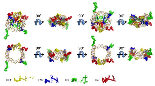
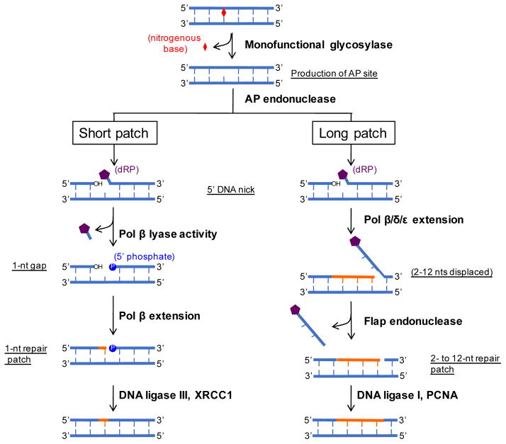
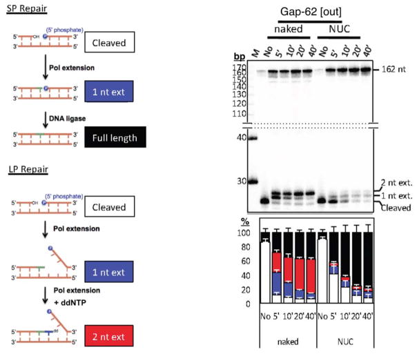
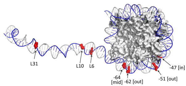
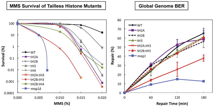
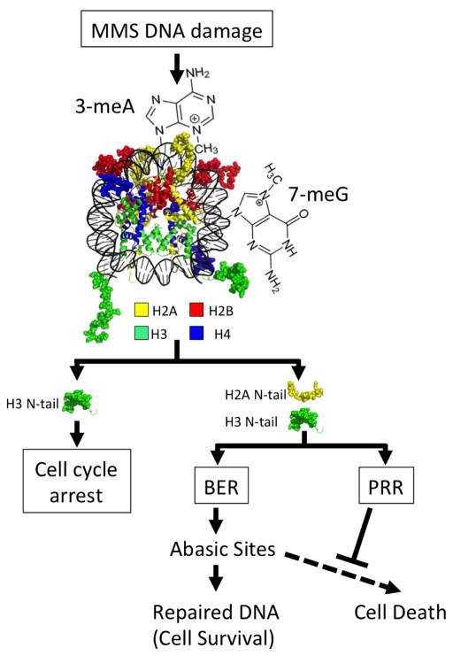
Chromatin is a significant barrier to many DNA damage response (DDR) factors, such as DNA repair enzymes, that process DNA lesions to reduce mutations and prevent cell death; yet, paradoxically, chromatin also has a critical role in many signaling pathways that regulate the DDR. The primary level of DNA packaging in chromatin is the nucleosome core particle (NCP), consisting of DNA wrapped around an octamer of the core histones H2A, H2B, H3 and H4. Here, we review recent studies characterizing how the packaging of DNA into nucleosomes modulates the activity of the base excision repair (BER) pathway and dictates BER subpathway choice. We also review new evidence indicating that the histone amino-terminal tails coordinately regulate multiple DDR pathways during the repair of alkylation damage in the budding yeast Saccharomyces cerevisiae.


Figures

Figure 1
Figure 1
The NCP and histone N-tails. The NCP is rotated at 90° intervals to show the positions of the histone N-tails (top panel). To better view the locations of the histone N-tails in the context of nucleosomal DNA, the histone core domains have been omitted and rotated 90° (bottom panel). H2A (yellow) H2B (blue) H3 (green) H4 (red). Nucleosome images are derived from the Protein Data Bank (PDB) using PDB ID 3LZ0.
Figure 2
Figure 2
BER pathway. A non-helix distorting lesion is recognized by a monofunctional glycosylase that cleaves at the N-glycosydic bond, which releases the base. This creates an abasic site that is recognized by an AP endonuclease that creates a 5′ nick. This substrate is then processed by SP or LP repair. SP repair uses the lyase activity of pol to remove the dRP moiety. Pol extends 1 nt followed by DNA ligation by the DNA ligase III/XRCC1 complex. LP repair proceeds by pol,, to extend •2 nts. The displaced DNA is cleaved by flap endonuclease followed by DNA ligation via the coordinated efforts of DNA ligase I and PCNA. Adapted from Figure 1 of Liu & Wilson [47].
Figure 3
Figure 3
The schematic of the assay to analyze repair in naked or nucleosome substrates is shown on the left. A representative gel of a gap substrate located at position −62 from the nucleosome dyad axis is shown after the indicated repair times. “M” is the marker lane and “No” is the no treatment lane. The composite bar graph is shown below the gel (white, cleaved; blue, 1 nt extension; red, 2 nt extension; black, full-length). Figure was adapted from Figs. 3a and 3d of ref. .
Figure 4
Figure 4
Map of the various template lesions used in experiments such as those described in Figure 3. The locations of uracils (or gaps) are shown in red. The “L” indicates position of lesions in linker DNA from the edge of the NCP. The number at each lesion in the NCP represents its distance from the dyad center and brackets indicate its rotational position relative to the histone surface (as described in ref. 73). Nucleosome image from PDB ID 1ZBB.
Figure 5
Figure 5
Survival curves (left) and global genome BER (right) for the different N-tail deleted mutants as compared to the wild type and the BER-defective mag1Δ mutant is shown. Adapted from Figures 1 and 2 of ref. .
Figure 6
Figure 6
The N-tails of H2A and H3 coordinate base excision repair (BER) and postreplication repair (PRR) to mitigate the cytotoxic effects of BER intermediates, such as abasic sites. The N-tails of H2A and H3 are highlighted in the model to show their role in different branches of the DNA damage response to methyl methanesulfonate (MMS). The N-tails of H2A and H3 are important for coordinately regulating BER, by modulating expression of Mag1, and PRR. Stimulating the PRR pathway could help suppress the cytotoxic effects of BER intermediates, such as abasic sites, during DNA replication. Nucleosome image from PDB ID IKX5.

Similar articles

Cited by

References

    1. Friedberg EC, Walker GC, Siede W, Wood RD, Schultz RA, Ellenberger T. DNA repair and mutagenesis. 2. ASM Press; Washington, D.C: 2006.
    1. Bernstein C, Prassad AR, Nfonsam V, Bernstein H. DNA Damage, DNA Repair and Cancer. In: Chen CC, editor. New Research Directions. Intech, open access; 2013. pp. 413–465.
    1. Kennedy SR, Loeb LA, Herr AJ. Somatic mutations in aging, cancer and neurodegeneration. Mech Ageing Dev. 2012;133:118–126. - PMC - PubMed
    1. Wyatt MD, Pittman DL. Methylating agents and DNA repair responses: Methylated bases and sources of strand breaks. Chem Res Toxicol. 2006;19:1580–1594. - PMC - PubMed
    1. Hinz JM, Czaja W. Facilitation of base excision repair by chromatin remodeling. DNA repair. 2015 - PMC - PubMed

Publication types

LinkOut - more resources