. 2020 Mar;31(3):535-543.
doi: 10.1007/s00192-019-04063-z. Epub 2019 Aug 6.

Aging effects on pelvic floor support: a pilot study comparing young versus older nulliparous women

Affiliations

Aging effects on pelvic floor support: a pilot study comparing young versus older nulliparous women

Carolyn W Swenson et al. Int Urogynecol J. 2020 Mar.

Abstract

Introduction and hypothesis: We sought to determine age-related changes to the pelvic floor in the absence of childbirth effects.

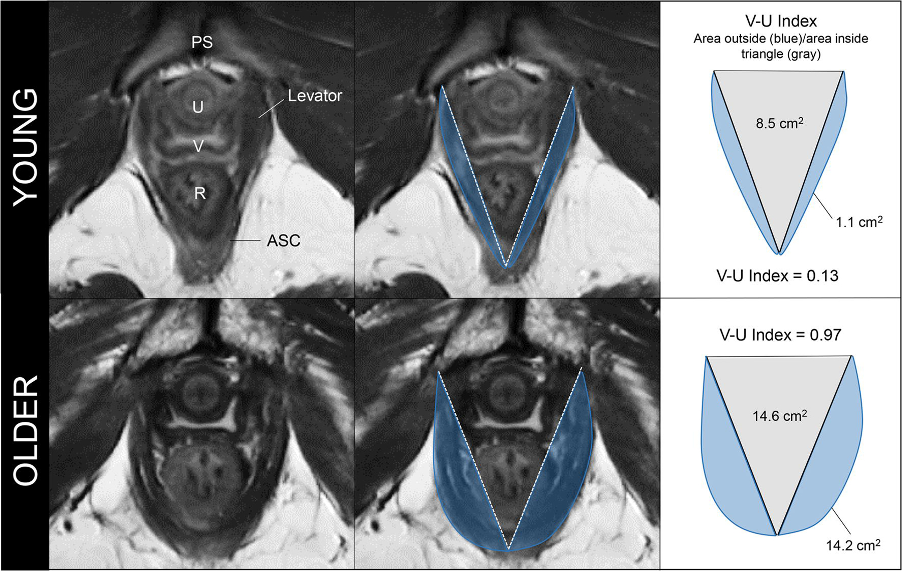
Methods: A case-control study was conducted from June 2017 to August 2018 comparing two groups of nulliparous women: <40 years old and ≥ 70 years old. Clinical evaluation included POP-Q, instrumented speculum testing, and handgrip strength. Dynamic 3D-stress MRI was performed on all women to obtain genital and levator hiatus (LH) lengths, LH area, and levator bowl volume. LH shape was quantified using a novel measure called the "V-U index." Pubovisceral muscle (PVM) cross-sectional area (CSA) was also measured. Bivariate comparisons between the two groups were made for all variables. Effect sizes were calculated for MRI measurements.

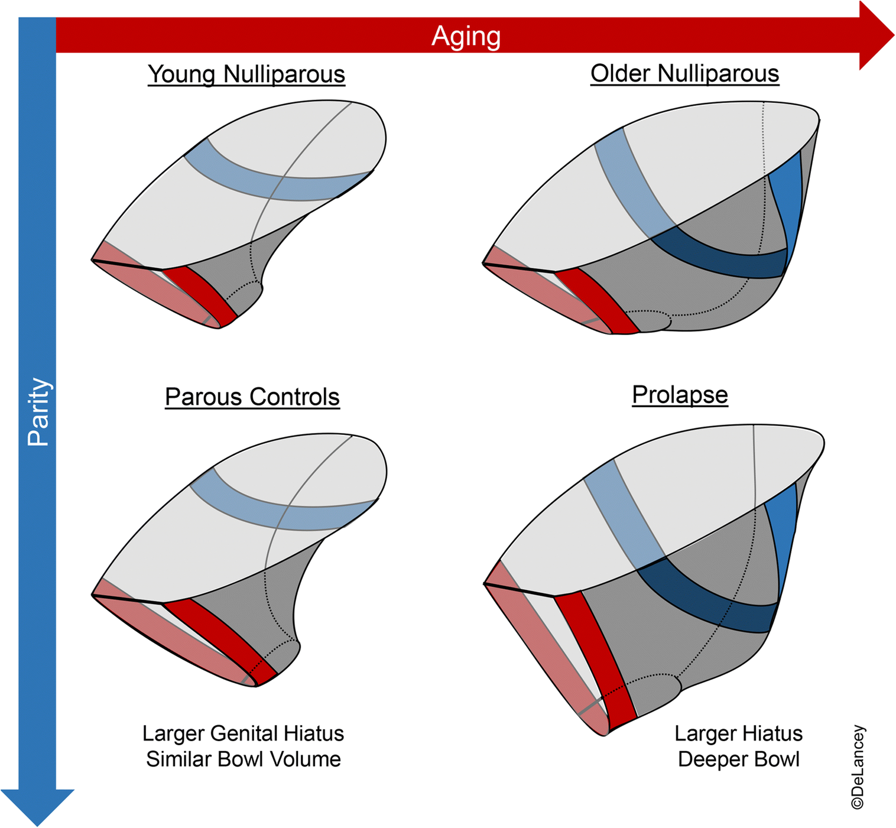
Results: Twelve young and 9 older nulliparous women were included. Levator bowl volume at rest was 83% larger in older women (108.0 ± 34.5 cm3 vs 59.2 ± 19.3 cm3, p = 0.001, d = 1.82). MRI genital hiatus at rest was larger among the older group (2.7 ± 0.6 cm vs 3.5 ± 0.6 cm, p = 0.007, d = 1.34). V-U index, a measure of LH shape where 0 = "V" and 1 = "U," differed between groups indicating a more "U"-like shape among older women (0.71 ± 0.23 vs 0.35 ± 0.18, p = 0.001, d = 1.72). Handgrip strength was lower in the older vs young group (23.2 ± 5.2 N vs 33.4 ± 5.2 N, p < 0.0001); however, the Kegel augmentation force and PVM CSA were similar (3.2 ± 1.1 N vs 3.3 ± 2.2 N, p = 0.89, and 0.8 ± 0.3 cm2 vs 0.7 ± 0.2 cm2, p = 0.23 respectively).

Conclusions: Levator bowl volume at rest was over 80% larger among older women, reflecting a generalized posterior distension with age.

Keywords: Aging; Genital hiatus; Levator area; Levator bowl volume; MRI; Pelvic organ support.


Conflict of interest statement

Financial disclaimer/conflict of interest: None

Figures

Figure 1.
Figure 1.. Mid-sagittal MRI measures obtained at rest and during maximal Valsalva
Pubic symphysis (PS), uterus (Ut), genital hiatus (GH), and perineal body (PB) are labeled. Pelvic inclination correction system (PICS) uses a standardized reference line 34° caudal to the sacrococcygeal to inferior pubic point (SCIPP) line to control for pelvic inclination during Valsalva. Asterisk (*) marks the location used to measure mid-paravaginal height. For the figure, this is shown in the mid-sagittal plane; however, the actual measures were obtained from the lateral margins of the anterior vaginal wall in the para-sagittal planes.
Figure 2.
Figure 2.. MRI levator areas and volume comparing a young (top row) versus older (bottom row) nulliparous woman
Left panel shows mid-sagittal levator area bordered by the genital hiatus (GH), sacrococcygeal to inferior pubic point (SCIPP) line, levator hiatus (LH), levator plate (LP), and anal sphincter complex (ASC). PS-pubic symphysis. Middle panel shows 3D levator bowl volume. Right panel shows the PICS levator hiatus area divided into pubic and posterior portions. U- urethra, V-vagina, R-rectum.
Figure 3.
Figure 3.. Method for calculating V-U Index and comparison between a young (top row) and older (bottom row) nulliparous woman
For ease of understanding, axial images are displayed; however, the V-U Index was actually measured in the PICS plane (same plane in which PICS LH area was measured). Left panel: axial MRI with pubic symphysis (PS), urethra (U), vagina (V), rectum (R), and anal sphincter complex (ASC). Notice the “V” shape of the levators in the young woman and the “U” shape in the older woman. Middle panel: at the pubic symphysis, a V is traced from the origin of the levators on each side to the midpoint of the levators behind the rectum. The posterior portion of the levator hiatus area is outlined. Right panel: The V-U index is calculated—index values closest to 0 indicate a “V”-shaped levator hiatus and those closest to 1 indicate a “U”-shaped levator hiatus.
Figure 4.
Figure 4.
Levator bowl volumes
Figure 5.
Figure 5.
Proposed conceptual model for pelvic floor changes resulting from aging and vaginal parity

References

    1. Dieter AA, Wilkins MF, Wu JM (2015) Epidemiological trends and future care needs for pelvic floor disorders. Curr Opin Obstet Gynecol 27(5):380–384. doi:10.1097/GCO.0000000000000200 - DOI - PMC - PubMed
    1. Swift SE (2000) The distribution of pelvic organ support in a population of female subjects seen for routine gynecologic health care. Am J Obstet Gynecol 183(2):277–285. doi:10.1067/mob.2000.107583 - DOI - PubMed
    1. Faulkner JA, Larkin LM, Claflin DR, Brooks SV (2007) Age-related changes in the structure and function of skeletal muscles. Clin Exp Pharmacol Physiol 34(11):1091–1096. doi:10.1111/j.1440-1681.2007.04752.x - DOI - PubMed
    1. Blomquist JL, Munoz A, Carroll M, Handa VL (2018) Association of Delivery Mode With Pelvic Floor Disorders After Childbirth. JAMA 320(23):2438–2447. doi:10.1001/jama.2018.18315 - DOI - PMC - PubMed
    1. Mant J, Painter R, Vessey M (1997) Epidemiology of genital prolapse: observations from the Oxford Family Planning Association Study. Br J Obstet Gynaecol 104(5):579–585 - PubMed