

Adenocarcinoma arising from an ectopic pancreas in the duodenum: a case report
- PMID: 31388774
- PMCID: PMC6684697
- DOI: 10.1186/s40792-019-0684-8
Adenocarcinoma arising from an ectopic pancreas in the duodenum: a case report
Abstract
Background: The malignant transformation of an ectopic pancreas in the duodenum is extremely rare. Herein, we report a case of an adenocarcinoma that arose from an ectopic pancreas. We also reviewed 14 cases of malignant transformations arising from an ectopic pancreas in the duodenum that were previously published.
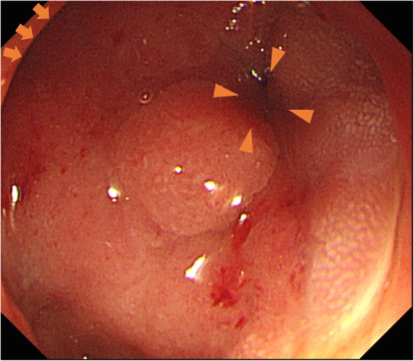
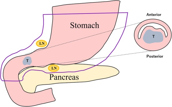
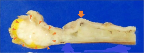
Case presentation: An 81-year-old man with a 1-month history of vomiting was admitted to our institution. Esophagogastroduodenoscopy (EGD) and computed tomography (CT) scans revealed an obstruction at the first part of the duodenum. A distal gastrectomy was performed for diagnostic and therapeutic purposes. The histopathological examination of the resected specimen showed adenocarcinoma that arose from an ectopic pancreas (Heinrich type 1). The patient is alive without relapse at 18 months of follow-up.
Conclusions: Adenocarcinoma that arises from an ectopic pancreas should be considered when an obstruction is identified in the duodenum.
Keywords: Cancer-induced vomiting; Distal gastrectomy; Duodenal adenocarcinoma; Ectopic pancreas.
Conflict of interest statement
The authors declare that they have no competing interests.
Figures


References
-
- Mulholland KC, Wallace WD, Epanomeritakis E, Hall SR. Pseudocyst formation in gastric ectopic pancreas. JOP. 2004;5:498–501. - PubMed
-
- Ishikawa O, Ishiguro S, Ohhigashi H, Sasaki Y, Yasuda T, Imaoka S, et al. Solid and papillary neoplasm arising from an ectopic pancreas in the mesocolon. Am J Gastroenterol. 1990;85:597–601. - PubMed
LinkOut - more resources
Full Text Sources
