. 2019 Oct;23(10):6885-6896.
doi: 10.1111/jcmm.14572. Epub 2019 Aug 7.

Truncated O-glycans promote epithelial-to-mesenchymal transition and stemness properties of pancreatic cancer cells

Affiliations

Truncated O-glycans promote epithelial-to-mesenchymal transition and stemness properties of pancreatic cancer cells

Divya Thomas et al. J Cell Mol Med. 2019 Oct.

Abstract

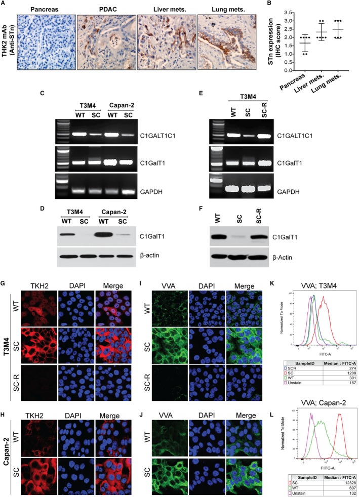
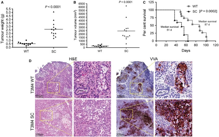
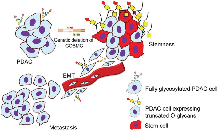
Aberrant expression of Sialyl-Tn (STn) antigen correlates with poor prognosis and reduced patient survival. We demonstrated that expression of Tn and STn in pancreatic ductal adenocarcinoma (PDAC) is due to hypermethylation of Core 1 synthase specific molecular chaperone (COSMC) and enhanced the malignant properties of PDAC cells with an unknown mechanism. To explore the mechanism, we have genetically deleted COSMC in PDAC cells to express truncated O-glycans (SimpleCells, SC) which enhanced cell migration and invasion. Since epithelial-to-mesenchymal transition (EMT) play a vital role in metastasis, we have analysed the induction of EMT in SC cells. Expressions of the mesenchymal markers were significantly high in SC cells as compared to WT cells. Equally, we found reduced expressions of the epithelial markers in SC cells. Re-expression of COSMC in SC cells reversed the induction of EMT. In addition to this, we also observed an increased cancer stem cell population in SC cells. Furthermore, orthotopic implantation of T3M4 SC cells into athymic nude mice resulted in significantly larger tumours and reduced animal survival. Altogether, these results suggest that aberrant expression of truncated O-glycans in PDAC cells enhances the tumour aggressiveness through the induction of EMT and stemness properties.

Keywords: COSMC; EMT; PDAC; core-1 synthase; stem cells; truncated O-glycans.


Conflict of interest statement

The authors of this manuscript declare no conflicts of interest.

Figures

Figure 1
Figure 1
STn antigen enhances PDAC tumorigenicity: A, IHC analysis of STn antigen expression in human RAP PDAC tumour tissues (normal pancreas, primary, liver mets and lung mets) using TKH2 monoclonal antibody. The images were taken at a magnification of 20×. B, IHC intensity score was compared against each tumour samples (n = 6). C, PCR amplification of COSMC/C1GALT1C1 and Core 1 synthase/C1GALT1 in T3M4 and Capan‐2 (WT and SC) cells. D, Western blotting of Core 1 synthase (C1GalT1) in T3M4 and Capan‐2 (WT and SC) cells. E, PCR amplification of COSMC/C1GALT1C1 and Core 1 synthase/C1GALT1 gene in T3M4 WT, SC and COSMC re‐expression (SC‐R) cells. Amplification of GAPDH was used as an internal loading control. F, Western blotting of Core 1 synthase (C1GalT1) in T3M4 WT, SC and SC‐R cells. Immunofluorescence analysis of STn antigen expression by TKH2 antibody in T3M4 WT, SC and SC‐R cells (G) and Capan‐2 WT and SC cells (H). Alexa fluor 488‐conjugated secondary antibody was used (images are represented with pseudo red‐colour). Magnification: 63×; Scale bar: 10 µm. Immunofluorescence analysis of VVA in T3M4 WT, SC and SC‐R cells (I) and Capan‐2 WT and SC cells (J). Alexa fluor 488‐conjugated secondary antibody was used. Magnification: 63×; Scale bar: 10 µm. Analysis of VVA by flow cytometry in T3M4 WT, SC and SC‐R cells (K) and Capan‐2 WT and SC cells (L)
Figure 2
Figure 2
Truncated O‐glycans enhances PDAC cell invasiveness and induction of EMT. A, Migration assay. T3M4 WT and SC cells (5 × 104) that migrate through trans well inserts were fixed and counted five different fields under the light microscope (20×) (n = 3). B, Matrigel Invasion assay. T3M4 WT and SC cells (5 × 104) that invaded through matrigel‐coated Boyden chambers were fixed and counted five different fields under the microscope (20×) (n = 3). P value < 0.05 considered as statistically significant. C, Western blotting of T3M4 and Capan‐2 (WT and SC) cell lysates with epithelial marker proteins E‐cadherin, claudin and occludin. D, Western blotting of T3M4 and Capan‐2 (WT and SC) cell lysate with MMP2, MMP9 and α‐SMA. E, Western blotting of T3M4 and Capan‐2 (WT and SC) cell lysate with mesenchymal marker proteins N‐cadherin, vimentin and transcription factors Snail and Slug. F, Western blotting of epithelial marker proteins E‐cadherin, claudin and occludin in T3M4 WT, SC and SC‐R. G, Western blotting of MMP2, MMP9 and α‐SMA in T3M4 WT, SC and SC‐R cells. H, Western blotting of mesenchymal marker proteins N‐cadherin, vimentin and transcription factors Snail and slug in T3M4 WT, SC and SC‐R cells. Detection of β‐actin and GAPDH served as loading control
Figure 3
Figure 3
Truncated O‐glycans induce stemness in PDAC cells. A, T3M4 cells (WT and SC, 1 × 106 cells/mL) were stained with Hoechst 33342 with or without verapamil and analysed by flow cytometry. The side population (SP) cells, which appear in the absence of verapamil, are outlined and shown as a percentage of the total cell population (left panel). The graphical representation of respective side cell population in T3M4 WT and SC cells (n = 3) (right panel). B, Tumour sphere forming assay. T3M4 (WT, SC and SC‐R) and Capan‐2 (WT and SC) Cells cultured in ultra‐low attachment plates with 3dGRO™ medium were analysed for the ability to form tumour sphere. COMC knockout enhanced tumour sphere formation in PDAC cells. C, Flow cytometry analysis of CD44 expression. T3M4 (WT, SC and SC‐R) and Capan‐2 (WT and SC) cells (1 × 106 cells/mL) were incubated with rabbit anti‐CD44 antibody and stained with Alexa fluor 488‐conjugated anti‐rabbit IgG and analysed in a flow cytometer. D, Flow cytometry of CD133 expression. T3M4 (WT, SC and SC‐R) and Capan‐2 (WT and SC) cells (1 × 106 cells/mL) were incubated with phycoerythrin‐conjugated CD133 and analysed in flow cytometer. The graphical representation of the expression of CD 44 and CD133 in PDAC cells (n = 3) (bottom panel). P value < .05 considered as statistically significant
Figure 4
Figure 4
Truncated O‐glycans induced tumour malignancy. Orthotopic implantation of T3M4 WT and COSMC deleted cells (0.25 × 106/30 µL PBS) in pancreas of athymic nude mice (n = 13). After 28 days of post‐injection, the tumour weight (g) (A) and tumour volume (B) was measured. C, Survival of tumour‐bearing animals. Orthotopic implantation of T3M4 WT and SC cells into the pancreas of athymic nude mice (n = 15/group). The Kaplan‐Meier curve represents the animal survival time from the beginning of tumour cell implantation. P value of <0.05 was considered statistically significant. D, Haematoxylin and eosin staining of the T3M4 (WT and SC) cells implanted mouse pancreas tumour tissues (left panel) and IHC analysis of STn antigen expression by VVA lectin staining (right panel)
Figure 5
Figure 5
Analysis of EMT in orthotopic PDAC tumours. A, IHC analysis of E‐cadherin in T3M4 WT and SC cells implanted mouse pancreas tumour tissues. The mean histoscore of E‐cadherin expression was compared between WT and SC cells implanted mouse tumour tissues (n = 5). B, IHC analysis of N‐cadherin in T3M4 WT and SC cells implanted mouse pancreas tumour tissues. The mean histoscore of N‐cadherin expression was compared between WT and SC cells implanted mouse tumour tissues (n = 5). C, Western blotting of E‐cadherin and N‐cadherin expressions in T3M4 WT and SC cells implanted mouse pancreas tumour tissues. Detection of β‐actin served as loading control. P value of <0.05 was considered statistically significant. D, IHC analysis of the expression of CD133 in T3M4 WT and SC cell implanted mouse tumour tissues. The mean histoscore of CD133 expression was compared between WT and SC cells implanted mouse tumour tissues (n = 3). P value of <0.05 was considered statistically significant
Figure 6
Figure 6
Proposed mechanism of truncated O‐glycans induced tumorigenicity in PDAC cells. Genetic deletion of COSMC results in the aberrant expression of Tn/STn antigen on mucin and other glycoproteins in PDAC cells. Such truncated O‐glycan‐expressing PDAC cells increase tumorigenicity and metastasis through the induction of EMT. Also, truncation of O‐glycans enhanced the cellular plasticity and stemness in PDAC cells

Similar articles

Cited by

References

    1. Ferlay J, Soerjomataram I, Dikshit R, et al. Cancer incidence and mortality worldwide: sources, methods and major patterns in GLOBOCAN 2012. Int J Cancer. 2015;136:E359‐E386. - PubMed
    1. Yonezawa S, Tachikawa T, Shin S, Sato E. Sialosyl‐Tn antigen. Its distribution in normal human tissues and expression in adenocarcinomas. Am J Clin Pathol. 1992;98:167‐174. - PubMed
    1. Kim GE, Bae HI, Park HU, et al. Aberrant expression of MUC5AC and MUC6 gastric mucins and sialyl Tn antigen in intraepithelial neoplasms of the pancreas. Gastroenterology. 2002;123:1052‐1060. - PubMed
    1. Davidson B, Berner A, Nesland JM, et al. Carbohydrate antigen expression in primary tumors, metastatic lesions, and serous effusions from patients diagnosed with epithelial ovarian carcinoma: evidence of up‐regulated Tn and Sialyl Tn antigen expression in effusions. Hum Pathol. 2000;31:1081‐1087. - PubMed
    1. Noda M, Okayama H, Tachibana K, et al. Glycosyltransferase gene expression identifies a poor prognostic colorectal cancer subtype associated with mismatch repair deficiency and incomplete glycan synthesis. Clin Cancer Res. 2018;24:4468‐4481. - PubMed

Publication types

MeSH terms