

Diagnostic accuracy of dynamic contrast-enhanced perfusion MRI in stratifying gliomas: A systematic review and meta-analysis
- PMID: 31389669
- PMCID: PMC6745862
- DOI: 10.1002/cam4.2369
Diagnostic accuracy of dynamic contrast-enhanced perfusion MRI in stratifying gliomas: A systematic review and meta-analysis
Abstract
Background: T1-weighted dynamic contrast-enhanced (DCE) perfusion magnetic resonance imaging (MRI) has been broadly utilized in the evaluation of brain tumors. We aimed at assessing the diagnostic accuracy of DCE-MRI in discriminating between low-grade gliomas (LGGs) and high-grade gliomas (HGGs), between tumor recurrence and treatment-related changes, and between primary central nervous system lymphomas (PCNSLs) and HGGs.
Methods: We performed this study based on the Preferred Reporting Items for Systematic Reviews and Meta-Analysis of Diagnostic Test Accuracy Studies criteria. We systematically surveyed studies evaluating the diagnostic accuracy of DCE-MRI for the aforementioned entities. Meta-analysis was conducted with the use of a random effects model.
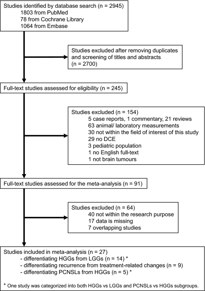
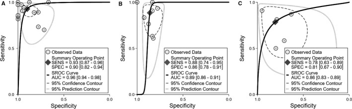
Results: Twenty-seven studies were included after screening of 2945 possible entries. We categorized the eligible studies into three groups: those utilizing DCE-MRI to differentiate between HGGs and LGGs (14 studies, 546 patients), between recurrence and treatment-related changes (9 studies, 298 patients) and between PCNSLs and HGGs (5 studies, 224 patients). The pooled sensitivity, specificity, and area under the curve for differentiating HGGs from LGGs were 0.93, 0.90, and 0.96, for differentiating tumor relapse from treatment-related changes were 0.88, 0.86, and 0.89, and for differentiating PCNSLs from HGGs were 0.78, 0.81, and 0.86, respectively.
Conclusions: Dynamic contrast-enhanced-Magnetic resonance imaging is a promising noninvasive imaging method that has moderate or high accuracy in stratifying gliomas. DCE-MRI shows high diagnostic accuracy in discriminating between HGGs and their low-grade counterparts, and moderate diagnostic accuracy in discriminating recurrent lesions and treatment-related changes as well as PCNSLs and HGGs.
Keywords: dynamic contrast-enhanced MRI; gliomas; lymphoma; meta-analysis; perfusion.
© 2019 The Authors. Cancer Medicine published by John Wiley & Sons Ltd.
Conflict of interest statement
None declared.
Figures
Similar articles
-
Intergrating conventional MRI, texture analysis of dynamic contrast-enhanced MRI, and susceptibility weighted imaging for glioma grading.Acta Radiol. 2019 Jun;60(6):777-787. doi: 10.1177/0284185118801127. Epub 2018 Sep 23. Acta Radiol. 2019. PMID: 30244590
-
Grading diffuse gliomas without intense contrast enhancement by amide proton transfer MR imaging: comparisons with diffusion- and perfusion-weighted imaging.Eur Radiol. 2017 Feb;27(2):578-588. doi: 10.1007/s00330-016-4328-0. Epub 2016 Mar 22. Eur Radiol. 2017. PMID: 27003139
-
Diffusional kurtosis imaging for differentiating between high-grade glioma and primary central nervous system lymphoma.J Magn Reson Imaging. 2016 Jul;44(1):30-40. doi: 10.1002/jmri.25090. Epub 2015 Nov 20. J Magn Reson Imaging. 2016. PMID: 26588793
-
The role of APT imaging in gliomas grading: A systematic review and meta-analysis.Eur J Radiol. 2020 Dec;133:109353. doi: 10.1016/j.ejrad.2020.109353. Epub 2020 Oct 16. Eur J Radiol. 2020. PMID: 33120241
-
The diagnostic value of intravoxel incoherent motion imaging in differentiating high-grade from low-grade gliomas: a systematic review and meta-analysis.Br J Radiol. 2021 May 1;94(1121):20201321. doi: 10.1259/bjr.20201321. Br J Radiol. 2021. PMID: 33876653 Free PMC article.
Cited by
-
Dynamic contrast-enhanced magnetic resonance imaging parameter changes as an early biomarker of tumor responses following radiation therapy in patients with spinal metastases: a systematic review.Radiat Oncol J. 2023 Dec;41(4):225-236. doi: 10.3857/roj.2023.00290. Epub 2023 Oct 27. Radiat Oncol J. 2023. PMID: 38185927 Free PMC article.
-
Hemodynamic Imaging in Cerebral Diffuse Glioma-Part B: Molecular Correlates, Treatment Effect Monitoring, Prognosis, and Future Directions.Cancers (Basel). 2022 Mar 5;14(5):1342. doi: 10.3390/cancers14051342. Cancers (Basel). 2022. PMID: 35267650 Free PMC article. Review.
-
MRI biomarkers in neuro-oncology.Nat Rev Neurol. 2021 Aug;17(8):486-500. doi: 10.1038/s41582-021-00510-y. Epub 2021 Jun 20. Nat Rev Neurol. 2021. PMID: 34149051 Review.
-
Analyzing magnetic resonance imaging data from glioma patients using deep learning.Comput Med Imaging Graph. 2021 Mar;88:101828. doi: 10.1016/j.compmedimag.2020.101828. Epub 2020 Dec 2. Comput Med Imaging Graph. 2021. PMID: 33571780 Free PMC article. Review.
-
Glioma Type Prediction with Dynamic Contrast-Enhanced MR Imaging and Diffusion Kurtosis Imaging-A Standardized Multicenter Study.Cancers (Basel). 2024 Jul 25;16(15):2644. doi: 10.3390/cancers16152644. Cancers (Basel). 2024. PMID: 39123372 Free PMC article.
References
-
- Louis DN, Perry A, Reifenberger G, et al. The 2016 World Health Organization classification of tumors of the central nervous system: a summary. Acta Neuropathol. 2016;131:803‐820. - PubMed
-
- Stupp R, Mason WP, van den Bent MJ, et al. Radiotherapy plus concomitant and adjuvant temozolomide for glioblastoma. N Engl J Med. 2005;352:987‐996. - PubMed
-
- Yang I, Aghi MK. New advances that enable identification of glioblastoma recurrence. Nat Rev Clin Oncol. 2009;6:648‐657. - PubMed
Publication types
MeSH terms
Substances
Grants and funding
- BRC345/NS/SB/101410/The National Institute for Health Research (NIHR) University College London Hospitals Biomedical Research Centre (UCLH BRC)/International
- The NIHR Collaboration for Leadership in Applied Health Research and Care North Thames at Bart's Health NHS Trust (NIHR CLAHRC North Thames)/International
LinkOut - more resources
Full Text Sources
Medical
