. 2019 Sep;8(12):5716-5734.
doi: 10.1002/cam4.2455. Epub 2019 Aug 7.

Nonconserved miR-608 suppresses prostate cancer progression through RAC2/PAK4/LIMK1 and BCL2L1/caspase-3 pathways by targeting the 3'-UTRs of RAC2/BCL2L1 and the coding region of PAK4

Affiliations

Nonconserved miR-608 suppresses prostate cancer progression through RAC2/PAK4/LIMK1 and BCL2L1/caspase-3 pathways by targeting the 3'-UTRs of RAC2/BCL2L1 and the coding region of PAK4

Xu Zhang et al. Cancer Med. 2019 Sep.

Abstract

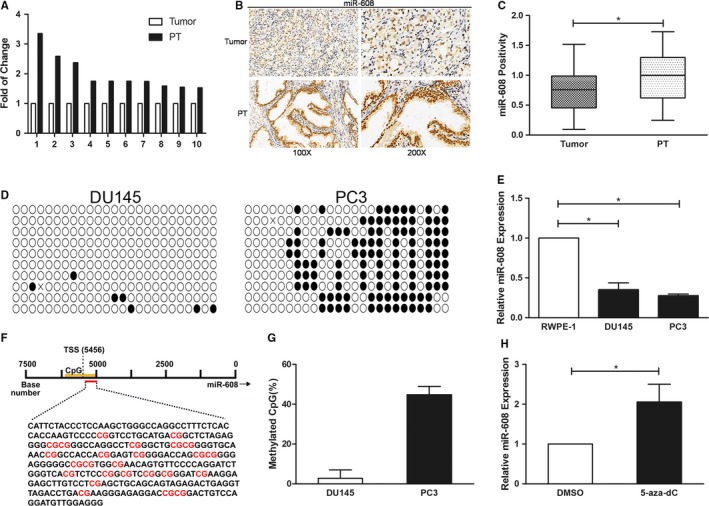
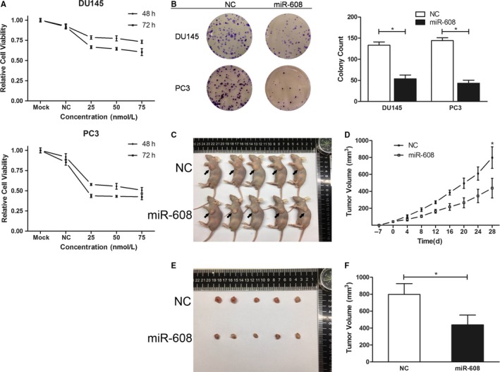
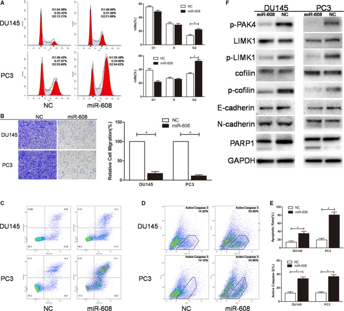
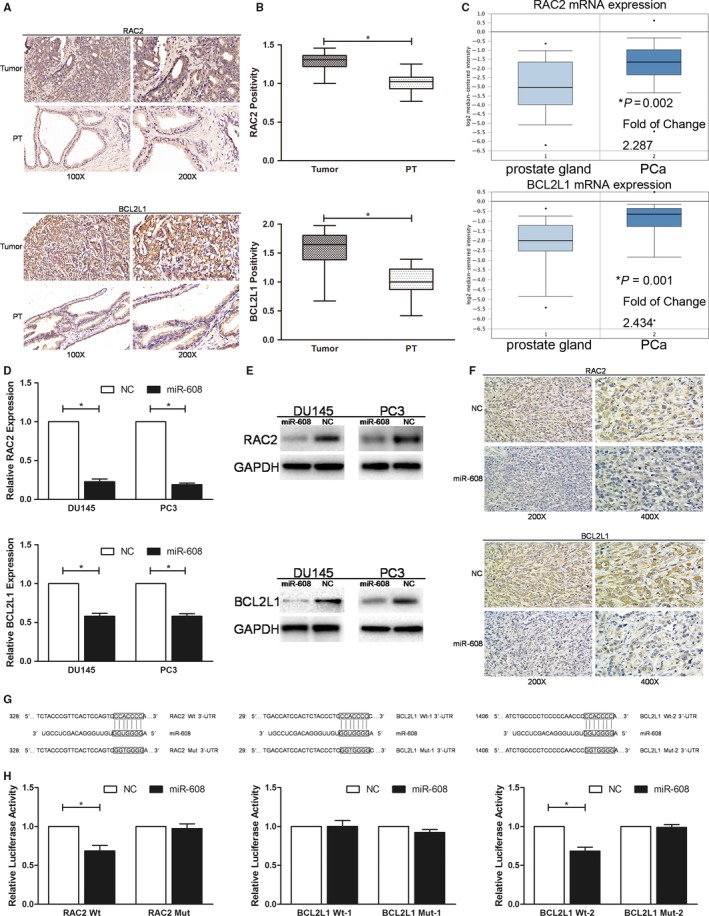
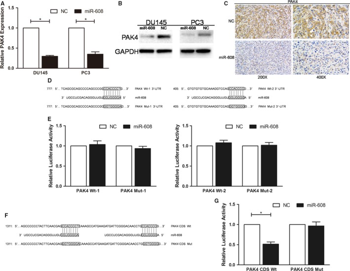
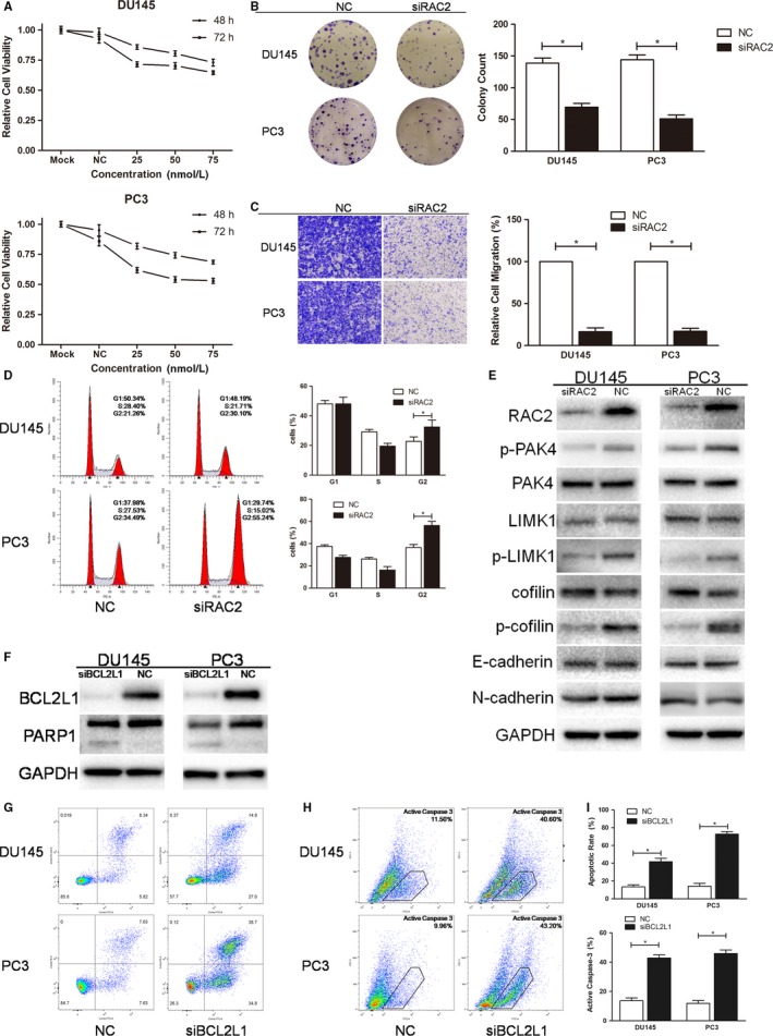
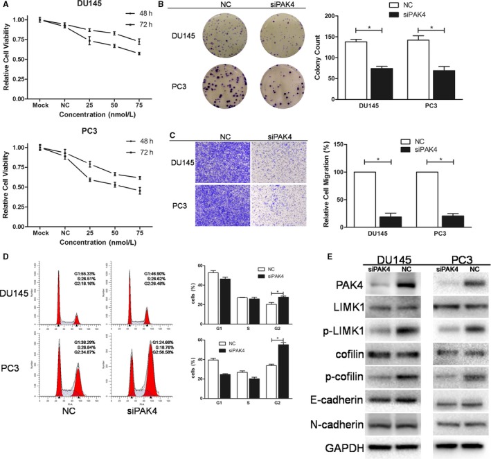
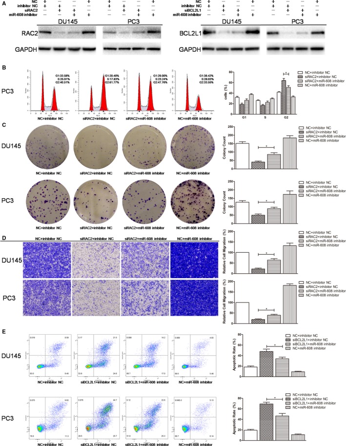
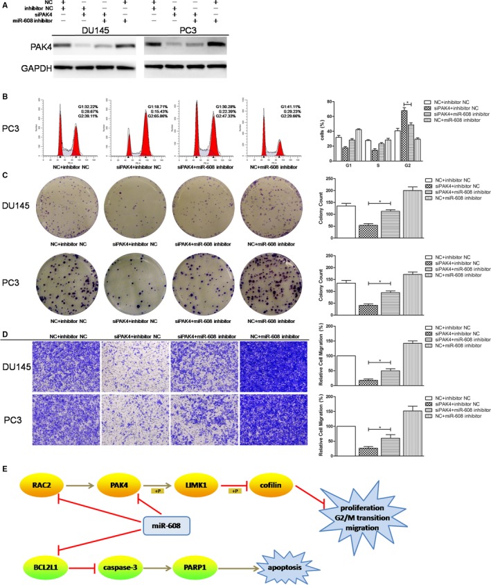
The aim of this study is to investigate the functions and mechanisms of miR-608 in prostate cancer (PCa). CISH and qRT-PCR analysis demonstrated that miR-608 was low expressed in PCa tissues and cells, which was partly attributed to the methylation of CpG island adjacent to the transcription start site (TSS) of miR-608 gene. Intracellular miR-608 overexpression inhibited in vivo PCa tumor growth, and suppressed PCa cell proliferation, G2/M transition, and migration in vitro, which was independent of EMT-associated mechanisms. Then RAC2, a GTPase previously deemed hematopoiesis-specific but now discovered to exist and play important roles in PCa, was verified by western blot and dual-luciferase reporter assays to mediate the effects of miR-608 through RAC2/PAK4/LIMK1/cofilin pathway. MiR-608 also promoted the apoptosis of PCa cells through BCL2L1/caspase-3 pathway by targeting the 3'-UTR of BCL2L1. Moreover, PAK4, the downstream effector of RAC2, was found to be targeted by miR-608 at the mRNA coding sequence (CDS) instead of the canonical 3'-UTR. Knocking down RAC2, PAK4, or BCL2L1 with siRNAs reproduced the antiproliferative, mitosis-obstructive, antimigratory and proapoptotic effects of miR-608 in PCa cells, which could be attenuated by downregulating miR-608. In conclusion, miR-608 suppresses PCa progression, and its activation provides a new therapeutic option for PCa.

Keywords: BCL2L1; G2/M arrest; RAC2; PAK4; microRNA-608; prostate cancer.


Conflict of interest statement

The authors declare no conflict of interest.

Figures

Figure 1
Figure 1
MiR‐608 is low expressed in PCa tissues and is partly correlated with CpG‐island methylation. A, Fold of change of miR‐608 expression in 10 peritumoral tissues (PT) compared to the corresponding PCa tissues. B, Representative results of miR‐608 CISH in PCa TMA. C, Statistical analysis of miR‐608 CISH in PCa TMA. D, The BSP results of the identified CpG island near miR‐608 TSS. The black and white dots represent the methylated and unmethylated CpGs respectively. E, MiR‐608 tailing qRT‐PCR in three prostate cell lines. U6 served as the internal control. F, The DNA sequence of the identified CpG island near miR‐608 TSS with 25 CpGs denoted in red. G, Statistical analysis of the methylation rate of the CpG island in both PCa cell lines. H, The expression of miR‐608 in PC3 cells after 5‐aza‐2′‐deoxycytidine treatment. Error bars represent SD from three independent experiments. *P < .05
Figure 2
Figure 2
MiR‐608 overexpression suppresses PCa cell proliferation both in vivo and in vitro. A, Representative results of cell viability assay. MiR‐608 mimic of different concentrations was transfected into PCa cells. Error bars represent SD from four replicates. B, Colony formation assay was performed to assess the influence of miR‐608 on PCa cell proliferation. Error bars represent SD from three independent experiments. *P < .05. C, The nude mice sacrificed for the in vivo tumorigenesis experiment. Arrows point to the subcutaneous xenograft tumors. D, The growth curves of the PC3 xenograft tumors of either miR‐608 or NC treated nude mice. Error bars represent SD of the volumes of five xenograft tumors. *P < .05. E, The harvested PC3 xenograft tumors. F, Statistical analysis of the volumes of the harvested xenograft tumors. Error bars represent SD of the volumes of five xenograft tumors. *P < .05
Figure 3
Figure 3
MiR‐608 overexpression induces G2/M arrest, inhibits the migration, and promotes the apoptosis of PCa cells. A, Flow cytometry cell cycle assay. The cell cycle distribution of miR‐608 mimic‐transfected PCa cells was analyzed. B, Transwell migration assay. Cells were observed at 200× magnification. C and D, Flow cytometry apoptosis and active caspase‐3 assay. Apoptotic cells produced by miR‐608 overexpression are in upper right and lower right quadrants. E, Statistical analyses of the apoptosis assay and the active caspase‐3 assay results. F, The expression of miR‐608‐associated proteins after miR‐608 overexpression in PCa cells. Error bars represent SD from three independent experiments. *P < .05
Figure 4
Figure 4
MiR‐608 directly targets RAC2 and BCL2L1 3′‐UTRs. A, Representative results of RAC2/BCL2L1 IHC in PCa TMA which contained 60 pairs of PCa tissues and peritumoral tissues (PT). B, Statistical analysis of RAC2/BCL2L1 IHC in PCa TMA. C, Oncomine analysis of RAC2/BCL2L1 mRNA expression. The results are from Wallace's microarray data sets, which include 69 cases of PCa and 20 cases of normal prostate tissues. D, qRT‐PCR and (E) western blot analysis were applied to determine the expression of RAC2/BCL2L1 after miR‐608 overexpression in PCa cells. F, Representative results of RAC2/BCL2L1 IHC in the sections of the xenograft tumors of either miR‐608 or NC treated nude mice. G, Segments of RAC2/BCL2L1 3′‐UTR containing either the wild‐type (RAC2 Wt/BCL2L1 Wt‐1/BCL2L1 Wt‐2) or the mutant‐type (RAC2 Mut/BCL2L1 Mut‐1/BCL2L1 Mut‐2) miR‐608 binding sites. H, Dual‐luciferase reporter assay. The relative luciferase activity is presented as the ratio of the activity of firefly luciferase to that of Renilla luciferase. Error bars represent SD from three independent experiments. *P < .05
Figure 5
Figure 5
MiR‐608 targets the PAK4 CDS. A, qRT‐PCR and (B) western blot analysis were applied to determine the expression of PAK4 after miR‐608 overexpression in PCa cells. C, Representative results of PAK4 IHC in the sections of the xenograft tumors of either miR‐608 or NC treated nude mice. D, Segments of PAK4 3′‐UTR containing either the wild‐type (PAK4 Wt‐1 and PAK4 Wt‐2) or the mutant‐type (PAK4 Mut‐1 and PAK4 Mut‐2) miR‐608 binding sites. E, Dual‐luciferase reporter assay. MiR‐608 mimic had no effects on the relative luciferase activities of PAK4 3′‐UTR Wt vector‐transfected HEK 293T cells. F, Segments of PAK4 CDS containing either the wild‐type (PAK4 CDS Wt) or the mutant‐type (PAK4 CDS Mut) miR‐608 binding sites. G, Dual‐luciferase reporter assay. The relative luciferase activity is presented as the ratio of the activity of firefly luciferase to that of Renilla luciferase. Error bars represent SD from three independent experiments. *P < .05
Figure 6
Figure 6
Knocking down RAC2/BCL2L1 with RAC2/BCL2L1 siRNA pool mimics the effects of miR‐608 in PCa cells. A, Representative results of cell viability assay. RAC2 siRNA of different concentrations was transfected into PCa cells. Error bars represent SD from four replicates. B, Colony formation assay was performed to assess the influence of RAC2 siRNA on PCa cell proliferation. C, Transwell migration assay. Cells were observed at 200× magnification. D, Flow cytometry cell cycle assay. The cell cycle distribution of RAC2 siRNA‐transfected PCa cells was analyzed. E and F, Western blot analysis. RAC2/BCL2L1 siRNA pool knocked down RAC2/BCL2L1 and the downstream proteins in PCa cells. G and H, Flow cytometry apoptosis and active caspase‐3 assay. Apoptotic cells produced by BCL2L1 knock down are in upper right and lower right quadrants. I, Statistical analyses of the apoptosis assay and the active caspase‐3 assay results. Error bars represent SD from three independent experiments. *P < .05
Figure 7
Figure 7
Knocking down PAK4 with PAK4 siRNA pool mimics the effects of miR‐608 in PCa cells. A, Representative results of cell viability assay. PAK4 siRNA of different concentrations was transfected into PCa cells. Error bars represent SD from four replicates. B, Colony formation assay was performed to assess the influence of PAK4 siRNA on PCa cell proliferation. C, Transwell migration assay. Cells were observed at 200× magnification. D, Flow cytometry cell cycle assay. The cell cycle distribution of PAK4 siRNA‐transfected PCa cells was analyzed. E, Western blot analysis. PAK4 siRNA pool knocked down PAK4 and the downstream proteins in PCa cells. Error bars represent SD from three independent experiments. *P < .05
Figure 8
Figure 8
RAC2/BCL2L1 rescue experiments. A, Western blot analysis. MiR‐608 inhibitor re‐elevated the expression of RAC2/BCL2L1 in RAC2/BCL2L1 knocked‐down PCa cells. B, Flow cytometry cell cycle assay. MiR‐608 inhibitor abolished the G2/M arrest caused by RAC2 knock down in PCa cells. C, Colony formation assay. MiR‐608 inhibitor partially recovered the proliferation of RAC2 knocked‐down PCa cells. D, Transwell migration assay. MiR‐608 inhibitor restimulated the migration of RAC2 knocked‐down PCa cells. E, Flow cytometry apoptosis assay. MiR‐608 inhibitor significantly reduced the apoptosis caused by BCL2L1 knock down in PCa cells. Error bars represent SD from three independent experiments. *P < .05
Figure 9
Figure 9
PAK4 rescue experiments. A, Western blot analysis. MiR‐608 inhibitor re‐elevated the expression of PAK4 in PAK4 knocked‐down PCa cells. B, Flow cytometry cell cycle assay. MiR‐608 inhibitor abolished the G2/M arrest caused by PAK4 knock down in PCa cells. C, Colony formation assay. MiR‐608 inhibitor partially recovered the proliferation of PAK4 knocked‐down PCa cells. D, Transwell migration assay. MiR‐608 inhibitor restimulated the migration of PAK4 knocked‐down PCa cells. E, Summary of the mechanisms of miR‐608 in PCa. Error bars represent SD from three independent experiments. *P < .05

Similar articles

Cited by

References

    1. Bray F, Ferlay J, Soerjomataram I, Siegel RL, Torre LA, Jemal A. Global cancer statistics 2018: GLOBOCAN estimates of incidence and mortality worldwide for 36 cancers in 185 countries. CA Cancer J Clin. 2018;68:394‐424. - PubMed
    1. Crunkhorn S. TRIAL WATCH Pioneering RNAi therapy shows antitumour activity in humans. Nat Rev Drug Discovery. 2013;12:178‐178. - PubMed
    1. Joo MK, Yhee JY, Kim SH, Kim K. The potential and advances in RNAi therapy: chemical and structural modifications of siRNA molecules and use of biocompatible nanocarriers. J Control Release. 2014;193:113‐121. - PubMed
    1. Gregory RI, Shiekhattar R. MicroRNA biogenesis and cancer. Cancer Res. 2005;65:3509‐3512. - PubMed
    1. Soifer HS, Rossi JJ, Saetrom P. MicroRNAs in disease and potential therapeutic applications. Mol Ther. 2007;15:2070‐2079. - PubMed

Publication types

MeSH terms