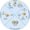
Brain Organoids as Tools for Modeling Human Neurodevelopmental Disorders
- PMID: 31389776
- PMCID: PMC6863377
- DOI: 10.1152/physiol.00005.2019
Brain Organoids as Tools for Modeling Human Neurodevelopmental Disorders
Abstract
Brain organoids recapitulate in vitro the specific stages of in vivo human brain development, thus offering an innovative tool by which to model human neurodevelopmental disease. We review here how brain organoids have been used to study neurodevelopmental disease and consider their potential for both technological advancement and therapeutic development.
Keywords: disease modeling; neurodevelopmental disease; organoids; stem cells.
Figures


References
-
- Abrahams BS, Geschwind DH. Advances in autism genetics: on the threshold of a new neurobiology. Nat Rev Genet 9: 341–355, 2008. doi: 10.1038/nrg2346 . A corrigendum for this article is available at http://dx.doi.org/10.1038/nrg2861. - DOI - PMC - PubMed
-
- Abud EM, Ramirez RN, Martinez ES, Healy LM, Nguyen CHH, Newman SA, Yeromin AV, Scarfone VM, Marsh SE, Fimbres C, Caraway CA, Fote GM, Madany AM, Agrawal A, Kayed R, Gylys KH, Cahalan MD, Cummings BJ, Antel JP, Mortazavi A, Carson MJ, Poon WW, Blurton-Jones M. iPSC-derived human microglia-like cells to study neurological diseases. Neuron 94: 278–293.e9, 2017. doi: 10.1016/j.neuron.2017.03.042. - DOI - PMC - PubMed
-
- Ariani F, Hayek G, Rondinella D, Artuso R, Mencarelli MA, Spanhol-Rosseto A, Pollazzon M, Buoni S, Spiga O, Ricciardi S, Meloni I, Longo I, Mari F, Broccoli V, Zappella M, Renieri A. FOXG1 is responsible for the congenital variant of Rett syndrome. Am J Hum Genet 83: 89–93, 2008. doi: 10.1016/j.ajhg.2008.05.015. - DOI - PMC - PubMed
Publication types
MeSH terms
Grants and funding
LinkOut - more resources
Full Text Sources
Other Literature Sources

