Review
. 2019 Oct 1;115(12):1732-1756.
doi: 10.1093/cvr/cvz203.

Non-coding RNAs in cardiovascular cell biology and atherosclerosis

Affiliations
Review

Non-coding RNAs in cardiovascular cell biology and atherosclerosis

Francesca Fasolo et al. Cardiovasc Res. .

Abstract

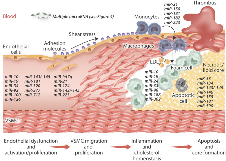
Atherosclerosis underlies the predominant number of cardiovascular diseases and remains a leading cause of morbidity and mortality worldwide. The development, progression and formation of clinically relevant atherosclerotic plaques involves the interaction of distinct and over-lapping mechanisms which dictate the roles and actions of multiple resident and recruited cell types including endothelial cells, vascular smooth muscle cells, and monocyte/macrophages. The discovery of non-coding RNAs (ncRNAs) including microRNAs, long non-coding RNAs, and circular RNAs, and their identification as key mechanistic regulators of mRNA and protein expression has piqued interest in their potential contribution to atherosclerosis. Accruing evidence has revealed ncRNAs regulate pivotal cellular and molecular processes during all stages of atherosclerosis including cell invasion, growth, and survival; cellular uptake and efflux of lipids, expression and release of pro- and anti-inflammatory intermediaries, and proteolytic balance. The expression profile of ncRNAs within atherosclerotic lesions and the circulation have been determined with the aim of identifying individual or clusters of ncRNAs which may be viable therapeutic targets alongside deployment as biomarkers of atherosclerotic plaque progression. Consequently, numerous in vivo studies have been convened to determine the effects of moderating the function or expression of select ncRNAs in well-characterized animal models of atherosclerosis. Together, clinicopathological findings and studies in animal models have elucidated the multifaceted and frequently divergent effects ncRNAs impose both directly and indirectly on the formation and progression of atherosclerosis. From these findings' potential novel therapeutic targets and strategies have been discovered which may pave the way for further translational studies and possibly taken forward for clinical application.

Keywords: Atherosclerosis; Endothelial cells; Macrophages; Non-coding RNA; Vascular smooth muscle cells; microRNA.


Figures

Figure 1
Figure 1
Overview of the cellular regulation of long non-coding RNAs (lncRNAs). LncRNA may act both within nuclear and cytoplasmic compartments. Within the nucleus, they contribute to shaping chromatin structure and accessibility via recruitment of chromatin modifiers (A); they can regulate transcription rate by modulating transcription factor availability at transcription start sites (B); they can control RNA splicing by directing the splicing machinery (C); they can also work as scaffolding structures through provision of components to aid the formation of specific subnuclear bodies (D); they regulate the shuttling of proteins between cellular compartments (E). In the cytoplasm, lncRNAs can regulate mRNA turnover by guiding the degradation machinery to specific transcripts (F); they can also dictate translational regulation through actions such as blocking ribosome binding to RNA (G), or as ‘molecular sinks’ to sequester different factors (microRNAs and proteins) from their site of action (H).
Figure 2
Figure 2
Overview of the cellular regulation of circular RNAs (circRNAs). CircRNAs may can exert actions within the nucleus, the cytoplasm, or as secreted molecules. Within the nucleus, they can contribute to transcriptional (A) and splicing (B) regulation. Within the cytoplasm, circRNAs can affect translational regulation through actions such as blocking ribosome binding to RNA or alternatively, they can be translated into small pepdtides (C); they can also serve as ‘molecular sinks’ to sequester different factors (microRNAs and proteins) from their site of action (D). CircRNAs can be secreted within exosomes and therefore participate in intercellular communication, while their presence within bodily fluids suggests they may be able to be potentially exploited as biomarkers.
Figure 3
Figure 3
Proposed roles of long non-coding and circular RNAs in atherosclerotic plaque development, progression and stability. The association of long non-coding RNAs (lncRNAs) and circular RNAs (circRNAs) are shown during the different stages of atherosclerotic plaque development. Atherogenesis is initially characterized by substantial alterations in the inner arterial surface: stress stimuli (nitric oxide, hypoxia, oxidative stress, shear stress…) trigger endothelial cell (EC) activation. The activated endothelium express numerous adhesion molecules (such as VCAM-1 and depicted as green circles) promoting the recruitment of monocytes (in green) from the blood stream and, at the same time, stimulating vascular smooth muscle cell (VSMC) migration and proliferation. VSMCs acquire a synthetic phenotype and contribute to the formation of the protective fibrous cap (in pink) by secreting differing extracellular matrix (ECM) proteins. However, perpetual monocyte recruitment, their differentiation into macrophages and their associated accrual of lipids (such as modified LDL) results in macrophage foam cell formation. During atherosclerotic plaque progression, foam cell macrophages undergo apoptosis and drive the formation of the necrotic/lipid-rich core (depicted in yellow), while VSMC apoptosis and dysregulated proteolysis drives thinning of the protective fibrous cap, both of which characterizes advanced unstable plaques.
Figure 4
Figure 4
MicroRNA expression in human atherosclerotic plaques and circulating blood. This diagram illustrates the dysregulated microRNAs identified through profiling approaches within atherosclerotic plaques, circulating plasma samples, and peripheral blood mononuclear cells (PBMCs). Coloured boxes indicate the patient cohorts from within which the dysregulated microRNAs were identified. MicroRNA depicted by yellow highlighting have been verified in two independent studies, while microRNA with red highlighting have been independently reported to be up- and down-regulated.
Figure 5
Figure 5
Proposed roles microRNAs in atherosclerotic plaque development, progression, and stability. The association of microRNAs are shown during the different stages of atherosclerotic plaque development. Atherogenesis is initially characterized by substantial alterations in the inner arterial surface: stress stimuli (nitric oxide, hypoxia, oxidative stress, shear stress…) trigger endothelial cell (EC) activation. The activated endothelium express numerous adhesion molecules (such as VCAM-1 and depicted as green circles) promoting the recruitment of monocytes (in green) from the blood stream and, at the same time, stimulating vascular smooth muscle cell (VSMC) migration and proliferation. VSMCs acquire a synthetic phenotype and contribute to the formation of the protective fibrous cap (in pink) by secreting differing extracellular matrix (ECM) proteins. However, perpetual monocyte recruitment, their differentiation into macrophages and their associated accrual of lipids (such as modified LDL) results in macrophage foam cell formation. During atherosclerotic plaque progression, foam cell macrophages undergo apoptosis and drive the formation of the necrotic/lipid-rich core (depicted in yellow), while VSMC apoptosis and dysregulated proteolysis drives thinning of the protective fibrous cap, both of which characterizes advanced unstable plaques.

References

    1. Skalen K, Gustafsson M, Rydberg EK, Hulten LM, Wiklund O, Innerarity TL, Boren J. Subendothelial retention of atherogenic lipoproteins in early atherosclerosis. Nature 2002;417:750–754. - PubMed
    1. Kolodgie FD, Burke AP, Nakazawa G, Virmani R. Is pathologic intimal thickening the key to understanding early plaque progression in human atherosclerotic disease? Arterioscler Thromb Vasc Biol 2007;27:986–989. - PubMed
    1. Leitinger N. Oxidized phospholipids as modulators of inflammation in atherosclerosis. Curr Opin Lipidol 2003;14:421–430. - PubMed
    1. Hansson GK. Inflammation, atherosclerosis, and coronary artery disease—reply. N Engl J Med 2005;353:429–430. - PubMed
    1. Allahverdian S, Chaabane C, Boukais K, Francis GA, Bochaton-Piallat ML. Smooth muscle cell fate and plasticity in atherosclerosis. Cardiovasc Res 2018;114:540–550. - PMC - PubMed

Publication types

MeSH terms