

Development and Application of Carbonyl Sulfide-Based Donors for H2S Delivery
- PMID: 31390174
- PMCID: PMC7047812
- DOI: 10.1021/acs.accounts.9b00315
Development and Application of Carbonyl Sulfide-Based Donors for H2S Delivery
Abstract
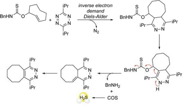
In addition to nitric oxide and carbon monoxide, hydrogen sulfide (H2S) has been recently recognized as an important biological signaling molecule with implications in a wide variety of processes, including vasodilation, cytoprotection, and neuromodulation. In parallel to the growing number of reports highlighting the biological impact of H2S, interest in developing H2S donors as both research tools and potential therapeutics has led to the growth of different H2S-releasing strategies. Many H2S investigations in model systems use direct inhalation of H2S gas or aqueous solutions of NaSH or Na2S; however, such systems do not mimic endogenous H2S production. This stark contrast drives the need to develop better sources of caged H2S. To address these limitations, different small organosulfur donor compounds have been prepared that release H2S in the presence of specific activators or triggers. Such compounds, however, often lack suitable control compounds, which limits the use of these compounds in probing the effects of H2S directly. To address these needs, our group has pioneered the development of carbonyl sulfide (COS) releasing compounds as a new class of H2S donor motifs. Inspired by a commonly used carbamate prodrug scaffold, our approach utilizes self-immolative thiocarbamates to access controlled release of COS, which is rapidly converted to H2S by the ubiquitous enzyme carbonic anhydrase (CA). In addition, this design enables access to key control compounds that release CO2/H2O rather than COS/H2S, which enables delineation of the effects of COS/H2S from the organic donor byproducts. In this Account, we highlight a library of first-generation COS/H2S donors based on self-immolative thiocarbamates developed in our lab and also highlight challenges related to H2S donor development. We showcase the release of COS in the presence of specific triggers and activators, including biological thiols and bio-orthogonal reactants for targeted applications. We also demonstrate the design and development of a series of H2O2/reactive oxygen species (ROS)-triggered donors and show that such compounds can be activated by endogenous levels of ROS production. Utilizing approaches in bio-orthogonal activation, we establish that donors functionalized with an o-nitrobenzyl photocage can enable access to light-activated donors. Similar to endogenous production by cysteine catabolism, we also prepared a cysteine-selective COS donor activated by a Strongin ligation mechanism. In efforts to help delineate potential differences in the chemical biology of COS and H2S, we also report a simple esterase-activated donor, which demonstrated fast COS-releasing kinetics and inhibition of mitochondrial respiration in BEAS-2B cells. Additional investigations revealed that COS release rates and cytotoxicity correlated directly within this series of compounds with different ester motifs. In more recent and applied applications of this H2S donation strategy, we also highlight the development of donors that generate either a colorimetric or fluorescent optical response upon COS release. Overall, the work described in this Account outlines the development and initial application of a new class of H2S donors, which we anticipate will help to advance our understanding of the rapidly emerging chemical biology of H2S and COS.
Figures


References
Publication types
MeSH terms
Substances
Grants and funding
LinkOut - more resources
Full Text Sources
Other Literature Sources
Miscellaneous
