. 2019 Aug 6;30(2):385-401.e9.
doi: 10.1016/j.cmet.2019.06.017.

Generation of Human Fatty Livers Using Custom-Engineered Induced Pluripotent Stem Cells with Modifiable SIRT1 Metabolism

Affiliations

Generation of Human Fatty Livers Using Custom-Engineered Induced Pluripotent Stem Cells with Modifiable SIRT1 Metabolism

Alexandra Collin de l'Hortet et al. Cell Metab. .

Abstract

The mechanisms by which steatosis of the liver progresses to non-alcoholic steatohepatitis and end-stage liver disease remain elusive. Metabolic derangements in hepatocytes controlled by SIRT1 play a role in the development of fatty liver in inbred animals. The ability to perform similar studies using human tissue has been limited by the genetic variability in man. We generated human induced pluripotent stem cells (iPSCs) with controllable expression of SIRT1. By differentiating edited iPSCs into hepatocytes and knocking down SIRT1, we found increased fatty acid biosynthesis that exacerbates fat accumulation. To model human fatty livers, we repopulated decellularized rat livers with human mesenchymal cells, fibroblasts, macrophages, and human SIRT1 knockdown iPSC-derived hepatocytes and found that the human iPSC-derived liver tissue developed macrosteatosis, acquired proinflammatory phenotype, and shared a similar lipid and metabolic profiling to human fatty livers. Biofabrication of genetically edited human liver tissue may become an important tool for investigating human liver biology and disease.

Keywords: NAFLD; NASH; SIRT1; cellular engineering; hepatic differentiation; human fatty liver; human iPSCs; liver metabolism.


Conflict of interest statement

COMPETING INTERESTS STATEMENT

A.S.-G., is inventor on a patent application that involves some of the perfusion technology used in this work (WO/2011/002926); K.H., J.G.-L., and A.S.-G. have an international patent related to this work that describes methods of preparing artificial organs and related compositions for transplantation and regeneration (WO/2015/168254). A.C.-H, K.T., J.G.-L., K.H.; Y.W., B.P., A.S.-G. has a provisional international patent application that describes hepatic differentiation of human pluripotent stem cells and liver repopulation (PCT/US2018/018032). A.C.-H, K.T., J.G.-L., K.H.; Y.W., B.P., I.J.F., A.S.-G. has a provisional international patent application that describes the use of human induced pluripotent stem cells for highly genetic engineering (PCT/US2017/044719). A.C.-H, K.T., J.G.-L., Y.W., I.J.F., and A.S.-G., are co-founders and have a financial interest in Von Baer Wolff, Inc. a company focused on biofabrication of autologous human hepatocytes from stem cells technology and programming liver failure and their interests are managed by the Conflict of Interest Office at the University of Pittsburgh in accordance with their policies.

Figures

Figure 1.
Figure 1.. Generation of custom engineered human induced pluripotent stem cells.
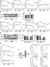
(A) Immunohistochemical staining micrographs of SIRT1 show cytoplasmic and nuclear decrease of SIRT1 expression in NASH human livers (n=4) compared to Normal human livers (n=3). Western blot analysis and quantification of SIRT1 normalized to β-Actin in Normal (n=3) and NASH (n=3) human livers (P=0.400, Mann-Whitney test). Quantitative gene expression analysis of SIRT1 expression normalized to actb in Normal (n=4) and NASH (n=4) human livers (P=0.200, Mann-Whitney test). (B) Quantitative gene expression analysis of puromycin selection cassette gene normalized to actb on human fibroblasts transduced with lentiviral vector for –iRFP (hFib-iRFP) or –iKD-SIRT1 (hFibiKD-SIRT1) and non transduced human fibroblasts (hFib) as control (*P=0.0338, *P=0.0130, Kruskal-Wallis test and Dunnett’s multiple comparisons). Quantitative gene expression analysis SIRT1 normalized to actb in hFib-iRFP and hFib-iKD-SIRT1 in the presence or absence of doxycycline (*P=0.0422, Kruskal-Wallis test and Dunnett’s multiple comparisons). Western blot analysis and quantification of SIRT1 normalized to GAPDH on hFib-iRFP (n=4) and hFib-iKD-SIRT1 (n=4) with and without doxycycline treatment (*P=0.0395, Kruskal-Wallis test and Dunnett’s multiple comparisons). (C) Immunofluorescence micrographs of SIRT1 in hFF-iKD-SIRT1 with and without doxycycline treatment, human fetal hepatocytes and human adult hepatocytes were used as controls. Light Red fluorescence and bright light micrographs were used to analyze hFib-iRFP with and without doxycycline treatment. hFib-iRFP (n=4) hFib-iKD-SIRT1 (n=4), human Fetal hepatocytes (n=4), Adult primary hepatocytes untreated (n=3). (D) Light Red fluorescence micrographs of hiPS-iRFP colony after doxycycline exposure for 48h. DAPI was used as counterstaining. Quantitative gene expression analysis of SIRT1 expression normalized to actb show knockdown of SIRT1 in hiPS-iKD-SIRT1-#17 but not in hiPS-IRFP-#3 after exposure to docycycline (*P = 0.0360, *P=0.0140, Kruskal-Wallis test and Dunnett’s multiple comparisons). Western blot analysis and quantification of SIRT1 normalized to GAPDH in hiPS-IRFP-#3 (n=4) and hiPS-iKDSIRT1-# 17 (n=4) with and without doxycycline exposure for 48h (*P=0.0222, Kruskal-Wallis test and Dunnett’s multiple comparisons). (E) Immunofluorescence micrographs of pluripotency markers Nanog, Oct4, TRA-1–60 and SSEA-4 in hiPS-IRFP-#3 and hiPS-iKD-SIRT1-#17. Quantitative gene expression analysis of pluripotency markers c-myc, Lin28 and Oct3/4 normalized to actb shows that hiPS-IRFP and hiPS-iKD-SIRT1 express bona fide pluripotency markers comprable to human Embryonic Stem (hES) cells. hiPS-IRFP#3 with (n=4) and without (n=4) doxycycline, hiPS-iKDSIRT1# 17 with (n=4), and without DOX (n=4), human embryonic stem cells (n=3) were included as controls. hiPS-IRFP-#3 and hiPS-iKD-SIRT1-#17 both carry a normal female karyotype by G-banding analysis.
Figure 2.
Figure 2.. Characterization of hepatocyte-directed differentiation of custom engineered human iPS cells.
(A) Schematic illustration of hiPS differentiation into hepatocytes highlighting the three main stages of differentiation by sequential addition of defined medium protocols containing Activin-A, BMP-4 and FGF2 (Stage 1), Activin-A (Stage 2), and Dimethyl Sulfoxide (DMSO) and Hepatocyte Growth Factor (HGF) (Stage 3). Immunofluorescence micrographs of SOX17 show efficient definitive endoderm (DE) induction with more than 80% SOX17 expressing cells (in both hiPS-iRFP and hiPSiKD-SIRT1 after definitive endoderm induction-stage 2. (B) Immunofluorescence micrographs of hiPSiRFP- and hiPS-iKD-SIRT1 after hepatocyte differentiation-stage 3 show protein expression of human albumin, and adult isoform of HNF4α comparable to primary adult hepatocytes (>70% and >85% respectively) and no expression of α-fetoprotein (AFP), freshly isolated human fetal and adult hepatocytes were used as controls. (C) Quantitative gene expression analysis of definitive endoderm (DE) and hepatocyte differentiated cells (iHeps) from hiPS-iRFP and hiPS-iKD-SIRT1. Quantitative qPCR is shown for genes encoding hepatocyte nuclear factor-4α (HNF4α, iHep-TagRFP vs. iHep-iKD-SIRT1 p>0.9999; iHep-TagRFP vs. Adult Heps p>0.9999; iHep-iKD-SIRT1 vs. Adult Heps >0.9999); Forkhead Box A2 (FOXA2, iHep-TagRFP vs. iHep-iKD-SIRT1 p>0.9999; iHep-TagRFP vs. Adult Heps p=0.8783; iHep-iKD-SIRT1 vs. Adult Heps >0.9999); Forkhead Box A1 (FOXA1, iHep-TagRFP vs. iHep-iKD-SIRT1 p>0.9999; iHep-TagRFP vs. Adult Heps p>0.9999; iHep-iKD-SIRT1 vs. Adult Heps >0.9999); hepatocyte nuclear factor-1 (HNF1, iHep-TagRFP vs. iHep-iKD-SIRT1 p>0.9999; iHep-TagRFP vs. Adult Heps p>0.9999; iHep-iKD-SIRT1 vs. Adult Heps >0.9999), CCAAT enhancer binding protein alpha (Cebpα, iHep-TagRFP vs. iHep-iKD-SIRT1 p>0.9999; iHep-TagRFP vs. Adult Heps p>0.9999; iHep-iKD-SIRT1 vs. Adult Heps >0.9999); α-fetoprotein (AFP, iHep-TagRFP vs. iHep-iKD-SIRT1 p>0.9999; iHep-TagRFP vs. Adult Heps p>0.9999; iHep-iKD-SIRT1 vs. Adult Heps >0.9999); Peroxisome proliferator-activated receptor alpha (PPARα, iHep-TagRFP vs. iHep-iKD-SIRT1 p>0.9999; iHep-TagRFP vs. Adult Heps p>0.9999; iHep-iKD-SIRT1 vs. Adult Heps >0.9999); liver × receptor (LXR, iHep-TagRFP vs. iHep-iKD-SIRT1 p>0.9999; iHep-TagRFP vs. Adult Heps p>0.9999; iHep-iKD-SIRT1 vs. Adult Heps >0.9999); retinoid × receptor (RXR, iHep-TagRFP vs. iHep-iKD-SIRT1 p>0.9999; iHep-TagRFP vs. Adult Heps p>0.9999; iHep-iKD-SIRT1 vs. Adult Heps p=0.6357); Sterol regulatory elementbinding protein 1 (Srebp1c, iHep-TagRFP vs. iHep-iKD-SIRT1 p>0.9999; iHep-TagRFP vs. Adult Heps p>0.9999; iHep-iKD-SIRT1 vs. Adult Heps >0.9999); Epidermal Growth Factor Receptor (EGFR, iHep-TagRFP vs. iHep-iKD-SIRT1 p>0.9999; iHep-TagRFP vs. Adult Heps p>0.9999; iHe-piKD-SIRT1 vs. Adult Heps >0.9999); ATP Binding Cassette Subfamily B Member 1 (ABCB1, iHep-TagRFP vs. iHep-iKD-SIRT1 p>0.9999; iHep-TagRFP vs. Adult Heps p>0.9999; iHep-iKD-SIRT1 vs. Adult Heps >0.9999); Stearoyl-CoA Desaturase (SCD, iHep-TagRFP vs. iHep-iKD-SIRT1 p>0.9999; iHep-TagRFP vs. Adult Heps p>0.9999; iHep-iKD-SIRT1 vs. Adult Heps >0.9999); Elongation of very long chain fatty acids protein 6 (ELOVL6, iHep-TagRFP vs. iHep-iKD-SIRT1 p>0.9999; iHep-TagRFP vs. Adult Heps p>0.9999; iHep-iKD-SIRT1 vs. Adult Heps >0.9999); Fatty acid synthase (FASN, iHep-TagRFP vs. iHep-iKD-SIRT1 p>0.9999; iHep-TagRFP vs. Adult Heps p>0.9999; iHep-iKD-SIRT1 vs. Adult Heps >0.9999); Acetyl-CoA carboxylase (ACC, iHep-TagRFP vs. iHep-iKD-SIRT1 p>0.9999; iHep-TagRFP vs. Adult Heps p=0.7625; iHep-iKD-SIRT1 vs. Adult Heps >0.9999). DE-iRFP (n=3) DE-iKD-SIRT1 (n=3) iHeps-iRFP (n=4) iHep-iKD-SIRT1 (n=4), human adult hepatocytes (HAH) (n=3) were included as controls (Kruskal-Wallis test and Dunnett’s multiple comparisons was used to compared iHeps groups and Adult Heps groups).
Figure 3.
Figure 3.. SIRT1 knockdown in custom engineered human iHeps induces deregulation of lipid homeostasis.
(A) Quantitative gene expression analysis for SIRT1 in hiPS-iRFP and hiPS-iKD-SIRT1 at different stages of hepatic differentiation after exposure to doxycycline. Gene expression was normalized to actb. Definitive endoderm (stage 2)(From left to right: *P=0.0360, *P=0.0140, Kruskal-Wallis test and Dunnett’s multiple comparisons), Hepatocyte induction (stage 3) (*P=0.0504, **P = 0.029, Kruskal-Wallis test and Dunnett’s multiple comparisons) (iRFP-DOX vs iKD-SIRT1-DOX P>0.999). Freshly isolated human adult hepatocytes were used as control. (B) Western blot analysis of SIRT1 in iHeps-iRFP and hiPS-iKD-SIRT1 with and without doxycycline exposure. For normalization GAPDH was used. Human adult hepatocytes were used as control. (C) Light red fluorescence micrographs demonstrate inducible red fluorescent protein (iRFP) in hiPS-iRFP after doxycycline exposure at different stages of hepatic differentiation. (D) Intracellular triglycerides content (μg/106 cells) shows a 2.3 fold increase in iHeps-iKD-SIRT1 when exposed to doxycycline and free fatty acids whereas iHeps-iRFP in similar conditions, iHep-iKD-SIRT1 +FFA and adult human hepatocytes (HAH) +FFA/DOX show respectively 1.60, 1.3 and 1.4 fold increase. (From left to right: *P=0.0493, *P=0.0297, Kruskal-Wallis test and Dunnett’s multiple comparisons). (E) Nile Red staining micrographs and quantification of lipid accumulation demonstrating increased lipid content in iHeps with SIRT1 knockdown when challenged with free fatty acids (FFA) compared to controls iHeps-iRFP in similar conditions and iHep-iKD-SIRT1 (From left to right: *P= 0.025, **P=0.0098, *P= 0.0303, Kruskal-Wallis test and Dunnett’s multiple comparisons). (F) Lipid peroxidation with (malondialdehyde) quantification and 8-isoprostane ELISA assay in iHeps with doxycycline and free fatty acids (FFA) (From left to right: MDA: *P= 0.0409, *P=0.0202, **P= 0.0044, *P=0.489, 8-isoprostane: *P=0.028, *P= 0.042, Kruskal-Wallis test and Dunnett’s multiple comparisons). (G-H) Total cholesterol, Low-density lipoproteins (LDL), very low-density lipoproteins (VLDL) concentration and high-density lipoproteins (HDL) (ng/106 cells) in iHeps-IRFP, iHeps-IKD-SIRT1 and human adult hepatocytes (HAH) with and without doxycycline and free fatty acids (FFA) exposure. (From left to right: Total cholesterol, *P=0.0389, *P= 0.0455, *P=0.0389, Kruskal-Wallis test and Dunnett’s multiple comparisons; LDL/VLDL & HDL, P>0.99, Kruskal-Wallis test and Dunnett’s multiple comparisons). iHeps-iRFP and iHep-iKD-SIRT1 untreated (n=4 each), iHeps-iRFP and iHep-iKD-SIRT1 treated with FFA (n=3 each), iHeps-iRFP and iHep-iKD-SIRT1 treated with FFA/DOX (n=4 each) and human adult hepatocytes (n=3 for each condition) were included as controls.
Figure 4.
Figure 4.. SIRT1 knockdown in custom engineered human iHeps induces downregulation of lipolysis and increase of de novo lipogenesis.
(A) Quantitative gene expression analysis of de novo lipogenesis related genes; sterol regulatory element-binding protein 1 (Srebp1c) (From left to right: *P=0.04909, **P=0.0062, *P=0.0166, Kruskal-Wallis test and Dunnett’s multiple comparisons), elongation of very long chain fatty acids protein 6 (ELOVL6) (From left to right: *P=0.0433, *P=0.0221 *P=0.0151, Kruskal-Wallis test and Dunnett’s multiple comparisons), Fatty acid synthase (FASN) (P=0.3686, P>0.99, P>0.99 not statistically different, Kruskal-Wallis test and Dunnett’s multiple comparisons), Stearoyl-CoA Desaturase (SCD) (P>0.999 not statistically different, Kruskal-Wallis test and Dunnett’s multiple comparisons) in iHeps-iKD-SIRT1 and controls iHeps-iRFP and human adult hepatocytes (HAH) in the presence or absence of doxycycline and free fatty acids (FFA). actb expression was used for normalization. (B) Western blot analysis and quantification of Srebp1c normalized to GAPDH in iHeps-iRFP and hiPS-iKD-SIRT1 in the presence or absence of doxycycline to induce SIRT1-shRNA (SIRT1 knockdown) or red fluorescent protein (RFP). Human adult hepatocytes (HAH) were used as control (From left to right: *P=0.0150, *P=0.0463, Kruskal-Wallis test and Dunnett’s multiple comparisons). (C) Quantitative gene expression analysis of genes encoding peroxisome proliferator-activated receptor alpha (PPARα) (From left to right: **P=0.0013, *P=0.0350; **P=0.0048, Kruskal-Wallis test and Dunnett’s multiple comparisons), peroxisome proliferator-activated receptor gamma coactivator 1-alpha (PGC1a) (From left to right: **P=0.0013, *P=0.0409, *P=0.0293, Kruskal-Wallis test and Dunnett’s multiple comparisons), Acyl-CoA Dehydrogenase Medium Chain (ACADM) (From left to right: **P=0.0048, *P=0.0480, *P=0.0123, Kruskal-Wallis test and Dunnett’s multiple comparisons) and cluster of differentiation 36 (CD36) (From left to right: *P=0.0373, *P=0.0293, *P=0.0222, Kruskal-Wallis test and Dunnett’s multiple comparisons) in iHeps-iKD-SIRT1 and controls iHeps-iRFP and human adult hepatocytes (HAH) in the presence or absence of doxycycline and free fatty acids (FFA). actb expression was used for normalization. (D) Western blot analysis and quantification of PGC1a and PPARα normalized to GAPDH in iHeps-iRFP and hiPS-iKDSIRT1 in the presence or absence of doxycycline. Human adult hepatocytes (HAH) were used as control (From left to right: PGC1a, *P=0.0436, *P=0.0411; PPARα, *P=0.0257, *P=0.0376 Kruskal-Wallis test and Dunnett’s multiple comparisons). (E) Quantitative gene expression analysis of genes encoding ABC lipid transporters 1 and 11 (From left to right: ABCB1, *P=0.0286, *P=0.0221, *P=0.0222; ABCB11, *P=0.0303, **P=0.0055, *P=0.0283, Kruskal-Wallis test and Dunnett’s multiple comparisons) in iHeps-iKD-SIRT1 and controls iHeps-iRFP and human adult hepatocytes (HAH) in the presence or absence of doxycycline and free fatty acids (FFA). actb expression was used for normalization. (F) Mapping of carbon atom transitions using uniformly labeled 13-C6-glucose. Total metabolite levels show a significant increase in palmitate and stearate pools of iHep-iKD-SIRT1 +DOX cells. Isotopomer spectral analysis shows a significant upregulation in absolute lipid synthesis rate increases with flux of de novo palmitate synthesis and for de novo stereate synthesis (From left to right: *P=0.0456, *P=0.0332, *P=0.0308, *P=0.0457). iHeps-iRFP and iHep-iKD-SIRT1 treated with FFA (n=3 each), iHeps-iRFP and iHep-iKD-SIRT1 treated with FFA/DOX (n=4 each for A-E; n=3 each for F) and human adult hepatocytes (n=3 for each condition) were included as controls.
Figure 5.
Figure 5.. Bioengineering of human iPS-derived fatty liver tissue with modifiable SIRT1 metabolism.
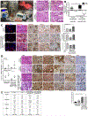
(A) Photograph of the organ perfusion and culture organ system constituted by the organ culture chamber, perfusion pump, cell infusion pump and bubble trap. Photograph of recellularized liver matrix with iHeps, human microvascular endothelial cells, mesenchymal cells and fibroblast. Also shown are haematoxylin and eosin staining of engineered human liver tissue–iRFP and –iKD-SIRT1 in the presence of doxycycline. Human normal and fatty livers were used as controls. Asterisks (*) indicate large vacuoles of triglyceride fat with compression and displacement of the nuclei to the periphery of affected hepatocytes consistent with macrovesicular steatosis. (B) Quantitative gene expression analysis of pro-inflammatory marker CD80 and anti-inflammatory marker CD163 (*P=0.0286, Mann-Whitney test) (n=4) in co-cultured iHeps-iKD-SIRT1 or iHeps-iRFP with human primary macrophages in the presence or absence of doxycycline and free fatty acids (FFA). (C) Histological analysis of engineered human fatty liver tissue –iKD-SIRT1 (doxycycline and free fatty acids treated) in the presence or absence of human primary macrophages (n=5). Oil Red O staining show macrovesicular steatosis. Also shown are immunohistochemistry analysis of CD68, NFκB p65, MCP-1 and IL-6 showing increased parenchymal inflammation with addition of human macrophages and the absence of SIRT1 as demonstrated by immunohistochemistry quantification (*P= 0.0116, **P=0.0059, Kruskal-Wallis test and Dunnett’s multiple comparisons). Human fatty livers (n=3) were included as controls. (D) Quantitative gene expression analysis of FGF21 and Selenoprotein-P (From left to right: FGF21, *P=0.0178; Selenoprotein-P, *P=0.0318, *P=0.0213, Kruskal-Wallis test and Dunnett’s multiple comparisons). Hematoxylin and eosin (H&E) staining of engineered human fatty liver tissue –iKD-SIRT1 (free fatty acids treated) in the presence (n=7) or absence of Doxycycline (n=6) in comparison to human normal livers and human NASH livers (n=5). Immunohistochemistry analysis and quantification of FGF21 and Selenoprotein-P in human fatty liver tissues –iKD-SIRT1 −/+ doxycycline compared to human normal and NASH livers. Immunohistochemistry analysis of zonation markers Glutamine synthetase and E-cadherin in human fatty liver tissue –iKD-SIRT1 −/+ doxycycline compared to human normal and NASH liver. Immunohistochemistry analysis and quantification of Ki-67 exhibited a significant higher percentage of positive hepatocytes in human fatty liver tissue –iKD-SIRT1 −/+ doxycycline and human NASH liver compared to human normal liver. (From left to right: *P= 0.0410, *P=0.0247, Kruskal-Wallis test and Dunnett’s multiple comparisons). (E) Human NASH livers and human iPS-derived fatty liver tissues-iKD-SIRT1 with and without doxycycline were scored by Brunt scoring for histologic nonalcoholic fatty liver disease.
Figure 6.
Figure 6.. Transcription profiling analysis of human iPS-derived fatty liver tissue-iKD-SIRT1.
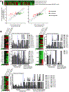
(A) Heat map analysis of fold expression data of genes related to transcription and metabolism of fatty acids in human NASH livers (n=3) and human iPS-derived fatty liver tissue-iKD-SIRT1 (n=6) compared to human normal livers (n=3). Also shown are scatter plots of genes differentially expressed in human iPS-derived fatty liver tissue-iKD-SIRT1 compared to human NASH livers or human normal livers. (B) Heat maps and RT-qPCR-based analysis of the expression of the indicated genes related to De Novo Lipogenesis (Fatty Acid Biosynthesis Regulation genes, Acyl-CoA Synthetases), β-oxidation (Acyl-CoA Dehydrogenases, Acyl-CoA Oxidases) and Fatty Acid Transport targets in human iPS-derived fatty liver tissue-iKD-SIRT1, human normal liver and human NASH liver.
Figure 7.
Figure 7.. Lipidomics and metabolomics profile of human iPS-derived fatty liver tissue-iKDSIRT1.
(A) Unsupervised clustering of untargeted lipidomics data from human normal liver (n=3), human NASH liver (n=3) and human iPS-derived fatty liver tissue-iKD-SIRT1 (n=6). (B) Bar charts representing fold changes of lipids peak area in human iPS-derived fatty liver tissue-iKD-SIRT1 compared to human normal livers or human NASH livers. (C) Venn diagram showing 255 upregulated lipids in both human NASH liver and human iPS-derived fatty liver tissue-iKD-SIRT1 compared to human Normal liver. Lipid levels were considered to be upregulated when fold-change >1.5 and p<0.01. Venn diagram showing 27 downregulated lipids in both human NASH liver and human iPS-derived fatty liver tissue-iKD-SIRT1 compared to human normal liver. Lipid levels were considered to be downregulated when fold-change is <1.5 and p<0.01. (D) Relative levels of lipids from glycerophospholipid, sphingolipid and glycerolipid classes of lipids in human normal liver, human NASH liver and human iPS-derived fatty liver tissue-iKD-SIRT1. (E) Scatter plots of different triglycerides levels in human iPS-derived fatty liver tissue-iKD-SIRT1 compared to human NASH livers or human normal livers (F) Metabolite set enrichment analysis of (left) Human iPS-derived fatty liver tissue-iKDSIRT1 (n=6 each) vs. Human normal livers (n=3) and (right) human NASH livers (n=3) vs. Human normal livers (n=3). 41 out of 50 (highlighted) pathways enriched in NASH livers were enriched in iPS-derived fatty liver tissue-iKD-SIRT1 (highlighted in yellow).

References

    1. Abul-Husn NS, Cheng X, Li AH, Xin Y, Schurmann C, Stevis P, Liu Y, Kozlitina J, Stender S, Wood GC, et al. (2018). A Protein-Truncating HSD17B13 Variant and Protection from Chronic Liver Disease. N Engl J Med 378, 1096–1106. - PMC - PubMed
    1. Achreja A, Zhao H, Yang L, Yun TH, Marini J, and Nagrath D (2017). Exo-MFA - A 13C metabolic flux analysis framework to dissect tumor microenvironment-secreted exosome contributions towards cancer cell metabolism. Metab Eng 43, 156–172. - PMC - PubMed
    1. Anjani K, Lhomme M, Sokolovska N, Poitou C, Aron-Wisnewsky J, Bouillot JL, Lesnik P, Bedossa P, Kontush A, Clement K, et al. (2015). Circulating phospholipid profiling identifies portal contribution to NASH signature in obesity. J Hepatol 62, 905–912. - PubMed
    1. Anstee QM, and Day CP (2013). The genetics of NAFLD. Nat Rev Gastroenterol Hepatol 10, 645–655. - PubMed
    1. Aoyama T, Peters JM, Iritani N, Nakajima T, Furihata K, Hashimoto T, and Gonzalez FJ (1998). Altered constitutive expression of fatty acid-metabolizing enzymes in mice lacking the peroxisome proliferator-activated receptor alpha (PPARalpha). J Biol Chem 273, 5678–5684. - PubMed

Publication types

LinkOut - more resources