. 2019 Dec 1;200(11):1392-1401.
doi: 10.1164/rccm.201903-0615OC.

Chronic E-Cigarette Use Increases Neutrophil Elastase and Matrix Metalloprotease Levels in the Lung

Affiliations

Chronic E-Cigarette Use Increases Neutrophil Elastase and Matrix Metalloprotease Levels in the Lung

Arunava Ghosh et al. Am J Respir Crit Care Med. .

Abstract

Rationale: Proteolysis is a key aspect of the lung's innate immune system. Proteases, including neutrophil elastase and MMPs (matrix metalloproteases), modulate cell signaling, inflammation, tissue remodeling, and leukocyte recruitment via cleavage of their target proteins. Excessive proteolysis occurs with chronic tobacco use and is causative for bronchiectasis and emphysema. The effect of e-cigarettes (vaping) on proteolysis is unknown.

Objectives: We used protease levels as biomarkers of harm to determine the impact of vaping on the lung.

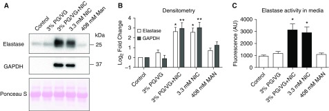
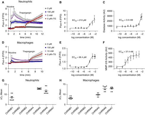
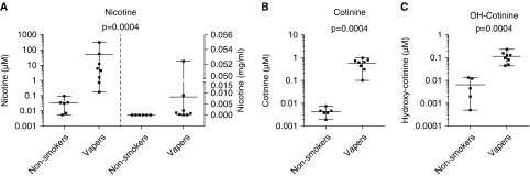
Methods: We performed research bronchoscopies on healthy nonsmokers, cigarette smokers, and e-cigarette users (vapers), and determined protease levels in BAL. In parallel, we studied the effects of e-cigarette components on protease secretion in isolated human blood neutrophils and BAL-derived macrophages. We also analyzed the nicotine concentration in induced sputum and BAL.

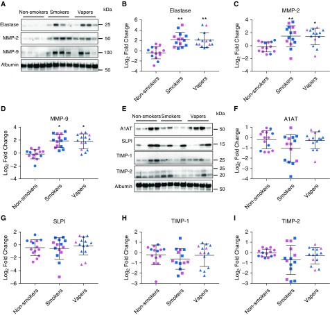
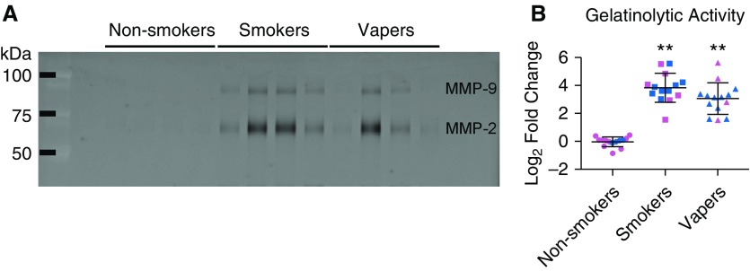
Measurements and Main Results: Neutrophil elastase, MMP-2, and MMP-9 activities and protein levels were equally elevated in both vapers' and smokers' BAL relative to nonsmokers. In contrast, antiprotease levels were unchanged. We also found that exposure of isolated neutrophils and macrophages to nicotine elicited dose-dependent increases in protease release. After vaping, measurable levels of nicotine were detectable in sputum and BAL, which corresponded to the half-maximal effective concentration values for protease release seen in immune cells.

Conclusions: We conclude that vaping induces nicotine-dependent protease release from resident pulmonary immune cells. Thus, chronic vaping disrupts the protease-antiprotease balance by increasing proteolysis in lung, which may place vapers at risk of developing chronic lung disease. These data indicate that vaping may not be safer than tobacco smoking.

Keywords: BAL; nicotine; protease; sputum; vaping.


Figures

Figure 1.
Figure 1.
Protease protein levels are equally increased in vapers’ and smokers’ BAL. (A) Representative Western blots showing increased protease protein in smokers’ and vapers’ BAL, along with albumin as loading control. Full blots are provided in Figure E1. (BD) Densitometric analysis of total protease levels normalized to albumin for neutrophil elastase (B), MMP-2 (matrix metalloprotease-2) (C), and MMP-9 (D). (E) Representative Western blots of BAL protease inhibitors. (FI) Densitometric analysis of BAL normalized to albumin control for A1AT (alpha-1 antitrypsin) (F), SLPI (G), TIMP1 (tissue inhibitor of metalloproteinases-1) (H), and TIMP2 (I). Values are log2 fold change in smokers and vapers compared with age-matched nonsmokers; *P ≤ 0.05 and **P ≤ 0.001. Pink, female subjects; blue, male subjects. Data are shown as mean ± SD. All n = 14 subjects per group. Data were analyzed using ANOVA followed by Tukey’s test. SLPI = secretory leukocyte protease inhibitor.
Figure 2.
Figure 2.
Total gelatinolytic activities are increased in vapers’ BAL. Gelatinolytic activity was measured in BAL samples by zymography. (A) Inverted image of representative zymogram showing increased MMP (matrix metalloprotease) activity in smokers’ and vapers’ BAL samples. (B) Quantification of gelatinolytic activity by MMP, shown as log2 fold change, compared with age-matched nonsmokers. Individual activities of MMP-2 and MMP-9 are shown in Figure E4. **P ≤ 0.001. Pink, female subjects; blue, male subjects. Data are shown as mean ± SD. All n = 14 subjects per group. Data were analyzed using ANOVA followed by Tukey’s test.
Figure 3.
Figure 3.
Nicotine, but not propylene glycol and vegetable glycerin (PG/VG), induces neutrophil elastase (NE) release from neutrophils. Neutrophils were isolated from peripheral blood and exposed to combinations of PG/VG, nicotine or mannitol (osmotic control) for 4 hours. (A) Representative Western blots of neutrophil secretions after incubation with the indicated e-liquid components, with Ponceau S staining as a loading control. (B) Densitometric analysis of NE and GAPDH normalized to Ponceau S staining shown as fold change compared with control. (C) NE activity in concentrated media, as measured using the fluorogenic substrate assay. *P ≤ 0.05 and **P ≤ 0.001. Data are shown as mean ± SEM. All n = 12 per group. Data were analyzed using ANOVA followed by Tukey’s test. AU = arbitrary units; MAN = mannitol; NIC = nicotine.
Figure 4.
Figure 4.
Nicotine causes dose-dependent increases in cytoplasmic Ca2+ and induces protease release from immune cells. Neutrophils and alveolar macrophages were loaded with the calcium indicator dye Fluo-4 direct and the change in fluorescence, as an indicator of cytoplasmic Ca2+, was recorded over time. The media was then sampled 4 hours after exposure to measure protease activity, as indicated by changes in cleavage of fluorogenic substrates specific to neutrophil elastase (NE) and MMP-2/9 (matrix metalloprotease-2/9). (A) Changes in Fluo4 fluorescence, as an indicator of cytoplasmic Ca2+ levels over time in peripheral blood neutrophils. (B and C) Dose responses to nicotine exposure in neutrophils for peak (2 min) increases in cytoplasmic Ca2+ levels and 3 hours secreted NE activity, respectively; n = 9 per group. Data were normalized to GAPDH expression; n = 8 per group. (D) Changes in Fluo-4 fluorescence, as an indicator of cytoplasmic Ca2+ levels, in BAL macrophages. (E and F) Dose responses to nicotine exposure in BAL macrophages for peak (2 min) increases in cytoplasmic Ca2+ levels after nicotine exposure and secreted NE activity, respectively; n = 9 per group. (G) Nicotinic acetylcholine receptor expression in peripheral blood neutrophils, as measured by quantitative PCR (qPCR). (H) Nicotinic acetylcholine receptor expression in alveolar macrophages, as measured by qPCR; n = 8 per group. *P ≤ 0.05 compared with control. Data are shown as mean ± SEM. Data were analyzed using Mann-Whitney U test (A and E) or fit to the equation Y = bottom + (top − bottom)/(1 + 10X − logIC50) (B, C, F, and G). AU = arbitrary unit; ΔCT = delta cycle threshold; CHRN = cholinergic receptors nicotinic subunit; EC = effective concentration; F/F0 = ratio of final to initial fluorescence; TG = thapsigargin.
Figure 5.
Figure 5.
Measurements of nicotine and its metabolites in vapers’ sputum. Vapers were asked to vape normally over 1 hour, and sputum was induced immediately afterwards. Metabolite levels were then measured by gas chromatography–mass spectrometry. (A–C) Graphs show concentrations of nicotine (A), cotinine (B), and hydroxycotinine (C), from control subjects (n = 6) and vapers (n = 8). Nicotine concentrations are shown as both micromolar (left) and milligrams per milliliter (right). Data are shown as mean with range along with individual data points and P values. Data were analyzed using the Mann-Whitney U test. OH-Cotinine = hydroxycotinine.

Comment in

References

    1. WHO. World health statistics 2018: monitoring health for the SDGs, sustainable development goals. Geneva, Switzerland: World Health Organization; 2018.
    1. Prochaska JJ, Benowitz NL. The past, present, and future of nicotine addiction therapy. Annu Rev Med. 2016;67:467–486. - PMC - PubMed
    1. Siegel MB, Tanwar KL, Wood KS. Electronic cigarettes as a smoking-cessation: tool results from an online survey. Am J Prev Med. 2011;40:472–475. - PubMed
    1. Kalkhoran S, Glantz SA. E-cigarettes and smoking cessation in real-world and clinical settings: a systematic review and meta-analysis. Lancet Respir Med. 2016;4:116–128. - PMC - PubMed
    1. Bullen C, Howe C, Laugesen M, McRobbie H, Parag V, Williman J, et al. Electronic cigarettes for smoking cessation: a randomised controlled trial. Lancet. 2013;382:1629–1637. - PubMed

Publication types

MeSH terms