. 2019 Sep 27;294(39):14319-14332.
doi: 10.1074/jbc.RA119.007692. Epub 2019 Aug 7.

The subcellular localization of type I p21-activated kinases is controlled by the disordered variable region and polybasic sequences

Affiliations

The subcellular localization of type I p21-activated kinases is controlled by the disordered variable region and polybasic sequences

Xiaowen Sun et al. J Biol Chem. .

Abstract

p21-activated kinases (PAKs) are serine/threonine kinase effectors of the small GTPases Rac and Cdc42 and major participants in cell adhesion, motility, and survival. Type II PAKs (PAK4, -5, and -6) are recruited to cell-cell boundaries, where they regulate adhesion dynamics and colony escape. In contrast, the type I PAK, PAK1, does not localize to cell-cell contacts. We have now found that the other type I PAKs (PAK2 and PAK3) also fail to target to cell-cell junctions. PAKs contain extensive similarities in sequence and domain organization; therefore, focusing on PAK1 and PAK6, we used chimeras and truncation mutants to investigate their differences in localization. We observed that a weakly conserved sequence region (the variable region), located between the Cdc42-binding CRIB domain and the kinase domain, inhibits PAK1 targeting to cell-cell junctions. Accordingly, substitution of the PAK1 variable region with that from PAK6 or removal of this region of PAK1 resulted in its localization to cell-cell contacts. We further show that Cdc42 binding is required, but not sufficient, to direct PAKs to cell-cell contacts and that an N-terminal polybasic sequence is necessary for PAK1 recruitment to cell-cell contacts, but only if the variable region-mediated inhibition is released. We propose that all PAKs contain cell-cell boundary-targeting motifs but that the variable region prevents type I PAK accumulation at junctions. This highlights the importance of this poorly conserved, largely disordered region in PAK regulation and raises the possibility that variable region inhibition may be released by cellular signals.

Keywords: Cdc42; adherens junction; cell adhesion; cell signaling; cell-cell contact; intrinsically disordered region; serine/threonine-protein kinase PAK1; serine/threonine-protein kinase PAK6; small GTPase; subcellular localization.


Conflict of interest statement

The authors declare that they have no conflicts of interest with the contents of this article

Figures
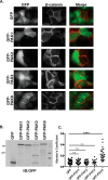
Figure 1.
Figure 1.
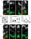
Type II but not type I PAKs target to cell–cell contacts. A, DU145 cells transiently expressing GFP-tagged type I PAKs (PAK1, PAK2, or PAK3) or PAK6 were plated on glass coverslips, fixed after 24 h, and stained with antibody to β-catenin. Scale bar, 10 μm. B, 5 × 10⁶ DU145 cells transiently transfected with GFP-PAK constructs were lysed in buffer X lysis buffer, diluted in sample buffer, and immunoblotted (IB) for GFP. C, colocalization of GFP and GFP-PAKs with β-catenin was assessed by calculating the Mander's coefficient. At least 15 nonoverlapping frames from at least three independent transfections were used in the analysis. Mean ± S.D. (error bars) is indicated. n.s., not significant; ****, p < 0.0001 in an ordinary one-way ANOVA test with Dunnett's correction for multiple comparisons.
Figure 2.
Figure 2.
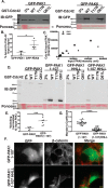
The variable region influences PAK1 localization. A, E, and I, schematic representations of the GFP-tagged PAK1/PAK6 chimeras used. The amino acid boundaries of the polybasic motif (PB) plus CRIB domain region, the VR, and kinase domain (Cat) are indicated. All GFP tags are positioned at the N terminus of the constructs. PAK1 is shown in yellow, and PAK6 is shown in blue. B, F, and J, DU145 cells were transiently transfected with GFP-tagged chimeric constructs on glass coverslips. 24 h later, cells were fixed and stained with antibody to β-catenin. Scale bar, 10 μm. C, G, and K, immunoblotting (IB) of transfected DU145 cell lysates for GFP. D, H, and L, colocalization of GFP-PAK chimeras with β-catenin was assessed by calculating the Mander's coefficient. At least 20 nonoverlapping frames from at least three independent transfections were used in the analysis. Mean ± S.D. (error bars) is indicated. n.s., not significant; ****, p < 0.0001 in an ordinary one-way ANOVA test with Dunnett's correction for multiple comparisons.
Figure 3.
Figure 3.
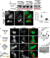
PAK1 variable region inhibits PAK1 from targeting to cell–cell junctions. A, DU145 cells were transiently transfected with GFP-tagged PAK1, PAK1 truncation mutants, or PAK6 on glass coverslips. 24 h later, cells were fixed and stained with antibody to β-catenin. Scale bar, 10 μm. B, immunoblotting (IB) of transfected DU145 cell lysates for GFP. C, colocalization of GFP-PAK1 truncations with β-catenin was assessed by calculating the Mander's coefficient in at least 20 nonoverlapping frames from at least three independent transfections. Mean ± S.D. (error bars) is indicated. n.s., not significant; ****, p < 0.0001 in an ordinary one-way ANOVA test with Dunnett's correction for multiple comparisons. D, localization of GFP-tagged PAK2 and PAK3 or their truncation mutants was assessed as in A. E, colocalization of GFP-PAK2/3 truncations with β-catenin was assessed by calculating the Mander's coefficient. At least 10 nonoverlapping frames from at least two independent transfections were used in the analysis. Mean ± S.D. is indicated. ****, p < 0.0001 in an ordinary one-way ANOVA test with Tukey's multiple-comparison test.
Figure 4.
Figure 4.
Neither PIX binding nor the inhibitory switch region inhibits PAK1 from localizing to cell–cell contacts. A, DU145 cells were transiently transfected with GFP-tagged PAK1 or the PAK1 (P191G/R192A) double mutant (PAK1-GA) on glass coverslips. 24 h post-transfection, cells were fixed and stained with antibody to β-catenin. Scale bar, 10 μm. B, colocalization of GFP-PAK1 and GFP-PAK1-GA with β-catenin was assessed by calculating the Mander's coefficient. At least 15 nonoverlapping frames from at least three independent transfections were used in the analysis. Mean ± S.D. (error bars) is indicated. An unpaired, two-tailed Student's t test was performed. n.s., not significant. C, bacterially purified GST, GST-tagged variable region of PAK1 (VR), or GST-VR containing the P191G R192A double mutation (VR-GA) loaded on GSH beads was incubated with or without DU145 cell lysates, fractionated by SDS-PAGE, and stained with Coomassie Blue. The result shown is representative of seven independent experiments. Bands of interest are marked with an arrowhead. D, samples indicated were taken from the experiment shown in C, analyzed on a separate gel, and immunoblotted (IB) for PIX or GIT. E, localization of GFP-PAK1 or the truncation mutant PAK1(1–149), which includes the inhibitory switch region and the kinase inhibitor segment, was assessed as in A. F, colocalization of GFP-PAK1 and mutant with β-catenin was assessed by calculating the Mander's coefficient. At least 15 nonoverlapping frames from at least three independent transfections were used in the analysis. Mean ± S.D. is indicated. ****, p < 0.0001 in an ordinary one-way ANOVA with Tukey's multiple-comparison test.
Figure 5.
Figure 5.
High Cdc42 binding is necessary for targeting. A, pulldown of GFP-tagged PAK1 or PAK6 from transiently transfected HEK293T cell lysates by bacterially purified GST-Cdc42, GST-Cdc42 T17N, or GST-Cdc42 Q61L. Samples were immunoblotted (IB) for GFP, and 3% input is shown. Ponceau staining was used to assess equal bead loading. B, PAK binding was quantified from four independent experiments (mean ± S.D. (error bars)). *, p < 0.05 in a paired, two-tailed Student's t test. C, representative binding curve of GFP-tagged PAK1 or PAK6 pulled down by GST-Cdc42. Input amounts were serially diluted with untransfected 293T cell lysates. D and E, Cdc42 binding of GFP-tagged PAK1, PAK1(1–107), or constructs containing inactivating H83L/H86L double mutations in the PAK1 CRIB domain (HHLL) was assessed as in A and quantified as in B from seven independent experiments. ***, p < 0.001. F, DU145 cells on glass coverslips were transiently transfected with GFP-tagged PAK1 mutants, and 24 h later, cells were fixed and stained for β-catenin. Scale bar, 10 μm. G, GFP-PAK colocalization with β-catenin was assessed by calculating the Mander's coefficient. At least 20 nonoverlapping frames from at least three independent transfections were used in the analysis. Mean ± S.D. is indicated. ****, p < 0.0001 in an unpaired, two-tailed Student's t test.
Figure 6.
Figure 6.
Polybasic is necessary for targeting once the inhibition from the variable region is released. A, pulldown of GFP-tagged PAK constructs from transiently transfected 293T cell lysates by bacterially purified GST-Cdc42, GST-Cdc42 T17N, or GST-Cdc42 Q61L. Samples were immunoblotted (IB) for GFP, and 3% input is shown. Ponceau staining was used to assess equal bead loading. B, DU145 cells were transiently transfected with GFP-tagged PAK1(1–107) or the PAK1 CRIB domain, and 24 h later, cells were fixed and stained with antibody to β-catenin. Scale bar, 10 μm. C, colocalization of GFP-PAK chimeras with β-catenin was assessed by calculating the Mander's coefficient. At least 15 nonoverlapping frames from at least three independent transfections were used in the analysis. Mean ± S.D. (error bars) is indicated. ****, p < 0.0001 in an ordinary one-way ANOVA test with Dunnett's correction for multiple comparisons. D–F, targeting of GFP-PAK constructs was assessed as in B. A schematic representation of the constructs with PAK1 shown in yellow and PAK6 shown in blue is provided. H, colocalization was assessed by Mander's coefficient as in C. I, immunoblotting of transfected DU145 cell lysates for GFP.
Figure 7.
Figure 7.
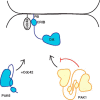
Schematic showing potential mechanisms for regulation of PAK recruitment to cell–cell contacts. Inactive PAK1 is thought to form an autoinhibited dimer (14), whereas PAK6 is inhibited in cis by binding of a pseudosubstrate sequence (18). PAKs are recruited to cell–cell contacts through direct interactions between their CRIB domain and Cdc42 and potentially through interactions of their polybasic (PB) motifs with the membrane. In PAK1, recruitment is blocked by the variable region, possibly through effects on PAK1 conformation or through interactions with other proteins.

References

    1. Field J., and Manser E. (2012) The PAKs come of age: celebrating 18 years of discovery. Cell Logist. 2, 54–58 10.4161/cl.22084 - DOI - PMC - PubMed
    1. Ha B. H., Morse E. M., Turk B. E., and Boggon T. J. (2015) Signaling, regulation, and specificity of the type II p21-activated kinases. J. Biol. Chem. 290, 12975–12983 10.1074/jbc.R115.650416 - DOI - PMC - PubMed
    1. Arias-Romero L. E., and Chernoff J. (2008) A tale of two Paks. Biol. Cell. 100, 97–108 10.1042/BC20070109 - DOI - PubMed
    1. Manser E., Leung T., Salihuddin H., Zhao Z. S., and Lim L. (1994) A brain serine/threonine protein kinase activated by Cdc42 and Rac1. Nature 367, 40–46 10.1038/367040a0 - DOI - PubMed
    1. Manser E., Chong C., Zhao Z. S., Leung T., Michael G., Hall C., and Lim L. (1995) Molecular cloning of a new member of the p21-Cdc42/Rac-activated kinase (PAK) family. J. Biol. Chem. 270, 25070–25078 10.1074/jbc.270.42.25070 - DOI - PubMed

Publication types

LinkOut - more resources