. 2019 Aug 8;4(15):e127098.
doi: 10.1172/jci.insight.127098.

Prohibitin is a prognostic marker and therapeutic target to block chemotherapy resistance in Wilms' tumor

Affiliations

Prohibitin is a prognostic marker and therapeutic target to block chemotherapy resistance in Wilms' tumor

Michael V Ortiz et al. JCI Insight. .

Abstract

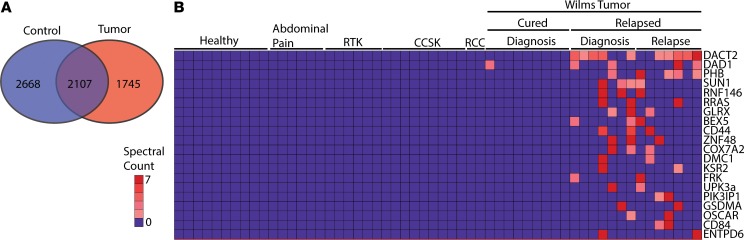
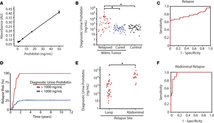
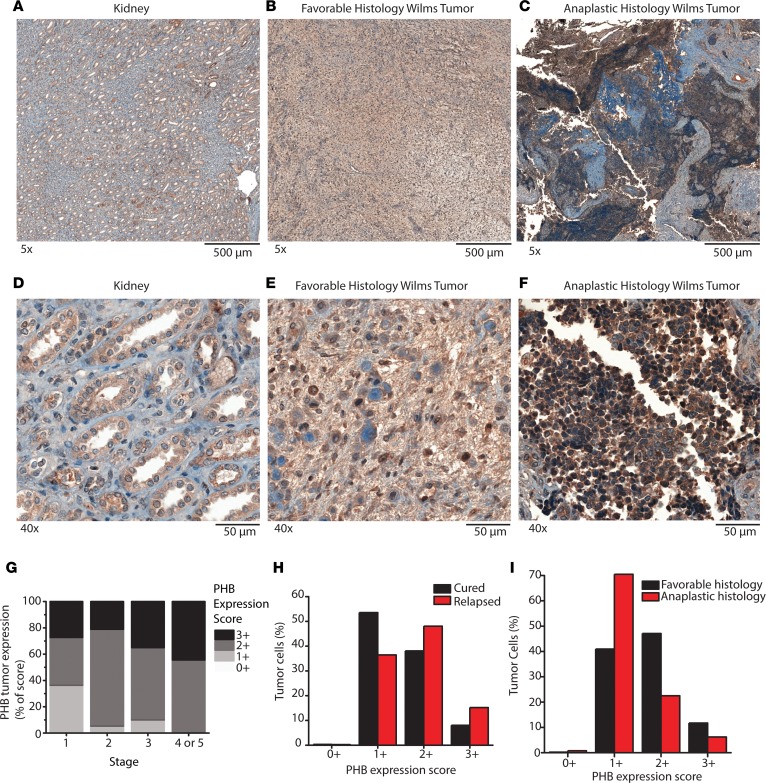
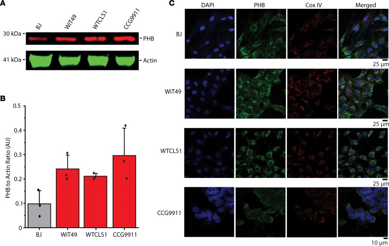
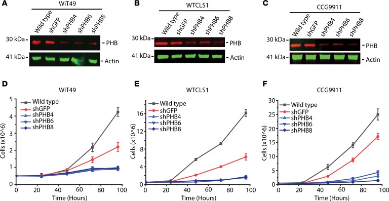
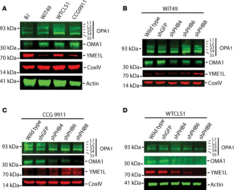
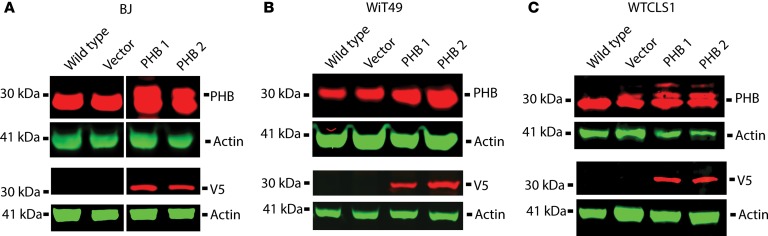
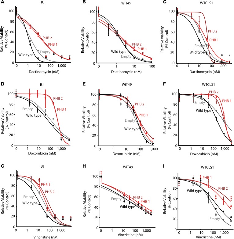
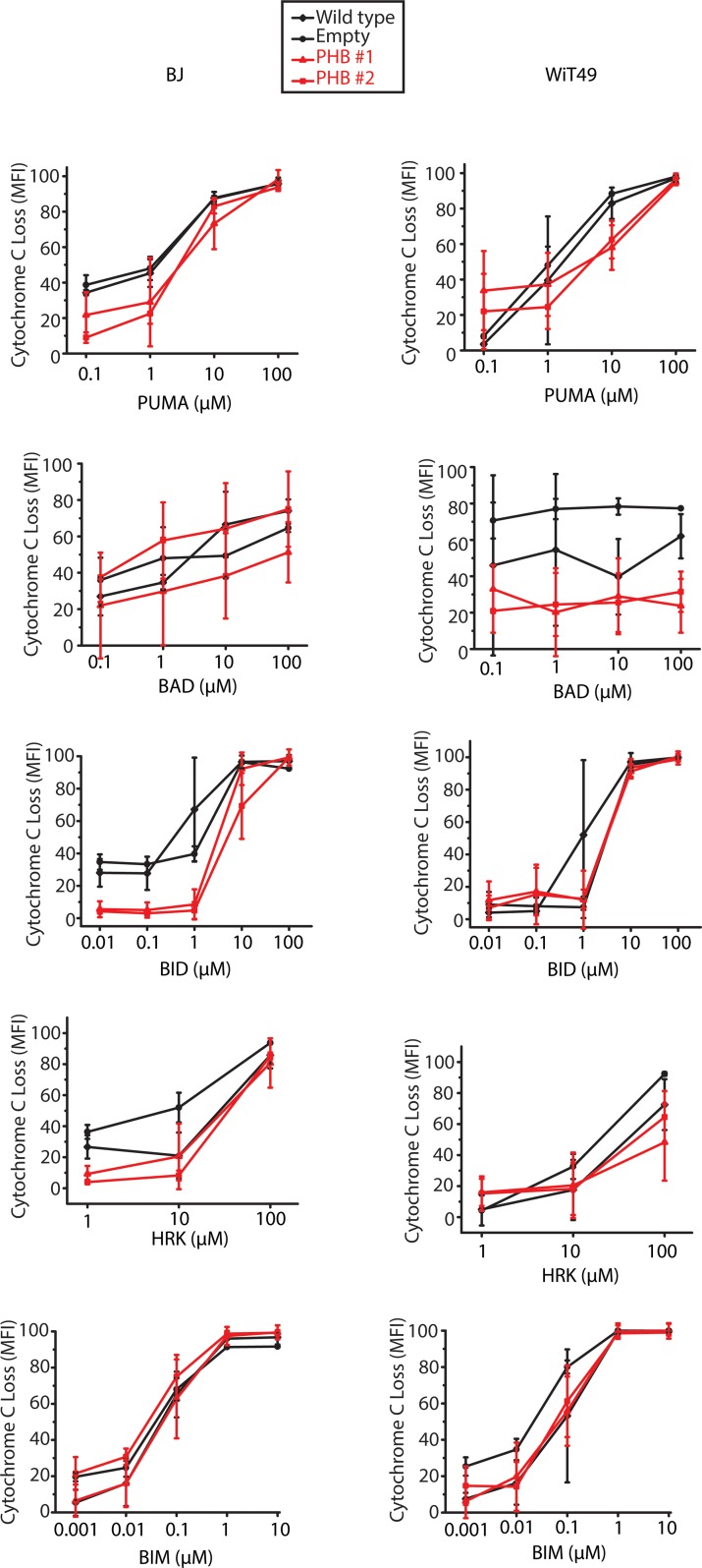
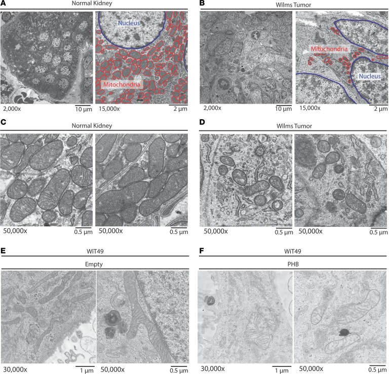
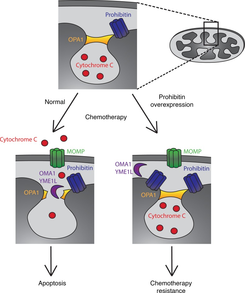
Wilms' tumor is the most common type of childhood kidney cancer. To improve risk stratification and identify novel therapeutic targets for patients with Wilms' tumor, we used high-resolution mass spectrometry proteomics to identify urine tumor markers associated with Wilms' tumor relapse. We determined the urine proteomes at diagnosis of 49 patients with Wilms' tumor, non-Wilms' tumor renal tumors, and age-matched controls, leading to the quantitation of 6520 urine proteins. Supervised analysis revealed specific urine markers of renal rhabdoid tumors, kidney clear cell sarcomas, renal cell carcinomas as well as those detected in patients with cured and relapsed Wilms' tumor. In particular, urine prohibitin was significantly elevated at diagnosis in patients with relapsed as compared with cured Wilms' tumor. In a validation cohort of 139 patients, a specific urine prohibitin ELISA demonstrated that prohibitin concentrations greater than 998 ng/mL at diagnosis were significantly associated with ultimate Wilms' tumor relapse. Immunohistochemical analysis revealed that prohibitin was highly expressed in primary Wilms' tumor specimens and associated with disease stage. Using functional genetic experiments, we found that prohibitin was required for the growth and survival of Wilms' tumor cells. Overexpression of prohibitin was sufficient to block intrinsic mitochondrial apoptosis and to cause resistance to diverse chemotherapy drugs, at least in part by dysregulating factors that control apoptotic cytochrome c release from mitochondrial cristae. Thus, urine prohibitin may improve therapy stratification, noninvasive monitoring of treatment response, and early disease detection. In addition, therapeutic targeting of chemotherapy resistance induced by prohibitin dysregulation may offer improved therapies for patients with Wilms' and other relapsed or refractory tumors.

Keywords: Apoptosis; Cancer; Cell Biology; Oncology.


Conflict of interest statement

Conflict of interest: AK is a consultant for Novartis.

Figures

Figure 1
Figure 1. High-accuracy mass spectrometry to profile the urine proteomes of childhood kidney tumors reveals markers of relapse and chemoresistance.
(A) Venn diagram demonstrates the distribution of proteins identified in the children without renal tumors (blue) compared with those with tumors (red) (n = 6520 proteins). (B) Heat map of top 20 proteins most highly enriched in Wilms’ tumor that relapsed (n = 56 samples). CCSK, clear cell sarcoma of the kidney; RCC, renal cell carcinoma; RTK, rhabdoid tumor of the kidney.
Figure 2
Figure 2. Elevated urine prohibitin at diagnosis is a specific biomarker of relapse in favorable-histology Wilms’ tumor.
(A) ELISA comparing known prohibitin levels (ng/mL) with measured absorbance via ELISA. (B) Diagnostic urine prohibitin levels (ng/mL) in patients with Wilms’ tumor who have favorable histology and relapsed (red, n = 49) are compared with those who were cured (blue, n = 50) and normal controls (black, n = 40). Exploratory simple logistic regression models determined that 998 ng/mL was the optimal cutoff point for urine prohibitin. Using Fisher’s exact test for distribution differences in dichotomized PHB among the 3 patient groups (relapsed, cured, control) revealed a statistically increased number of relapsed Wilms’ tumor with this cutoff threshold. OR of relapse for patients with diagnostic urine prohibitin greater than 998 ng/mL = 153 (95% CI, 19.6–1,000). (C) A receiver-operating characteristic curve demonstrates the prognostic power of diagnostic urine prohibitin to predict relapse in favorable-histology Wilms’ tumor at different sensitivity and specificity with an AUC of 0.78 (95% CI, 0.68–1.0). (D) Risk of relapse in patients with favorable-histology Wilms’ tumor are stratified by those with a diagnostic urine prohibitin greater than 998 ng/mL (red, n = 31) compared with those with a diagnostic urine prohibitin less than 998 ng/mL (blue, n = 68). (E) Diagnostic urine prohibitin levels in relapsed patients with favorable-histology Wilms’ tumor are stratified by site of relapse. (F) A receiver-operating characteristic curve demonstrates the prognostic power of diagnostic urine prohibitin to predict abdominal relapse in favorable-histology Wilms’ tumor at different sensitivity and specificity with an AUC of 0.96 (95% CI, 0.91–1.0).
Figure 3
Figure 3. Prohibitin is highly expressed in primary Wilms’ tumor samples.
(AF) PHB IHC staining was performed on formalin-fixed paraffin-embedded primary Wilms’ tumor samples and compared with adjacent normal kidneys. Original magnification, ×5 (AC); ×40 (DF). A and D include normal kidney. B and E include favorable-histology Wilms’ tumor. C and F include anaplastic histology Wilms’ tumor. (G) IHC was performed on a tissue microarray containing 59 primary Wilms’ tumor samples and graded from 0+ to 3+ in a blinded manner. Quantification of IHC from the tissue microarray is shown and stratified by initial tumor stage. (H and I) A second cohort of 38 patients with primary Wilms’ tumor was assessed (15 cured, 16 relapsed, 7 no information; 24 favorable, 14 anaplastic Wilms’ tumor). The expression of PHB was evaluated on a cell-by-cell basis using Halo imaging analysis software. A total of 24,862,509 cells were counted and scored from 0+ to 3+ based on PHB expression. In H, PHB expression in the cells of Wilms’ tumor that ultimately relapsed are compared with those that were cured. Whereas in I, PHB expression in the cells of Wilms’ tumor that had favorable histology are compared with those with anaplastic histology.
Figure 4
Figure 4. Prohibitin exhibits largely mitochondrial expression in Wilms’ tumor cell lines.
(A) Our in vitro studies of PHB included a control fibroblast cell line (BJ) as well as renal tumor cell lines (WiT49, WT-CLS1, and CCG9911). Endogenous PHB expression in the renal tumor cell line is shown with actin as a loading control. (B) PHB expression is compared in the different cell lines normalized to actin in Western blot triplicates. (C) Confocal fluorescence microscopy demonstrates that most cellular PHB (green) colocalizes with the inner mitochondrial membrane marker CoxIV (red) but not the nuclear marker DAPI (blue) in paraformaldehyde-fixed cells.
Figure 5
Figure 5. Prohibitin is required for Wilms’ tumor growth and survival.
(AC) Western blots of WT renal tumor cells (A, WiT49; B, WT-CLS1; C, CCG9911) as well as a nontargeting green fluorescence protein (shGFP) and 3 different shRNA hairpins targeting the PHB 3′UTR (shPHB4) and CDS (shPHB6 and shPHB8). Actin is used for whole cell loading control. (DF) Cell growth over time in renal tumor cells (D, WiT49; E, WT-CLS1; F, CCG9911), which are WT (black), compared with those transduced with nontargeting shGFP (red) and the 3 PHB targeting shRNA (blue).
Figure 6
Figure 6. Depletion of prohibitin results in alterations in mitochondrial intermembrane proteases and structural proteins involved in apoptosis and mitochondrial morphogenesis.
(A) Western blot of endogenous expression of OPA1, OMA1, and YME1L with CoxIV and actin as mitochondrial and whole cellular loading controls, respectively. (BD) Western blots of OPA1, OMA1, and YME1L in renal tumor cells (B, WiT49; C, WT-CLS1; D, CCG9911) comparing WT cells with nontargeting shGFP and 3 different shRNA hairpins targeting the PHB 3′UTR (shPHB4) and CDS (shPHB6 and shPHB8). Loading controls as shown.
Figure 7
Figure 7. Overexpression of prohibitin in Wilms’ tumor and control cell lines.
(AC) Western blots of PHB and V5 in control (A, BJ) and renal tumor cells (B, WiT49; C, WT-CLS1) comparing WT cells and those transduced with an empty vector with 2 different clones transduced with a PHB-expressing vector containing a V5 tag. Colors were reversed in V5 and bottom actin blots.
Figure 8
Figure 8. Overexpression of prohibitin results in resistance to diverse chemotherapy drugs in both Wilms’ tumor and control cells.
Dose response curve of BJ (A, D, and G), WiT49 (B, E, and H), and WT-CLS1 (C, F, and I) cells treated with dactinomycin (AC), doxorubicin (DF), or vincristine (GI) for 72 hours comparing WT cells (black), empty vector transduced cells (gray), as well as PHB-transduced cells (red, dark red).
Figure 9
Figure 9. BH3 profiling reveals globally decreased apoptotic priming in response to PHB overexpression.
Cytochrome c loss in response to treatment with different proapoptotic peptides comparing WT (black diamond), with empty (black circles), and 2 PHB overexpressing cell lines (red triangle, red square) in both BJ control fibroblasts and WiT49 Wilms’ tumor.
Figure 10
Figure 10. Primary Wilms’ tumor samples exhibit fewer mitochondria and predominantly a mitochondrial fission phenotype, as compared with adjacent normal kidneys.
(AD) Untreated favorable-histology Wilms’ tumor was immediately fixed following nephrectomy and imaged using a transmission electron microscope to evaluate mitochondrial morphology. The normal kidney (original magnification, ×2,000 [A, left]; ×50,000 [C]) is compared with the Wilms’ tumor (original magnification, ×2,000 [B, left]; ×50,000 [D]). A and B also highlight (original magnification, ×15,000, right) the size and number of the mitochondria (red) as compared with the nucleus (blue). (E and F) Characteristic mitochondria of Wilms’ tumor cells (WiT49) treated with an empty vector (E) compared with a PHB overexpressing vector (F).
Figure 11
Figure 11. PHB overexpression leads to failure to release cytochrome c from cristae junctions despite the presence of a MOMP complex.
Prohibitin (blue) forms a complex along the mitochondrial inner membrane that interacts with several key regulators of apoptosis, including the OPA1 complex (orange) located at cristae junctional openings. The majority of cytochrome c (red) is sequestered within these cristae and in response to apoptotic stimuli such as chemotherapy, the MOMP complex consisting of BAX and BAK is inserted into the mitochondrial outer membrane. Upon proteolysis of the OPA1 complex, generally via OMA1 or other intermembrane proteases (purple), cytochrome c is released into the cytosol where it results in the apoptotic cascade. Our model proposes that due to the overexpression of PHB, in response to apoptotic stimuli, intermembrane proteases are no longer able to access the OPA1 complex, thereby resulting in resistance to chemotherapy despite the presence of a MOMP pore in the outer mitochondrial membrane, ultimately resulting in failure to undergo apoptosis and chemotherapy resistance.

References

    1. Dome JS, et al. Advances in Wilms tumor treatment and biology: progress through international collaboration. J Clin Oncol. 2015;33(27):2999–3007. doi: 10.1200/JCO.2015.62.1888. - DOI - PMC - PubMed
    1. Termuhlen AM, et al. Twenty-five-year follow-up of childhood Wilms tumor: a report from the Childhood Cancer Survivor Study. Pediatr Blood Cancer. 2011;57(7):1210–1216. doi: 10.1002/pbc.23090. - DOI - PMC - PubMed
    1. Ooms AH, et al. Significance of TP53 mutation in Wilms tumors with diffuse anaplasia: a report from the children’s oncology group. Clin Cancer Res. 2016;22(22):5582–5591. doi: 10.1158/1078-0432.CCR-16-0985. - DOI - PMC - PubMed
    1. Grundy PE, et al. Loss of heterozygosity for chromosomes 1p and 16q is an adverse prognostic factor in favorable-histology Wilms tumor: a report from the National Wilms Tumor Study Group. J Clin Oncol. 2005;23(29):7312–7321. doi: 10.1200/JCO.2005.01.2799. - DOI - PubMed
    1. Grundy PE, Telzerow PE, Breslow N, Moksness J, Huff V, Paterson MC. Loss of heterozygosity for chromosomes 16q and 1p in Wilms’ tumors predicts an adverse outcome. Cancer Res. 1994;54(9):2331–2333. - PubMed

Publication types

MeSH terms