. 2019 Jun 6;10(7):1145-1159.
doi: 10.1039/c9md00102f. eCollection 2019 Jul 1.

Binding site characterization - similarity, promiscuity, and druggability

Affiliations

Binding site characterization - similarity, promiscuity, and druggability

Christiane Ehrt et al. Medchemcomm. .

Abstract

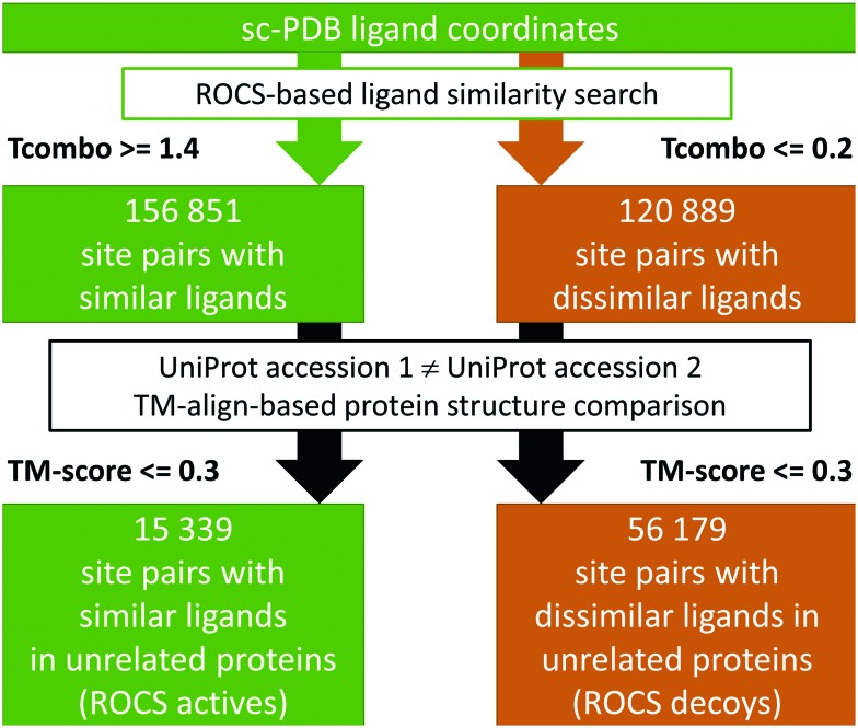
The elucidation of non-obvious binding site similarities has provided useful indications for the establishment of polypharmacology, the identification of potential off-targets, or the repurposing of known drugs. The concept underlying all of these approaches is promiscuous binding which can be analyzed from a ligand-based or a binding site-based perspective. Herein, we applied methods for the automated analysis and comparison of protein binding sites to study promiscuous binding on a novel dataset of sites in complex with ligands sharing common shape and physicochemical properties. We show the suitability of this dataset for the benchmarking of novel binding site comparison methods. Our investigations also reveal promising directions for further in-depth analyses of promiscuity and druggability in a pocket-centered manner. Drawbacks concerning binding site similarity assessment and druggability prediction are outlined, enabling researchers to avoid the typical pitfalls of binding site analyses.


Figures

Fig. 1
Fig. 1. The workflow for the generation of the ROCS dataset.
Fig. 2
Fig. 2. SMARTS patterns of maximum common substructures within the clusters derived from the ligands of the active site pairs in the ROCS dataset. The percentage of dataset compounds per cluster is given in parentheses.
Fig. 3
Fig. 3. Non-obvious binding site similarities. On the left, the binding site alignment of the human enzyme arginine N-methyltransferase 6 (PDB-ID ; 4hc4 chain A, green) and N-4 cytosine-specific methyltransferase from Proteus vulgaris (PDB-ID ; 1boo chain A, orange) is presented with SAH in ball-and-stick representation. The corresponding binding site residues are colored according to the structure. The figure was generated with UCSF Chimera. The interaction patterns of SAH in the binding sites of the complexes with the PDB-IDs ; 4hc4 and ; 1boo are shown on the right (the schemes were generated with LigandScout 4.065).
Fig. 4
Fig. 4. Evaluation of different binding site comparison tools with respect to the dataset of ROCS structures. Top: ROC curves for different binding site comparison methods for the initial purely ligand-based dataset. Bottom: ROC curves for the methods when only matches with a high SiteHopper PatchScore are taken into account as active site pairs. The name of the tool is colored according to its corresponding ROC curve. The binding site comparison tools are sorted in descending order with respect to their overall AUC. The scoring measures that yielded the highest AUC for both datasets were distance score d1, the similarity score TC, the SVA, and the DistanceScore for SiteAlign (thin red line), SMAP (thin blue line), ProBiS (thin purple line), and SiteEngine (thin orange line), respectively. TIFP AUC values significantly improved when using the hamming distance as the scoring measure in both datasets. Top: Shaper(PDB), VolSite/Shaper, and VolSite/Shaper(PDB) showed the highest AUC values when using the Tanimoto (fit) as the similarity score. Bottom: The SiteHopper AUC value improved slightly upon using the ColorTanimoto.
Fig. 5
Fig. 5. Examples of dissimilar and similar binding sites bound to ligands showing a high similarity in terms of the SiteHopper-derived PatchScore. Left panel: SiteHopper- (top) and ligand-based (bottom) alignment of the binding sites of tankyrase 2 (PDB-ID 4hkn, ligand-ID LU2) and dihydroflavonol-4-reductase (PDB-ID ; 2nnl, ligand-ID ERD). Despite their common shape and physicochemical properties, both ligands interact in a unique way with their respective targets. Right panel: Alignment of the metalloproteases E. coli peptide deformylase (PDB-ID ; 3k6l, ligand-ID 2BB) and human ADAMTS-1 (PDB-ID ; 2jih, ligand-ID 097) together with bound peptidomimetic compounds (top). The crucial common interactions are depicted separately with the corresponding ligand 2D representations. Blue and red arrows indicate hydrogen bond donor and acceptor functionalities of the protein residues.
Fig. 6
Fig. 6. Box plots of the scaled hydrophobicity (top) and polarity (center) descriptors derived from the analyses with fpocket (dark green boxes), VolSite (light green boxes), and DoGSite (yellow boxes) together with the obtained druggability scores (bottom) for the X-ray dataset. The cut-offs to distinguish between druggable and non-druggable sites are given as gray lines. The dashed line represents a stricter threshold for druggability predictions with fpocket.
Fig. 7
Fig. 7. Box plots of the DoGSite-derived scaled hydrophobicity (solid filled boxes) and polarity (hatch pattern-filled boxes) scores of the binding sites of ligands that bind to multiple binding sites which do not show a high similarity using SiteHopper (PatchScore < 0.82, mainly hydrophobic). For comparison purposes, the box plot of these properties is also given for all ligands of the sc-PDB. “Super-promiscuous” ligands as defined by an earlier study are highlighted in gray.

References

    1. Hu Y., Bajorath J. Drug Discov. Today. 2013;18(13–14):644–650. - PubMed
    1. Proschak E., Stark H., Merk D. J. Med. Chem. 2018;62(2):420–444. - PubMed
    1. Anighoro A., Bajorath J., Rastelli G. J. Med. Chem. 2014;57(19):7874–7887. - PubMed
    1. Haupt V. J., Daminelli S., Schroeder M. PLoS One. 2013;8(6):e65894. - PMC - PubMed
    1. Saqib U., Kelley T. T., Panguluri S. K., Liu D., Savai R., Baig M. S. Front. Pharmacol. 2018;9:1201. - PMC - PubMed