Review
. 2019 Jun 26:9:52.
doi: 10.1186/s13578-019-0317-8. eCollection 2019.

PKM2, function and expression and regulation

Affiliations
Review

PKM2, function and expression and regulation

Ze Zhang et al. Cell Biosci. .

Erratum in

Abstract

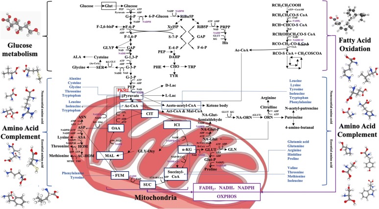
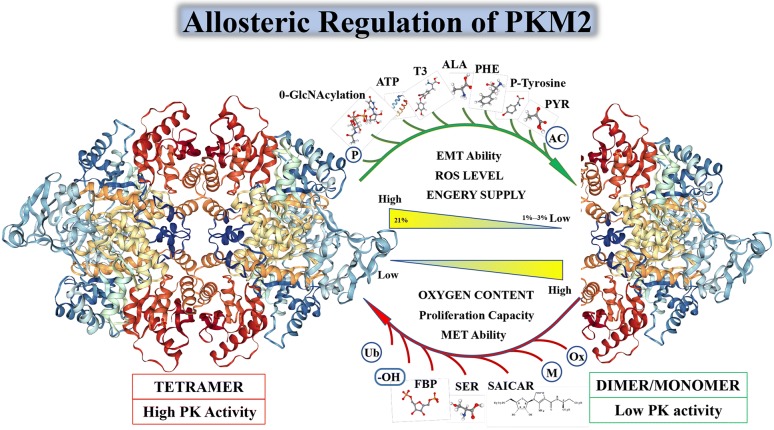
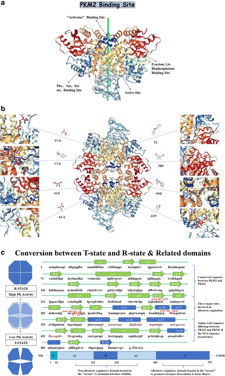
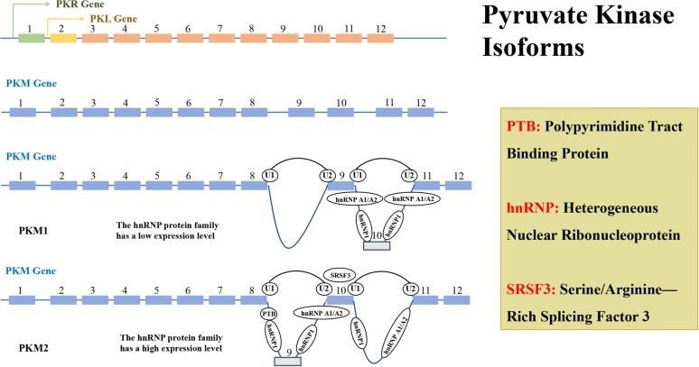
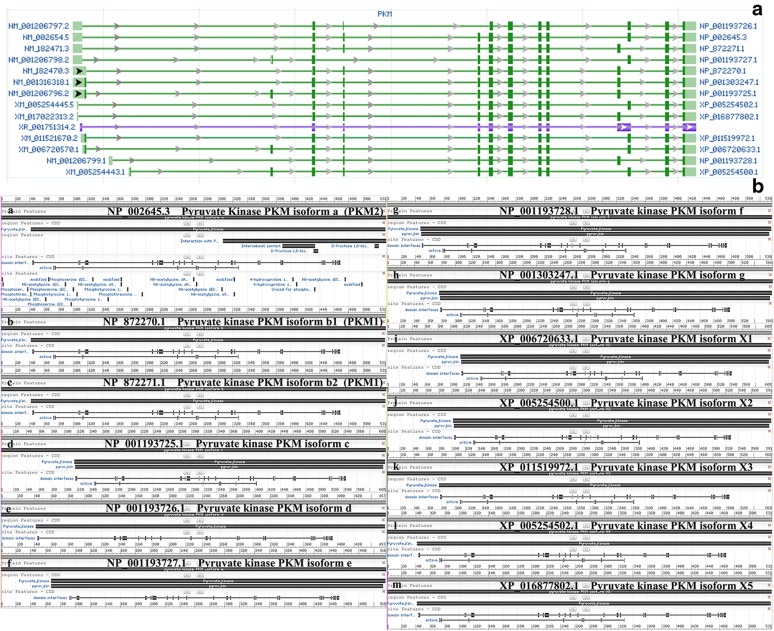
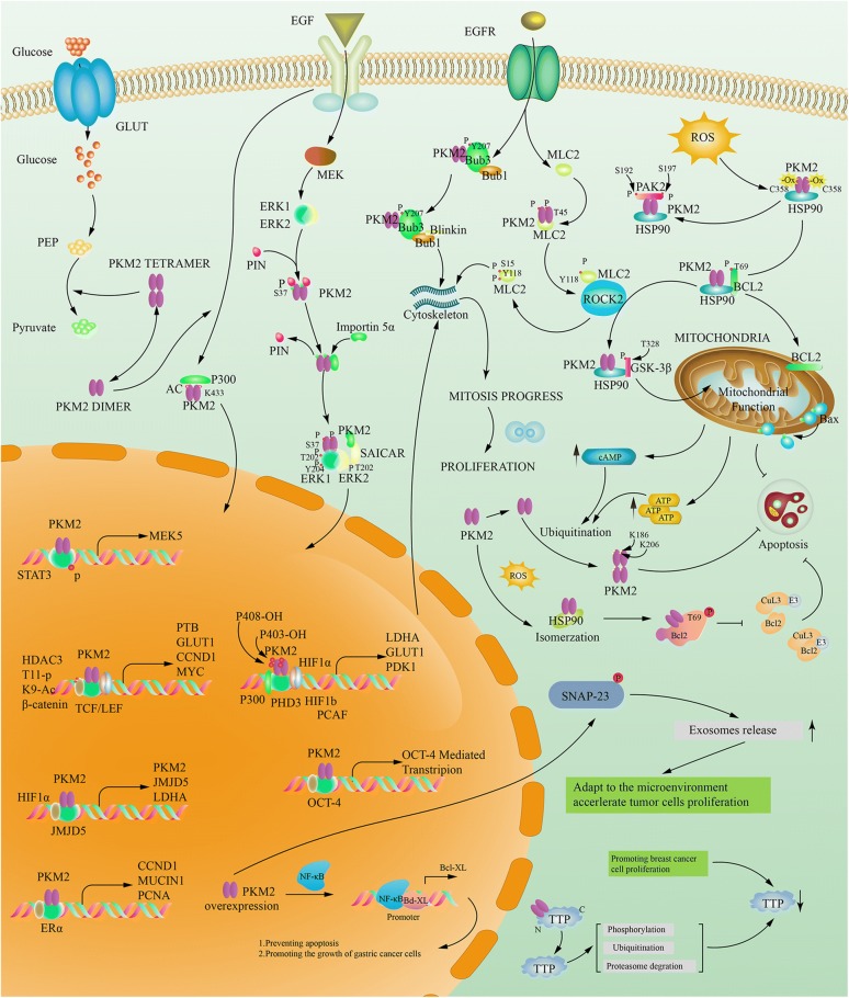
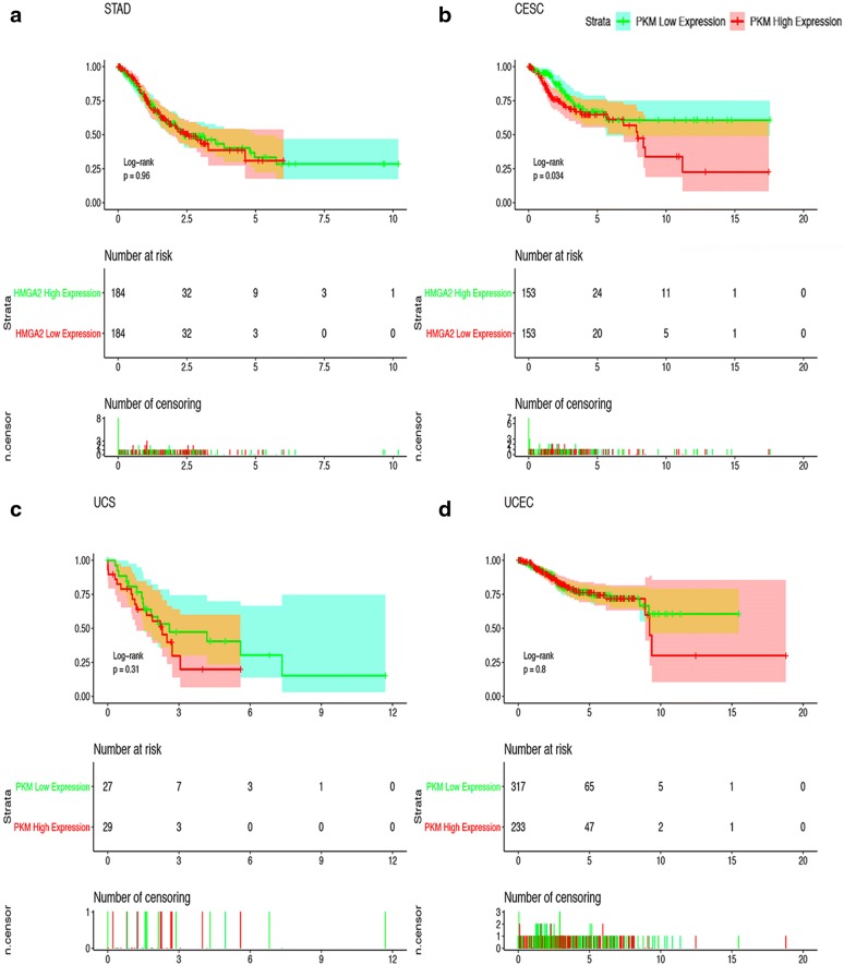
Pyruvate kinase (PK), as one of the key enzymes for glycolysis, can encode four different subtypes from two groups of genes, although the M2 subtype PKM2 is expressed mainly during embryonic development in normal humans, and is closely related to tissue repair and regeneration, with the deepening of research, the role of PKM2 in tumor tissue has received increasing attention. PKM2 can be aggregated into tetrameric and dimeric forms, PKM2 in the dimer state can enter the nuclear to regulate gene expression, the transformation between them can play an important role in tumor cell energy supply, epithelial-mesenchymal transition (EMT), invasion and metastasis and cell proliferation. We will use the switching effect of PKM2 in glucose metabolism as the entry point to expand and enrich the Warburg effect. In addition, PKM2 can also regulate each other with various proteins by phosphorylation, acetylation and other modifications, mediate the different intracellular localization of PKM2 and then exert specific biological functions. In this paper, we will illustrate each of these points.

Keywords: Cancer metabolism; Glycolysis; Pyruvate kinase; Warburg effect.


Conflict of interest statement

Competing interestsThe authors declare that they have no competing interests.

Figures

Fig. 1
Fig. 1
PKM2: Junction of Metabolic Networks and Signal Cascades. POST-Warburg effect, in glucose metabolism, tumor cells divide glucose metabolism into three separate parts of glycolysis, tricarboxylic acid cycle and oxidative phosphorylation (OXPHOS). The effect of PKM2 is currently considered to be the interception of glucose metabolism and the metabolic pathway is transferred to the pentose phosphate pathway (PPP), the uronic acid pathway (UAP), the polyol pathway (PYP), etc. for the synthesis of the subsequent five-carbon ribose and non-essential amino acids. The TCA circle is backed up by fatty acid metabolism and amino acid metabolism, and its main purpose is to provide raw materials for the synthesis of non-essential amino acids, and the secondary purpose is to supply REDOX equivalents [43]. Although the classic glutathione replenishment pathway is well known. But there are more similar pathways in tumor cells which marked with blue text in the picture. PKM2 can be replenished by many amino acids such as alanine (Ala), cysteine (Cys), glycine (Gly), threonine (Thr), tryptophan (Try), etc. Although Ac-CoA mainly relies on fatty acid metabolism for supply, it can also be replenished by some amino acids such as leucine, isoleucine, tryptophan etc. The tricarboxylic acid cycle (TCA) serves as the focal point for the metabolism of the three major metabolic processes, the amino acid is also the most abundant in its form of replenishment. For example, a variety of amino acids such as aspartic acid, arginine, glutamic acid, glutamine, histidine, isoleucine, methionine, phenylalanine, proline, tyrosine, threonine, valine etc. can complement the eight intermediate metabolites in the tricarboxylic acid cycle (TCA). In the three major metabolic processes, the REDOX equivalents produced will converge in the mitochondria and eventually promote the oxidative phosphorylation (OXPHOS) of the electron transport chain, while protecting the mitochondrial function of the tumor cells, also providing the cells with a large amount of adenosine triphosphate (ATP) required for survival as well [44]. REDOX equivalents marked with purple text in the picture
Fig. 2
Fig. 2
Relationship between PKM2 enzyme activity and spatial conformation. The transition between PKM2 dimers and tetramers is allosterically regulated by endogenous and exogenous activators and inhibitors. PKM2 has PK enzyme activity only when it serves as a tetramer. PKM2 is activated by the glycolytic intermediate products named fructose 1,6-bisphosphate (FBP). It can also be activated by the allosteric effects of serine and succinylaminoimidazolecarboxamide ribose-50 phosphate SDH succinate dehydrogenase (SAICAR) [54, 55]. The PK enzymatic activity of PKM2 can be inhibited by many endogenous inhibitors and cellular signaling events including 0-GlcNAcylation, pyruvate (PYR), P-tyrosine (P-TYR), phenylalanine (PHE), alanine (ALA), adenosine triphosphate (ATP), and thyroid hormone T3 [–58]. In addition, due to the number of related molecules involved in PKM2’s post-translational modification (PTM), I will not list them in Fig. 2, but in the form of Tables 2 and 3 in the fourth part of this article “Interaction of PKM2 with other proteins”
Fig. 3
Fig. 3
The Specific Site of PKM2 Allosteric Regulation and The Amino Acid Sequence of PKM2. It is now accepted that the PKM2 in the tetrameric state has an allosteric regulatory domain within its spatial structure, forming a pattern similar to the seesaw, when some allosteric regulators are inserted into the spatial structure involved in PKM2 allosteric regulation. After the domain (a), the tetramer PKM2 can be transferred from a compact state (R-state) to a loose state (T-state) and finally disassembled into a dimeric form [59]. When these allosteric regulators bind to PKM2, they will change the spatial conformation of PKM2, and affect the electrostatic force inside the molecule, and then affect the transition state of PKM2. The allosteric form a stable and compact PKM2 R-state to form a tetramer and perform PK enzyme activity. After allosteric adjustment the PKM2 forms a loose and unstable T-state, and eventually breaks the linked fragment in the tetramer to form a PKM2 dimer form with lower PK enzymatic activity. When PKM2 is allosteric to form a dimer, it will expose the active region inside the molecule, although the PK enzyme activity is low, it has protein activation activity [60]. In b we specifically list the each participating allosteric regulates the binding site of the small molecule and the binding site of the activator in the PKM2 protein spatial structure [61]. In Fig. 3c we simply describe the seesaw structure of PKM2 and specifically identify the specific spatial domains that participate in the seesaw pattern: α-9 and 10 and 11 and 13 and 14 and 15 and 18 and β-20 which marked with blue text in c. The residues at the active site which was mentioned in a are highlighted by red box in c. Residue Arg342 and Residue Lys342, which is responsible for active site “RGD” stabilization is colored in red. There is one point to be noted here: whether PKM2 has protein kinase activity or not, there is a negative attitude, however in some of the researchers’ experiments described later, phosphorylation was indeed found [62]
Fig. 4
Fig. 4
The Splicing of PMK2. The genes encoding pyruvate kinase can be divided into two types: PKLR and PKM. PKLR binds to the coding gene through a tissue-specific promoter, encoding two subtypes of PKL and PKR (green for PKR and yellow for PKL). PKM encodes PKM1 and PKM2 subtypes by alternative splicing of mutually exclusive exon 9 and 10, and a high expression level of PTB, hnRNP 1 & hnRNP A1/A2 are required for during the cleavage process of exon 9 of PKM2, while the cleavage process of exon 10 of PKM1 is not required. Transcription factor SRSF3 also plays an important role in the conversion of PKM2 and PKM1. Each pyruvate kinase subtype has a different tissue expression pattern
Fig. 5
Fig. 5
Expression Patterns of The Transcript and The Protein Subtypes of PKM. a That there are 14 different subtype sequences in the PKM gene transcript, and there are some differences between them, among which the PKM1 (No. 3 and No. 5) and PKM2 (No. 2) subtypes are more compared, b the coding sequence NO. 12 expresses PKM1 and the coding sequence expresses NO. 13 PKM2. There are only 23 amino acid residues between the two protein sequences. And there are few studies on other PKM protein subtypes, and the functions are not sure yet
Fig. 6
Fig. 6
PKM2 Interacting Protein. PKM2 can not only exert the activity of PK enzyme in the form of tetramer, but also can enter the nucleus as a transcription factor to mediate the transcription of other genes when convert to dimer, can also regulate each other in the cytoplasm and other proteins. It has an impact on many different biological effects
Fig. 7
Fig. 7
Kaplan–Meier Curves for Survival of Four Most Relevant Cancers. Kaplan–Meier curves for survival of four most relevant cancers according to PKM2 special transcript (NP-002645.3 refer to Fig. 5a and Table 1) expression in cancer tissues. Patients were divided into high and low PKM2 special transcript expression groups using the median value of PKM2 special transcript expression as the cutpoint. Survival analysis and subgroup analysis were performed based on Kaplan–Meier curves. One thing should be pointed out is that in some clinical studies, the researchers found that the effect of contrast PKM2 protein expression was significantly better than the copy number of the comparative mRNA. But considering that there is currently no database on protein expression, we only counted the data in TGCA

References

    1. Warburg O, Wind F, Negelein EN. The metabolism of tumors in the body. J Gen Physiol. 1927;8:519–530. doi: 10.1085/jgp.8.6.519. - DOI - PMC - PubMed
    1. Koppenol WH, Bounds PL, Dang CV. Otto Warburg’s contributions to current concepts of cancer metabolism. Nat Rev Cancer. 2011;11:325–337. doi: 10.1038/nrc3038. - DOI - PubMed
    1. Otto AM. Warburg effect(s)-a biographical sketch of Otto Warburg and his impacts on tumor metabolism. Cancer Metab. 2016;4:5. doi: 10.1186/s40170-016-0145-9. - DOI - PMC - PubMed
    1. Gupta V, Bamezai RNK. Human pyruvate kinase M2: a multifunctional protein. Protein Sci. 2010;19:2031–2044. doi: 10.1002/pro.505. - DOI - PMC - PubMed
    1. Israelsen WJ, et al. PKM2 isoform-specific deletion reveals a differential requirement for pyruvate kinase in tumor cells. Cell. 2013;155:397–409. doi: 10.1016/j.cell.2013.09.025. - DOI - PMC - PubMed

LinkOut - more resources