. 2019 Jul;7(3):292-299.
doi: 10.29252/beat-0703013.

Curcumin and Nanocurcumin Oral Supplementation Improve Muscle Healing in a Rat Model of Surgical Muscle Laceration

Affiliations

Curcumin and Nanocurcumin Oral Supplementation Improve Muscle Healing in a Rat Model of Surgical Muscle Laceration

Siamak Kazemi-Darabadi et al. Bull Emerg Trauma. 2019 Jul.

Abstract

Objective: To compare the effects of curcumin and nanocurcumin oral supplementation on the muscle healing rate of an animal model of surgical muscle laceration.

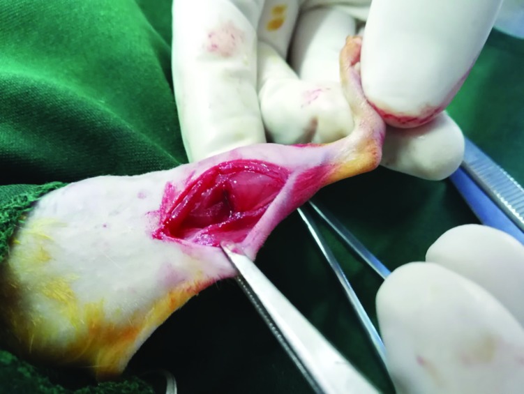
Methods: Thirty-two male adult rats were randomly divided into sham, control, curcumin, and nanocurcumin groups. Partial transection of the gastrocnemius muscle was made in the right limb of the control and treatment groups. The sham and control groups received normal saline, curcumin group received 500 mg/kg of curcumin and nanocurcumin group received 100 mg curcumin-loaded nanomicelles orally every day. They euthanized two weeks later and the specimens were stained by hematoxylin-eosin (H&E) and Masson's trichrome methods. Aspartate transaminase (AST) and creatine phosphokinase (CPK) were measured in blood samples.

Results: The percentage of collagen fibers in the nanocurcumin group was significantly lesser than the control and curcumin groups (p<0.001). Muscle fiber regeneration in the treatment groups was significantly higher than the control group (p<0.001). The blood vessels of the nanocurcumin group were significantly more than other groups (p<0.001). Plasma AST had a significant difference in the control group compared to the sham and nanocurcumin groups (p=0.026). The plasma CPK level of the control group was also significantly higher than other groups (p<0.001).

Conclusion: In conclusion, although oral curcumin supplementation has little effects because of its poor bioavailability, embedding it in nanoparticles could enhance its systemic effects in promoting the muscle healing process.

Keywords: Curcumin; Muscle laceration; Nano particles; Rat..


Conflict of interest statement

There is no conflict of interests to declare.

Figures

Fig. 1
Fig. 1
Partial transection of the rat’s right gastrocnemius muscle about 2.5 cm proximal to the calcaneus, while the tarsus was flexed in 90º angle and the stifle was extended
Fig. 2
Fig. 2
Transmission electron microscopy (TEM) image of the content of the curcumin-loaded nanomicelles shows the particles are less than 50nm in diameter
Fig. 3
Fig. 3
Fourier-transform infrared (FTIR) spectrum of the curcumin-loaded nanomicelles
Fig. 4
Fig. 4
Healed areas of the gastrocnemius muscle in experimental groups. A: Sham group. Normal muscle fibers. B: Negative control group. Abundant fibroblasts and fibrocytes along with irregular connective tissue containing collagen fibers. C: Curcumin group. Similar to the control group except that collagen fibers are more regular. D: Nanocurcumin group. Fibroblasts and fibrocytes are less than control and curcumin groups and the muscle fibers have better healing. (H&E,  200)
Fig. 5
Fig. 5
Healed areas of the gastrocnemius muscle in experimental groups. Muscle fibers are seen in red and collagen is seen blue (Masson’s trichrome, A: Sham group, B: Negative control group, C: Curcumin group, D: Nanocurcumin group, A and B:  800, C and D:  200)
Fig. 6
Fig. 6
Percentage of collagen, muscle fibers, and blood vessels in the healed area of the lacerated gastrocnemius muscle in experimental groups. Different letters (a, b, c, d) indicate significant difference among the groups (p<0.05)
Fig. 7
Fig. 7
Blood AST levels (U/L) in the experimental groups two weeks after muscle injury (mean ± SD). Different letters (a, b) indicate significant difference (p<0.05)
Fig. 8
Fig. 8
Blood CPK levels (U/L) in the experimental groups two weeks after muscle injury (mean ± SD). Different letters (a, b, c) indicate significant difference (p<0.05)

References

    1. Danna NR, Beutel BG, Campbell KA, Bosco JA 3rd. Therapeutic approaches to skeletal muscle repair and healing. Sports Health. 2014;6(4):348–55. - PMC - PubMed
    1. Covey DC. Combat orthopaedics: a view from the trenches. J Am Acad Orthop Surg. 2006;14(10 Spec No.):S10–7. - PubMed
    1. Järvinen TA, Järvinen M, Kalimo H. Regeneration of injured skeletal muscle after the injury. Muscles Ligaments Tendons J. 2014;3(4):337–45. - PMC - PubMed
    1. Fukushima K, Badlani N, Usas A, Riano F, Fu F, Huard J. The use of an antifibrosis agent to improve muscle recovery after laceration. Am J Sports Med. 2001;29(4):394–402. - PubMed
    1. Shen W, Li Y, Tang Y, Cummins J, Huard J. NS-398, a cyclooxygenase-2-specific inhibitor, delays skeletal muscle healing by decreasing regeneration and promoting fibrosis. Am J Pathol. 2005;167(4):1105–17. - PMC - PubMed

LinkOut - more resources