. 2019 Jul 1;6(6):470-478.
doi: 10.1002/mdc3.12801. eCollection 2019 Jul.

Volitional Suppression of Parkinsonian Resting Tremor

Affiliations

Volitional Suppression of Parkinsonian Resting Tremor

Rebekah L Blakemore et al. Mov Disord Clin Pract. .

Abstract

Background: We have observed in the clinic that a number of patients with Parkinson's disease (PD) can suppress their tremor at will for brief periods, by conscious mental processes. To our knowledge, the ability to consciously diminish one's resting tremor has not yet been reported nor assessed quantitatively.

Objective: To provide the first detailed systematic investigation of the phenomenon of voluntary tremor suppression in PD.

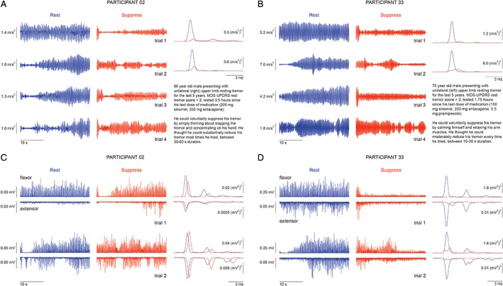
Methods: We examined changes in tremor characteristics during voluntary tremor suppression in 37 PD patients (on medication) presenting with rest tremor in their upper limb. We measured tremor oscillations with a triaxis accelerometer on the index finger of the most-affected hand (n = 27). With surface electromyography (EMG), we measured changes in neuromuscular activity of the forearm flexor digitorum superficialis and extensor digitorum muscles (n = 15). Participants completed four 1-minute trials, consisting of alternating consecutive 30-second periods of resting tremor and 30-second periods of attempted tremor suppression.

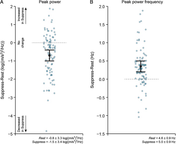
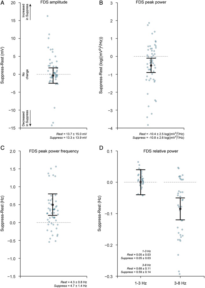
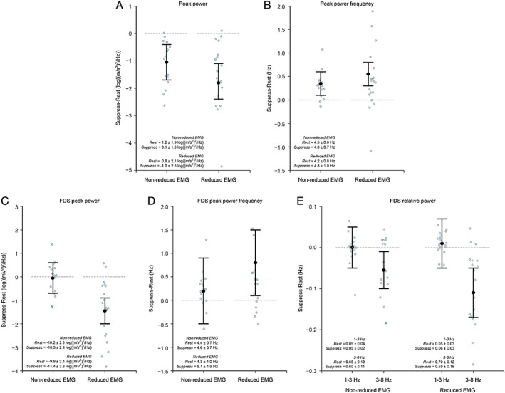
Results: Bayesian multilevel modeling revealed that attempted voluntary tremor suppression did indeed reduce tremor amplitude (peak power) of the acceleration signal and increased tremor frequency of the acceleration and EMG signals. Relative EMG power in the 3- to 8-Hz tremor band was also smaller. Tremor suppression was not by enhanced voluntary contraction of the relevant muscle pairs.

Conclusions: We present novel empirical evidence that PD resting tremor can be suppressed by an act of will, as evidenced by significant modulation of key neurophysiological tremor characteristics. These data highlight that it is possible to exert significant conscious control over parkinsonian resting tremor.

Keywords: Parkinson's disease; electromyography; tremor; volition; voluntary suppression.


Conflict of interest statement

This work was supported by a Neurological Foundation of New Zealand Project Grant (1540‐PG to R.L.B.) and the New Zealand Brain Research Institute. The authors declare that there are no conflicts of interest relevant to this work.

Figures

Figure 1
Figure 1
Example accelerometry and EMG recordings, together with brief clinical descriptions, from 2 PD participants (participant 02 [A,C] and participant 33 [B,D]) who could voluntarily suppress their resting tremor. (A,B) Accelerometry data during resting tremor (left traces within each panel) and voluntary tremor suppression (middle traces) for all four trials and the corresponding power spectra under rest (blue) and suppress (orange) instructions (right traces). (C,D) Flexor and extensor EMG recordings during resting tremor (left traces within each panel) and voluntary tremor suppression (middle traces) for the first two trials (related to the first two trials in A,B) and the corresponding power spectra under rest (blue) and suppress (orange) instructions (right panels). See also Supporting Information Figure S1 for representative data with an expanded time scale and unrectified EMG signal.
Figure 2
Figure 2
Changes in accelerometry‐derived tremor characteristics (primary analyses; n = 27) during resting tremor compared to during voluntary tremor suppression in Parkinson's disease. Suppression of tremor was associated with a decrease in peak power (A) and an increase in the frequency of peak power (B). Graphs plot the estimate for the mean population change (Suppress minus Rest), together with 95% uncertainty intervals. Data for individual trials (Suppress minus Rest) are also shown (blue circles). Data for peak power were log transformed before analysis. The horizontal line at value = 0 represents no change in the measure of interest between the Rest and Suppress instructions; data values below 0 indicate that the measure of interest was smaller in the Suppress compared to Rest instruction; data values above 0 indicate that the measure of interest was greater in the Suppress compared to Rest instruction. The group mean ± standard deviation for the Rest and Suppress instructions are included as text inserts.
Figure 3
Figure 3
Changes in EMG‐derived tremor characteristics (secondary analyses; n = 15) during resting tremor compared to during voluntary tremor suppression in PD. (A) Forearm flexor muscle FDS amplitude, (B) FDS peak power, (C) FDS peak power frequency, and (D) FDS relative power in the 1‐ to 3‐Hz and 3‐ to 8‐Hz bands. FDS amplitude remained similar between the Rest and Suppress instructions; however, FDS peak power was smaller, and the peak frequency of FDS oscillation increased during suppression of tremor. The majority of power was contained in the 3‐to 8‐Hz tremor band. Relative power in the tremor band was smaller during suppression of tremor, but similar between the Rest and Suppress instructions in the 1‐ to 3‐Hz low‐frequency band. Graphs plot the estimate for the mean population change (Suppress minus Rest), together with 95% uncertainty intervals. Data for individual trials (Suppress minus Rest) are also shown (blue circles). Data for peak power were log transformed before analysis. The horizontal line at value = 0 represents no change in the measure of interest between the Rest and Suppress instructions; data values below 0 indicate that the measure of interest was smaller in the Suppress compared to Rest instruction; data values above 0 indicate that the measure of interest was greater in the Suppress compared to Rest instruction. The group mean ± standard deviation for the Rest and Suppress instructions are included as text inserts.
Figure 4
Figure 4
Exploratory analyses of EMG data (n = 15) during resting tremor compared to during voluntary tremor suppression provide insight into strategies to voluntarily diminish resting tremor in PD. Trials were categorised as nonreduced or reduced EMG according to whether mean EMG activity was reduced during the Suppress compared to the Rest instruction (see text and Supporting Information for details). (A) Acceleration peak power, (B) acceleration peak power frequency, (C) FDS peak power, (D) FDS peak power frequency, and (E) FDS relative power. Suppression of tremor involving reduced EMG amplitude resulted in greater reductions in peak power and relative power in the 3‐ to 8‐Hz tremor band, as well as increased frequency of tremor oscillations. Graphs plot the estimate for the mean population change (Suppress minus Rest), together with 95% uncertainty intervals. Data for individual trials (Suppress minus Rest) are also shown (blue circles). Data for peak power were log transformed before analysis. The horizontal line at value = 0 represents no change in the measure of interest between the Rest and Suppress instructions; data values below 0 indicate that the measure of interest was smaller in the Suppress compared to Rest instruction; data values above 0 indicate that the measure of interest was greater in the Suppress compared to Rest instruction. The group mean ± standard deviation for the Rest and Suppress instructions are included as text inserts.

References

    1. Fahn S. Description of Parkinson's disease as a clinical syndrome. Ann N Y Acad Sci 2003;991:1–14. - PubMed
    1. Fishman PS. Paradoxical aspects of Parkinsonian tremor. Mov Disord 2008;23:168–173. - PubMed
    1. Bhatia KP, Bain P, Bajaj N, et al. Consensus statement on the classification of tremors. From the task force on tremor of the International Parkinson and Movement Disorder Society. Mov Disord 2018;33:75–87. - PMC - PubMed
    1. Duval C, Daneault JF, Hutchison WD, Sadikot AF. A brain network model explaining tremor in Parkinson's disease. Neurobiol Dis 2016;85:49–59. - PubMed
    1. Sturman MM, Vaillancourt DE, Verhagen Metman L, Bakay RAE, Corcos DM. Effects of subthalamic nucleus stimulation and medication on resting and postural tremor in Parkinson's disease. Brain 2004;127:2131–2143. - PubMed

LinkOut - more resources