. 2019 Oct;101(4):851-858.
doi: 10.4269/ajtmh.19-0208.

Application of Recombinant Angiostrongylus cantonensis Galectin-2 Protein for Serodiagnosis of Human Angiostrongyliasis by Immunoblotting

Affiliations

Application of Recombinant Angiostrongylus cantonensis Galectin-2 Protein for Serodiagnosis of Human Angiostrongyliasis by Immunoblotting

Chalermchai Somboonpatarakun et al. Am J Trop Med Hyg. 2019 Oct.

Abstract

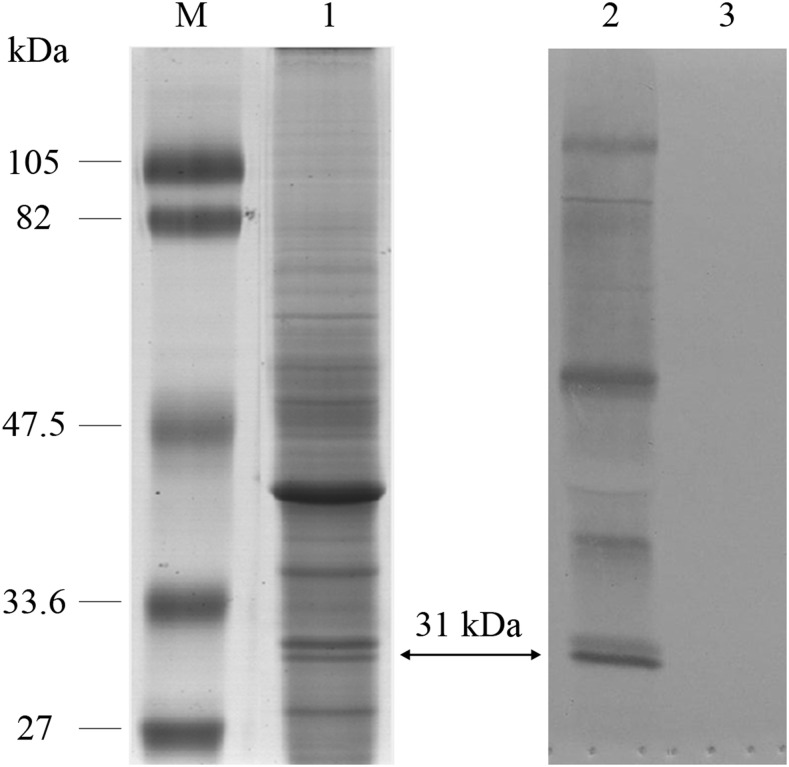
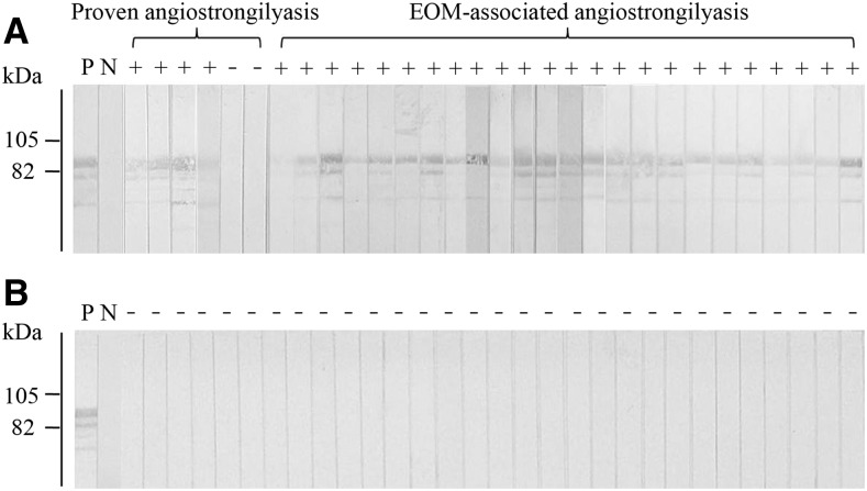
Angiostrongyliasis is a foodborne disease caused by a zoonotic nematode, Angiostrongylus cantonensis, which produces eosinophilic meningitis or meningoencephalitis (EOM) in humans. Definitive diagnosis is rarely possible because worms are almost never recovered from patients. Human disease can be diagnosed by clinical symptoms and serological tests. Presently, diagnosis is performed by serological detection of antibodies against specific somatic antigens (molecular mass 29-31 kDa) extracted from female worms. The life cycle of A. cantonensis must be maintained in the laboratory to provide a source of this diagnostic antigen. Here, we cloned and expressed recombinant A. cantonensis galectin-2 (rAcGal2) corresponding to a 31-kDa antigenic peptide. Recombinant protein was purified and used in immunoblot tests, which showed reactions with human serum panels consisting of six confirmed angiostrongyliasis and 24 clinically diagnosed cases of EOM-associated with angiostrongyliasis, 160 samples from patients with other parasitic infections, and 30 samples from normal healthy subjects. Accuracy, sensitivity, specificity, and positive and negative predictive values were 95.0%, 93.3%, 95.3%, 75.7%, and 98.9%, respectively. The test was nonreactive with sera of human gnathostomiasis and cysticercosis, two diseases that could present similar neurological symptoms. Recombinant AcGal2 has potential as a diagnostic antigen and could replace native parasite antigens in further development of an angiostrongyliasis serodiagnostic test kit.


Figures

Figure 1.
Figure 1.
SDS-PAGE analysis of Angiostrongylus cantonensis young adult female worm somatic extract (left) and representative immunoblotting reaction patterns (right). Lane M, molecular mass markers: lane 1, crude somatic extract of young adult female A. cantonensis stained with colloidal Coomassie G-250. Blots developed with pooled positive serum (lane 2) and pooled healthy control serum (lane 3). The arrows indicate the protein (on the left) and immunoreactive bands (on the right) at approximately 31 kDa.
Figure 2.
Figure 2.
The nucleotide and deduced amino acid sequences of recombinant galectin-2 protein derived from the 31-kDa antigenic protein of female Angiostrongylus cantonensis worms. The shaded regions indicate the amino acids comprising the carbohydrate recognition domains of the galectin superfamily. The bold italic letters indicate the predicted antigenic peptides. An asterisk indicates the stop codon. The underlines indicate antigenic peptide identified from liquid chromatography–tandem mass spectrometry.
Figure 3.
Figure 3.
Immunoblot characterization of purified fusion-tagged protein containing recombinant Angiostrongylus cantonensis galectin-2 (Gal2) and the fusion-tagged protein alone. (A) SDS-PAGE analysis of purified proteins stained with Coomassie Brilliant Blue. Lane 1: molecular weight markers; lanes 2 and 3: fusion-tagged proteins with and without recombinant Gal2, respectively. An arrow indicates the Gal2 fusion protein band at approximately 91 kDa, whereas an arrow head indicates 66-kDa band of the fusion-tagged protein alone. (B) Representative image of immunoblot analysis probed with anti-6× His-tagged antibody (a), pooled positive serum of angiostrongyliasis patients (b), and pooled negative serum of healthy controls (c). Lane 1: fusion-tagged protein-containing recombinant Gal2 and lane 2: fusion-tagged protein alone.
Figure 4.
Figure 4.
Representative immunoblotting patterns of recombinant Angiostrongylus cantonensis galectin-2 when reacted with angiostrongyliasis and healthy control sera. Immunoblot reactivity of the purified recombinant protein: (A) proved with individual sera from patients with confirmed angiostrongyliasis (proven angiostrongyliasis, N = 6) and eosinophilic meningitis or meningoencephalitis (EOM)–associated angiostrongyliasis (EOM-associated angiostrongyliasis, N = 24); (B) proved with healthy individuals. “P” and “N” indicate pooled positive and pooled negative control serum, respectively. “+” and “−” indicate positive and negative results, respectively.

References

    1. Cross JH, 1978. Clinical manifestations and laboratory diagnosis of eosinophilic meningitis syndrome associated with angiostrongyliasis. Southeast Asian J Trop Med Public Health 9: 161–170. - PubMed
    1. Pien FD, Pien BC, 1999. Angiostrongylus cantonensis eosinophilic meningitis. Int J Infect Dis 3: 161–163. - PubMed
    1. Tsai HC, et al. 2001. Eosinophilic meningitis caused by Angiostrongylus cantonensis: report of 17 cases. Am J Med 111: 109–114. - PubMed
    1. Wang QP, Lai DH, Zhu XQ, Chen XG, Lun ZR, 2008. Human angiostrongyliasis. Lancet Infect Dis 8: 621–630. - PubMed
    1. Eamsobhana P, Lim PE, Yong HS, 2013. Genetic diversity of the rat lungworm, Angiostrongylus cantonensis, the major cause of eosinophilic meningitis. Hawaii J Med Public Health 72: 15–17. - PMC - PubMed

Publication types

Supplementary concepts

LinkOut - more resources