. 2019 Aug 8;4(15):e128722.
doi: 10.1172/jci.insight.128722.

Cell-specific ablation of Hsp47 defines the collagen-producing cells in the injured heart

Affiliations

Cell-specific ablation of Hsp47 defines the collagen-producing cells in the injured heart

Hadi Khalil et al. JCI Insight. .

Abstract

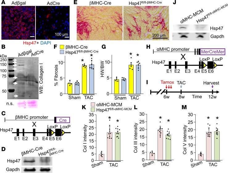
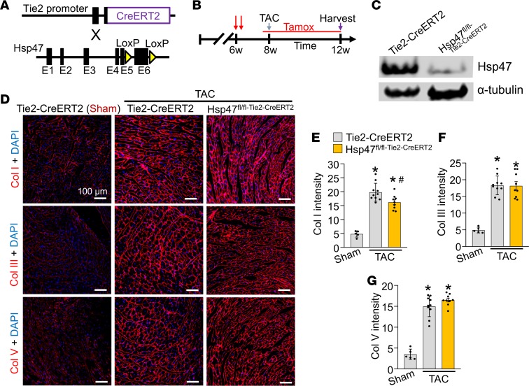
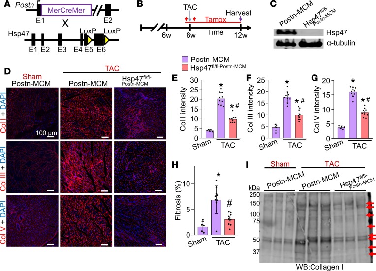
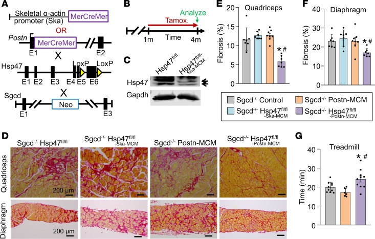
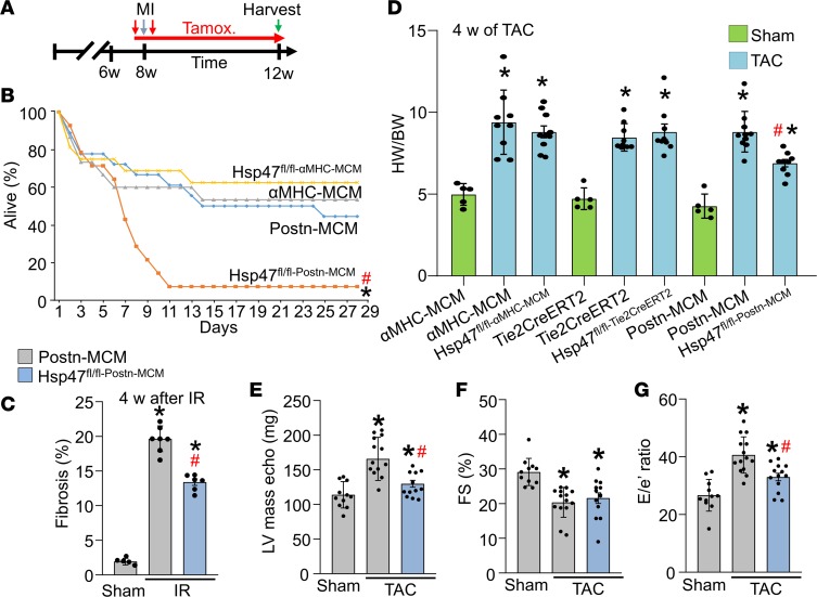
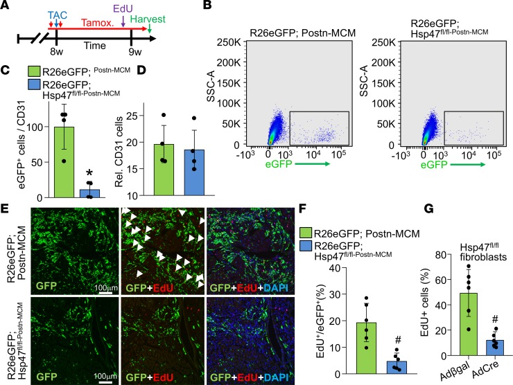
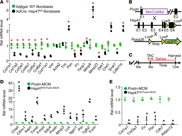
Collagen production in the adult heart is thought to be regulated by the fibroblast, although cardiomyocytes and endothelial cells also express multiple collagen mRNAs. Molecular chaperones are required for procollagen biosynthesis, including heat shock protein 47 (Hsp47). To determine the cell types critically involved in cardiac injury–induced fibrosis theHsp47 gene was deleted in cardiomyocytes, endothelial cells, or myofibroblasts. Deletion ofHsp47 from cardiomyocytes during embryonic development or adult stages, or deletion from adult endothelial cells, did not affect cardiac fibrosis after pressure overload injury. However, myofibroblast-specific ablation of Hsp47; blocked fibrosis and deposition of collagens type I, III, and V following pressure overload as well as significantly reduced cardiac hypertrophy. Fibroblast-specific Hsp47-deleted mice showed lethality after myocardial infarction injury, with ineffective scar formation and ventricular wall rupture. Similarly, only myofibroblast-specific deletion of Hsp47reduced fibrosis and disease in skeletal muscle in a mouse model of muscular dystrophy. Mechanistically, deletion of Hsp47 from myofibroblasts reduced mRNA expression of fibrillar collagens and attenuated their proliferation in the heart without affecting paracrine secretory activity of these cells. The results show that myofibroblasts are the primary mediators of tissue fibrosis and scar formation in the injured adult heart, which unexpectedly affects cardiomyocyte hypertrophy.

Keywords: Cardiovascular disease; Collagens; Fibrosis.


Conflict of interest statement

The authors have declared that no conflict of interest exists.

Figures

Figure 1
Figure 1. Cardiomyocyte-specific deletion of Hsp47 in the mouse heart does not block maladaptive fibrosis with TAC.
(A) Representative immunostaining for Hsp47 (red fluorescence) in cultured cardiac fibroblasts 72 hours after infection with Adβgal and AdCre. Nuclei are stained blue with DAPI. Scale bar: 100 μm. (B) Western blot showing levels of secreted collagen type I in the culture media of heart fibroblasts 72 hours after infection with either Adβgal or AdCre. White arrows show collagen isoforms. Molecular weight migration standard and sizes are also shown. Nonspecific (n.s.) Ponceau staining (pink) is shown as a processing and loading control. (C) Schematic representation of breeding βMHC-Cre–transgenic mice with Hsp47-loxP–targeted mice. (D) Western blot analysis for Hsp47 isolated from fractionated cardiomyocytes of the 2 genotypes of mice shown. Gapdh is shown as the loading control. (E and F) Picrosirius red–stained histological heart sections, and quantitation of the area of fibrosis (red) in hearts from the indicated genotypes of mice with the βMHC-Cre transgene after 4 weeks of TAC injury. Average fibrotic area ± SEM, n = 5–8 mice in each group, *P < 0.05 versus sham-operated βMHC-Cre mice. P values were calculated by 1-way ANOVA with Tukey’s post hoc test. Scale bar: 200 μm (G) Heart-weight-to-body-weight (HW/BW) ratio in mice after 4 weeks of TAC. n = 5–8 in each group. *P < 0.05 versus βMHC-Cre sham mice. (H) Breeding scheme of αMHC-MCM–transgenic mice with Hsp47-loxP–targeted mice. (I) Experimental regimen whereby mice were subjected to TAC injury or sham procedure for 4 weeks along with tamoxifen treatment by injection (vertical red arrows). (J) Western blot analysis for Hsp47 isolated from adult heart fractionated cardiomyocytes of the 2 genotypes shown. Gapdh is shown as a loading control. (K, L, and M) Quantitation of collagens type I, III, and V, respectively, from immunohistochemical heart images from WT αMHC-MCM mice versus Hsp47 cardiomyocyte-specific mice, as shown in Supplemental Figure 3. Ten random histological sections from each mouse heart were imaged and quantified from 5–10 mice each per group. *P < 0.05 versus αMHC-MCM Sham. P values were calculated with 1-way ANOVA with Tukey’s post hoc test.
Figure 2
Figure 2. Endothelial-specific deletion of Hsp47 in the heart.
(A) Schematic of breeding tamoxifen-inducible Tie2-CreERT2–transgenic mice with Hsp47-loxP–targeted mice. (B) Experimental regimen for mice subjected to TAC or a sham procedure for 4 weeks. Mice were injected at 6 weeks of age 2 times with tamoxifen and then put on tamoxifen chow before 8 weeks of age through harvesting at 12 weeks. (C) Western blot analysis for Hsp47 from endothelial cells isolated by FACS from the genotypes shown. Gapdh is a loading control. (DG) Immunohistochemical heart images stained (scale bar: 100 μm) and quantified for collagen type I, III, and V from Tie2-CreERT2–transgenic mice and Hsp47 endothelial-specific deleted mice. Mice were subjected to TAC as shown in B. Quantitation shows mean intensity of immunoreactivity taken from 10 random histological sections from 5–10 mice in each group. *P < 0.05 versus Tie2-CreERT2 Sham. #P < 0.05 versus Tie2-CreERT2 TAC. P values were calculated with 1-way ANOVA with Tukey’s post hoc test.
Figure 3
Figure 3. Myofibroblast-specific deletion of Hsp47 in the heart reduces myocardial fibrosis after TAC.
(A) Schematic representation of Postn-MerCreMer–targeted (MCM-targeted) mice crossed with Hsp47-loxP–targeted mice. (B) Experimental regimen of tamoxifen injections (red vertical arrows) and feed treatment (red horizontal line) in mice subjected to TAC for 4 weeks. (C) Western blot analysis for Hsp47 and α-tubulin from 500,000 EGFP+ cells isolated by FACS from hearts of the 2 genotypes of mice shown (R26eGFP reporter was also present). (DG) Representative immunohistochemistry (scale bar: 100 μm) of heart tissue sections and quantitation for collagen type I, III, and V from hearts of Postn-MCM control mice and Hsp47 myofibroblast-specific deleted mice using the Postn-MCM allele after 4 weeks of TAC. (H) Quantitation from Picrosirius red–stained histological sections in hearts from the indicated genotypes of mice after 4 weeks of TAC injury. *P < 0.05 versus Postn-MCM Sham. #P < 0.05 versus Postn-MCM TAC. n = 5–10 mice in each group. P values were calculated using a 1-way ANOVA with Tukey’s post hoc test. (I) Western blot analysis for collagen type I from heats of sham and TAC-operated mice using cardiac extracellular matrix–specific protein preparations from the indicated genotypes of mice. The red arrows show collagen isoforms. Position of molecular weight standards (kDa) are shown on the left.
Figure 4
Figure 4. Myofibroblast-specific but not myofiber-specific deletion of Hsp47 in skeletal muscle reduces muscular dystrophy–dependent tissue fibrosis.
(A) Schematic of the MCM cDNA driven by the human skeletal α-actin promoter (myofiber-specific) or the myofibroblast-specific Postn genetic locus to delete the Hsp47 gene with tamoxifen treatment. These lines were crossed into the δ-sarcoglycan–null (Sgcd–/–) background. (B) Experimental tamoxifen dosing regimen administered in the feed. (C) Western blot analysis for total Hsp47 protein using whole muscle protein lysates from the quadriceps of mice of the indicated genotypes. n = 6 mice per group. Gapdh is shown as a loading control. (D) Representative Picrosirius red–stained histological sections from quadriceps and diaphragm from 4-month-old mice of the indicated genotypes. Scale bar: 200 μm. (E and F) Quantitation of fibrosis from Picrosirius red–stained histological sections from quadriceps and diaphragm of 4-month-old mice of the indicated genotypes. n = 7 mice in each group. (G) Average time spent running on a treadmill of 4-month-old mice of the indicated genotypes. n = 6–10 in each group. Significance was determined using P values calculated by 1-way ANOVA with Tukey’s post hoc test. *P < 0.05 versus Sgcd–/–-control. #P < 0.05 versus Sgcd–/– Hsp47fl/fl-Ska-MCM. The legend applies to E, F and G.
Figure 5
Figure 5. Myofibroblast-specific Hsp47 deletion alters acute scar formation and the hypertrophic response.
(A) Experimental scheme whereby αMHC-MerCreMer–transgenic mice or Postn-MerCreMer allele–containing mice were subjected to myocardial infarction injury for 4 weeks with 2 injections (vertical red arrows) of tamoxifen treatment and then tamoxifen in the feed for 4 weeks (horizontal red arrow). (B) Kaplan-Meier plot of survival of the indicated genotypes of mice after MI injury. n = 11–13 mice in each group. (C) Quantitation of fibrosis from Picrosirius red–stained histological sections from hearts after 4 week of I/R injury of the indicated genotypes. n = 6 mice in each group. (D) Gravimetric assessed heart-weight-to-body-weight (HW/BW) ratios in mice of the indicated genotypes after 4 weeks of TAC. n = 5 sham mice, n = 9–10 TAC mice in each group. *P < 0.05 versus sham; #P < 0.05 versus Postn-MCM TAC. P values were calculated by 2-way ANOVA and Bonferroni post hoc test. (EG) Echocardiographic assessment of ventricular (LV) calculated mass, left ventricular fractional shortening (FS%) percentage, and early mitral inflow velocity to mitral annular early diastolic velocity ratio (E/e) in the indicated genotypes of mice after 4 weeks of TAC injury or a sham procedure. *P < 0.05 versus Postn-MCM sham. #P < 0.05 versus Postn-MCM TAC. P values were calculated with 1-way ANOVA with Tukey’s post hoc test. Number of mice used is shown in the scatter plots.
Figure 6
Figure 6. Myofibroblast-specific Hsp47 deletion reduces myofibroblasts in vivo and their proliferation.
(A) Experimental scheme whereby mice were subjected to TAC injury for 7 days. Mice received 2 i.p injections of tamoxifen and were fed tamoxifen-laden chow 48 hours before surgery and were then maintained on this chow until harvesting. Mice also received a single i.p EdU injection 4 hours before sacrifice at day 7 after TAC. (B) Representative flow cytometry plots of isolated EGFP+ interstitial cells (plotted as EGFP fluorescence signal on the x axis versus side scatter on the y axis) from hearts of the indicated genotypes of mice; 100,000 cells are displayed in the blots. (C) The ratio of total EGFP+ myofibroblasts normalized to CD31+ cells from the hearts of the indicated genotypes of mice after 1 week of TAC. Error bars represent SEM; n = 4 mice in each group. *P < 0.05 versus Postn-MCM; R26eGFP. P values were calculated with a Student’s t test. (D) Relative number of CD31+ cells in the interstitial fractions in hearts of the indicated genotypes of mice after 1 week of TAC. (E) Representative immunohistological images (scale bar: 100 μm) of EdU+ and EGFP+ interstitial cells at the time of harvest for mice treated, as shown in A. DAPI was used to show nuclei (blue). n = 5 mice in each group. (F) Quantitation of GFP+ cells that were also EdU+ in heart histological sections from mice subjected to TAC of the indicated genotype. P values were calculated with Student’s t test. #P < 0.05 versus R26eGFP Postn-MCM controls. (G) Quantitation of EdU+ Hsp47fl/fl cardiac fibroblasts over 24 hours in culture previously treated with AdCre or Adβgal infection. A total of 6 images were analyzed per group. P values were calculated with Student’s t test. #P < 0.05 versus Adβgal infection. Data shown are the mean ± SEM.
Figure 7
Figure 7. Hsp47 deletion in myofibroblasts reduces ECM-related gene expression and promotes an altered differentiated state.
(A) Adult primary heart fibroblasts were isolated from Hsp47-loxP–targeted mice infected with Adβgal (WT) or AdCre (deleted samples). Seventy-two hours after infection cells were washed and incubated in 2% serum containing DMEM media with 20 μM ascorbic acid for 24 hours before RNA isolation. The data are real-time PCR results showing the expression levels of the indicated genes. n = 4 separate experiments. *P < 0.05 versus Adβgal WT. (B) Schematic representation of the Postn-MCM mouse line crossed with the Hsp47-loxP site–containing gene–targeted line and the Rosa26 reporter line (R26eGFP). (C) Experimental scheme with TAC stimulation and tamoxifen with injection and laden food. (D and E) Quantification of selected mRNAs in Hsp47-deleted EGFP+ myofibroblasts isolated from hearts of Hsp47fl/flPostn-MCM/+R26eGFP/+ allele containing mice, 4 weeks after TAC injury. n = 3, *P < 0.5. P values were calculated with Student’s t test. Data shown are the mean ± SEM.

References

    1. Gordon MK, Hahn RA. Collagens. Cell Tissue Res. 2010;339(1):247–257. doi: 10.1007/s00441-009-0844-4. - DOI - PMC - PubMed
    1. Weber KT, Sun Y, Tyagi SC, Cleutjens JP. Collagen network of the myocardium: function, structural remodeling and regulatory mechanisms. J Mol Cell Cardiol. 1994;26(3):279–292. doi: 10.1006/jmcc.1994.1036. - DOI - PubMed
    1. Brilla CG, Maisch B, Weber KT. Myocardial collagen matrix remodelling in arterial hypertension. Eur Heart J. 1992;13 Suppl D:24–32. - PubMed
    1. Weber KT. Cardiac interstitium in health and disease: the fibrillar collagen network. J Am Coll Cardiol. 1989;13(7):1637–1652. doi: 10.1016/0735-1097(89)90360-4. - DOI - PubMed
    1. Sun Y, Weber KT. Infarct scar: a dynamic tissue. Cardiovasc Res. 2000;46(2):250–256. doi: 10.1016/S0008-6363(00)00032-8. - DOI - PubMed

Publication types

MeSH terms