

doi: 10.1097/CORR.0000000000000886.
Classifications in Brief: The Dejour Classification of Trochlear Dysplasia
Affiliations
- PMID: 31393338
- PMCID: PMC6999944
- DOI: 10.1097/CORR.0000000000000886
Item in Clipboard
Classifications in Brief: The Dejour Classification of Trochlear Dysplasia
Clin Orthop Relat Res.
2019 Oct.
No abstract available
Conflict of interest statement
All ICMJE Conflict of Interest Forms for authors and
Figures
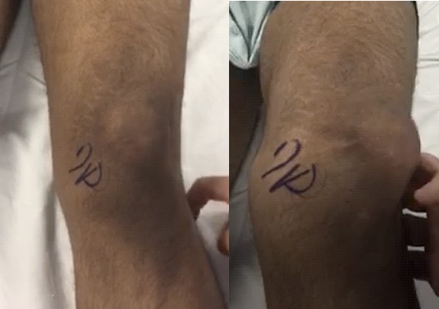
Clinical photographs of a patient with patellar dislocation on exam.
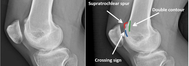
(A) Lateral knee radiograph with radiographic signs of trochlear dysplasia without labels. (B) Lateral knee radiograph with radiographic signs of trochlear dysplasia with labels: the blue line is the “Crossing sign,” representing the deepest point of the trochlear sulcus crossing the anterior border of the femoral condyles, the red line is a “Supratrochlear spur”, the prominence of the trochlea on the anterior aspect of the femoral cortex, and the green line is the “Double contour” line, the hypoplastic medial facet posterior to the lateral facet.
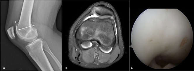
These images demonstrate trochlear dysplasia consistent with Dejour B trochlear dysplasia. (A) In this lateral knee radiograph, the asterisk (*) identifies the “Supratrochlear spur” and the arrow points to she “Crossing sign.” (B) This axial MRI shows a flattened trochlear groove. (C) This is an arthroscopic view of a flattened trochlea.
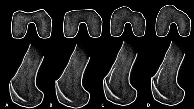
Illustrations of the Dejour classification on axial and lateral projection depicted as: (A) Dejour A, (B) Dejour B, (C) Dejour C, and (D) Dejour D.
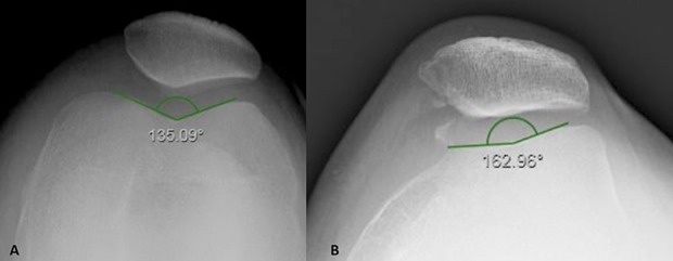
A sulcus angle is drawn on a Merchant view of the knee from the highest points of the medial and lateral condyle to the lowest point of the intercondylar notch. (A) This figure shows normal trochlear sulcus measurement (138° ± 6°). (B) In this image, a sulcus angle > 145° is indicative of trochlear dysplasia.
References
-
- Arendt EA, Dejour D. Patella instability: building bridges across the ocean a historic review. Knee Surg Sports Traumatol Arthrosc. 2013;21:279-293. - PubMed
-
- Biedert RM, Albrecht S. The patellotrochlear index: a new index for assessing patellar height. Knee Surg Sports Traumatol Arthrosc. 2006;14:707-712. - PubMed
-
- Bland JM, Altman DG. Statistical methods for assessing agreement between two methods of clinical measurement. Lancet. 1986;1:307-310. - PubMed
MeSH terms
LinkOut - more resources
Full Text Sources
