. 2019 Sep 6:17:601-614.
doi: 10.1016/j.omtn.2019.07.004. Epub 2019 Jul 19.

Suppression of Mutant Protein Expression in SCA3 and SCA1 Mice Using a CAG Repeat-Targeting Antisense Oligonucleotide

Affiliations

Suppression of Mutant Protein Expression in SCA3 and SCA1 Mice Using a CAG Repeat-Targeting Antisense Oligonucleotide

Eleni Kourkouta et al. Mol Ther Nucleic Acids. .

Abstract

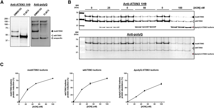
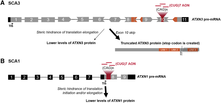
Spinocerebellar ataxia type 3 (SCA3) and type 1 (SCA1) are dominantly inherited neurodegenerative disorders that are currently incurable. Both diseases are caused by a CAG-repeat expansion in exon 10 of the Ataxin-3 and exon 8 of the Ataxin-1 gene, respectively, encoding an elongated polyglutamine tract that confers toxic properties to the resulting proteins. We have previously shown lowering of the pathogenic polyglutamine protein in Huntington's disease mouse models using (CUG)7, a CAG repeat-targeting antisense oligonucleotide. Here we evaluated the therapeutic capacity of (CUG)7 for SCA3 and SCA1, in vitro in patient-derived cell lines and in vivo in representative mouse models. Repeated intracerebroventricular (CUG)7 administration resulted in a significant reduction of mutant Ataxin-3 and Ataxin-1 proteins throughout the brain of SCA3 and SCA1 mouse models, respectively. Furthermore, in both a SCA3 patient cell line and the MJD84.2 mouse model, (CUG)7 induced formation of a truncated Ataxin-3 protein species lacking the polyglutamine stretch, likely arising from (CUG)7-mediated exon 10 skipping. In contrast, skipping of exon 8 of Ataxin-1 did not significantly contribute to the Ataxin-1 protein reduction observed in (CUG)7-treated SCA1154Q/2Q mice. These findings support the therapeutic potential of a single CAG repeat-targeting AON for the treatment of multiple polyglutamine disorders.

Keywords: CAG repeat; SCA1; SCA3; antisense oligonucleotide; exon skip; polyglutamine disorders.


Figures

Figure 1
Figure 1
(CUG)7 AON Induces Skipping of ATXN3 Exon 10 in Human SCA3 Fibroblasts (A) Scheme of repeat region in ATXN3 mRNA and exon 10 skip mRNA product; location of PCR primers for skip assessment and ddPCR primers and probes (in red) for skip level quantification is indicated. (B) Agarose gel analysis of ATXN3 transcript of SCA3 fibroblasts treated with increasing AON concentrations (10-50-200 nM), using PCR primers flanking exon 10. (C) Results of Sanger sequencing of 301 bp amplicon showing skip of exon 10 and conjoining of exon 9 to exon 11. (D) Absolute quantification of ATXN3 exon 10 skip levels by ddPCR. Data are presented as mean ± SEM of three technical replicates. H2O, no template control; L, SmartLadder Small Fragment (Eurogentec); NT, non-treated; RT, reverse transcriptase negative control.
Figure 2
Figure 2
(CUG)7 Reduces Levels of ATXN3 Protein and Results in Formation of the Truncated ΔpolyQ ATXN3 Isoform in Human SCA3 Fibroblasts (A) Validation of anti-ATXN3 antibody 1H9 and anti-polyglutamine (polyQ) antibody for specific detection of mutATXN3 in GM06153 protein lysate (input = 10 μg). (B) Western blot analysis of protein samples derived from (CUG)7-treated GM06153 fibroblasts, using 1H9 or anti-polyQ antibody (input = 10 μg). (C) Quantification of western blot band intensity, plotted as mutATXN3 and wtATXN3 protein reduction percentage and ΔpolyQ ATXN3 isoform percentage (calculated as the level of ΔpolyQ ATXN3 isoform signal divided by the sum of wtATXN3, mutATXN3, and ΔpolyQ ATXN3 isoform signal). Revert Total Protein whole-lane signal was used for normalization of amount of loaded protein per sample. Data are presented as mean ± SD of three or six technical replicates.
Figure 3
Figure 3
(CUG)-Induced Reduction of mutATXN3 Levels in the MJD84.2 SCA3 Mouse Model (A) Study design in the MJD84.2 mouse model, indicating treatment groups and sample size per treatment group. At first week of treatment, mice were 10–14 weeks old. (B) (CUG)7 distribution in right and left hemisphere regions of the MJD84.2 brain (n = 12–13 for right hemisphere groups and n = 5 for left hemisphere groups). Data are presented as mean ± SEM. The average difference in AON levels observed between left and right hemispheres of 19% was not significant, as assessed by two-tailed t test. (C) ddPCR analysis of exon 10 skipping levels in different brain regions. Data are presented as mean ± SEM. (D) Validation of anti-ATXN3 antibody in mouse brain, using 30 μg of cortex protein lysate as input (left panel), and representative example of western blot of some of the cerebellum samples of mice treated with VEH or (CUG)7, showing the emergence of the ΔpolyQ ATXN3 isoform after (CUG)7-treatment. (E) Mutant ATXN3 protein levels in various right hemisphere brain regions after treatment, as assessed by western blot analysis. Blot signals, normalized to Revert Total Protein whole-lane signal, are plotted on the right. Protein level data are presented as mean ± SEM. Percentages above bars indicate reduction in protein levels, compared with VEH set to 100%. Significance was assessed using a two-tailed t test comparing (CUG)7-treated mice with VEH-treated controls (*p < 0.05, **p < 0.01, ***p < 0.001).
Figure 4
Figure 4
Very Low Levels of ATXN1 Exon 8 Skipping Observed in Non-treated and (CUG)7-Treated Human SCA1 Fibroblasts (A) Scheme of repeat region on ATXN1 mRNA and exon 8 skip mRNA product; location of PCR primers for exon 8 skip detection on gel and of ddPCR primers and probes for quantification of skipping levels are indicated. (B) Agarose gel analysis of PCR products of ATXN1, using RNA extracted from SCA1 fibroblasts treated with 100 or 200 nM concentrations. (C) Results of Sanger sequencing of 183 bp amplicon showing skip of exon 8 and conjoining of exon 7 to exon 9. Non-coding strand, sequenced with reverse primer, is shown. (D) Absolute quantification of ATXN1 exon 8 skip levels by ddPCR. Data are presented as mean ± SEM of three technical replicates. One-way ANOVA followed by Dunnett’s multiple comparison post hoc test (*p < 0.05, **p < 0.01, compared with 0 nM AON). H2O, no template control; L, SmartLadder (Eurogentec); NT, non-treated, RT, reverse transcriptase negative control.
Figure 5
Figure 5
(CUG)7-Induced Reduction of mutATXN1 Levels in the SCA1154Q/2Q Mouse Model (A) Study design in the SCA1154Q/2Q mouse model, indicating treatment groups and sample size per treatment group. (B) (CUG)7 distribution in right hemisphere brain regions and spinal cord (SC) sections of the SCA1 mouse (n = 10–11). Data are presented as mean ± SEM. No significant differences in AON levels observed between different regions of mice treated with the same dosing regimen (one-way ANOVA and Tukey’s multiple comparison post hoc test). Unpaired two-tailed t test revealed significant differences between the low and high (CUG)7 dose in brainstem (**p < 0.01), cerebellum (*p < 0.05), and lumbar spinal cord (**p < 0.01). (C) Validation of anti-ATXN1 antibody in vivo, using 90 μg of cerebellum protein lysate as input. Blot was initially probed with anti-polyQ antibody 1C2, then stripped and reprobed with an anti-ATXN1 antibody (Origene) to confirm band identity. Secondary antibodies that fluoresce in different wavelengths were used in order to avoid false interpretations due to leftover signal. (D) ATXN1 protein levels in various right hemisphere brain regions and spinal cord sections after treatment, as assessed by western blot analysis. Protein level data of each mouse are plotted normalized to Revert Total Protein whole-lane signal and presented as mean ± SEM. Percentages above bars indicate reduction in protein levels, compared with VEH set to 100%. Significance was assessed using one-way ANOVA followed by Dunnett’s multiple comparison post hoc test (*p < 0.05, **p < 0.01, ***p < 0.001, compared with VEH). H, high-dose AON, 150 μg; L, low-dose AON, 75 μg; VEH, vehicle.
Figure 6
Figure 6
Proposed Mechanism of Action of (CUG)7 in SCA3 and SCA1 (A) (CUG)7 AONs bind to the (expanded) CAG repeat on exon 10 of the ATXN3 pre-mRNA. During pre-mRNA processing this can result in incorrect splicing of exon 10, leading to exclusion of exon 10 in mature mRNA and conjoining of exon 9 to exon 11. This junction results in generation of a stop codon in the beginning of exon 11, and therefore ATXN3 mRNA is translated into a truncated product that lacks regions encoded by exons 10 (i.e., polyQ stretch) and 11 (i.e., UIM 3). In addition, it is proposed that binding of (CUG)7 to the repeat on mature mRNA sterically hinders translation elongation, thereby conferring lower levels of ATXN3 protein. (B) (CUG)7 binds to the expanded CAG repeat on exon 8 of the ATXN1 (pre-)mRNA, causing steric hindrance of translation initiation and/or elongation, thereby lowering levels of ATXN1 protein. 5′ and 3′ UTRs and introns are in black; coding regions are in gray; exonic regions encoding UIMs and UIMs on protein are in orange. TIS, translation initiation site.

Similar articles

Cited by

References

    1. Bird T.D. Hereditary Ataxia Overview. In: Adam M.P., Ardinger H.H., Pagon R.A., Wallace S.E., editors. GeneReviews. University of Washington,; Seattle: 1993. pp. 1993–2019. - PubMed
    1. Paulson H.L., Shakkottai V.G., Clark H.B., Orr H.T. Polyglutamine spinocerebellar ataxias—from genes to potential treatments. Nat. Rev. Neurosci. 2017;18:613–626. - PMC - PubMed
    1. Monin M.L., Tezenas du Montcel S., Marelli C., Cazeneuve C., Charles P., Tallaksen C., Forlani S., Stevanin G., Brice A., Durr A. Survival and severity in dominant cerebellar ataxias. Ann. Clin. Transl. Neurol. 2015;2:202–207. - PMC - PubMed
    1. Krysa W., Sulek A., Rakowicz M., Szirkowiec W., Zaremba J. High relative frequency of SCA1 in Poland reflecting a potential founder effect. Neurol. Sci. 2016;37:1319–1325. - PMC - PubMed
    1. van de Warrenburg B.P., Sinke R.J., Verschuuren-Bemelmans C.C., Scheffer H., Brunt E.R., Ippel P.F., Maat-Kievit J.A., Dooijes D., Notermans N.C., Lindhout D. Spinocerebellar ataxias in the Netherlands: prevalence and age at onset variance analysis. Neurology. 2002;58:702–708. - PubMed

LinkOut - more resources