. 2019 Aug 7;11(8):1129.
doi: 10.3390/cancers11081129.

High Level Expression of MHC-II in HPV+ Head and Neck Cancers Suggests that Tumor Epithelial Cells Serve an Important Role as Accessory Antigen Presenting Cells

Affiliations

High Level Expression of MHC-II in HPV+ Head and Neck Cancers Suggests that Tumor Epithelial Cells Serve an Important Role as Accessory Antigen Presenting Cells

Steven F Gameiro et al. Cancers (Basel). .

Abstract

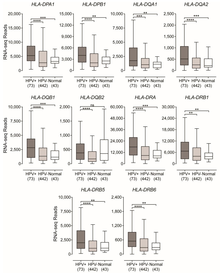
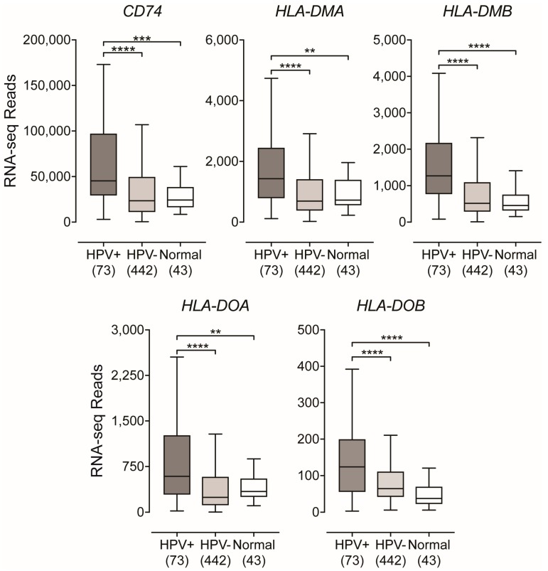
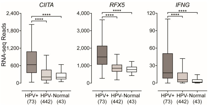
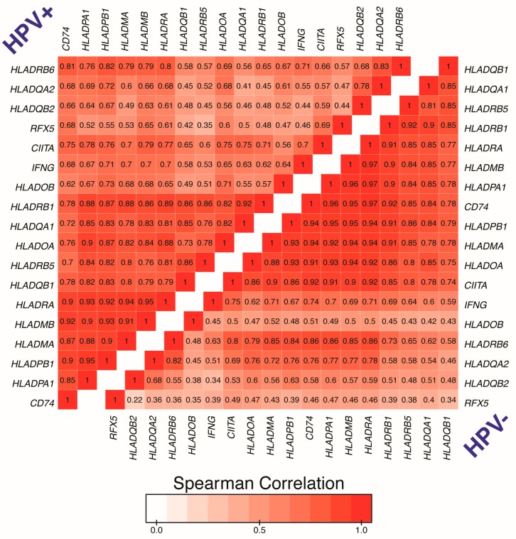
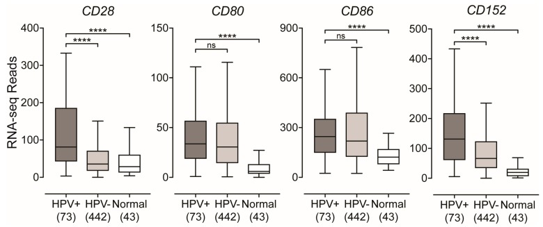
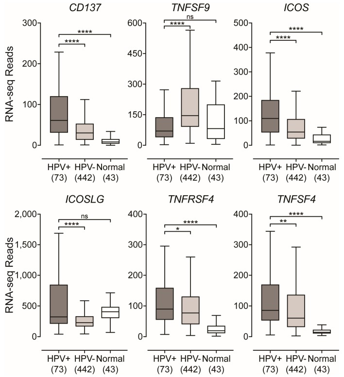
High-risk human papillomaviruses (HPVs) are responsible for a subset of head and neck squamous cell carcinomas (HNSCC). Expression of class II major histocompatibility complex (MHC-II) is associated with antigen presenting cells (APCs). During inflammation, epithelial cells can be induced to express MHC-II and function as accessory APCs. Utilizing RNA-seq data from over 500 HNSCC patients from The Cancer Genome Atlas, we determined the impact of HPV-status on the expression of MHC-II genes and related genes involved in their regulation, antigen presentation, and T-cell co-stimulation. Expression of virtually all MHC-II genes was significantly upregulated in HPV+ carcinomas compared to HPV- or normal control tissue. Similarly, genes that encode products involved in antigen presentation were also significantly upregulated in the HPV+ cohort. In addition, the expression of CIITA and RFX5-regulators of MHC-II-were significantly upregulated in HPV+ tumors. This coordinated upregulation of MHC-II genes was correlated with higher intratumoral levels of interferon-gamma in HPV+ carcinomas. Furthermore, genes that encode various co-stimulatory molecules involved in T-cell activation and survival were also significantly upregulated in HPV+ tumors. Collectively, these results suggest a previously unappreciated role for epithelial cells in antigen presentation that functionally contributes to the highly immunogenic tumor microenvironment observed in HPV+ HNSCC.

Keywords: MHC-II; T-cell; antigen presentation; co-stimulatory molecules; head and neck carcinoma; human papillomavirus; major histocompatibility complex; survival signals.


Conflict of interest statement

The authors declare no conflict of interest.

Figures

Figure 1
Figure 1
Expression of classical class II major histocompatibility complex (MHC-II) α- and β-chain genes in head and neck squamous cell carcinomas stratified by high-risk human papillomaviruses (HPV)-status. RNA-Seq by Expectation Maximization (RSEM)-normalized RNA-seq data for the indicated MHC-II genes was extracted from The Cancer Genome Atlas (TCGA) database for the head and neck squamous cell carcinoma (HNSC) cohort for HPV+, HPV−, and normal control tissues. Numbers in brackets refer to the number of samples included in each analysis. Statistical analysis was performed using a two-tailed non-parametric Mann–Whitney U test. ** p ≤ 0.01, *** p ≤ 0.001, **** p ≤ 0.0001, ns—not significant.
Figure 2
Figure 2
Expression of the invariant chain and MHC class II-like genes in head and neck carcinomas stratified by HPV-status. RSEM normalized RNA-seq data for the indicated genes involved in MHC-II-dependent antigen processing and presentation was extracted from the TCGA database for the HNSC cohort for HPV+, HPV−, and normal control tissues. Numbers in brackets refer to the number of samples included in each analysis. Statistical analysis was performed using a two-tailed non-parametric Mann–Whitney U test. ** p ≤ 0.01, *** p ≤ 0.001, **** p ≤ 0.0001, ns—not significant.
Figure 3
Figure 3
Expression of class II major histocompatibility complex transactivator (CIITA), regulatory factor X5 (RFX5), and interferon-gamma (IFNG) mRNA in head and neck carcinomas stratified by HPV-status. RSEM normalized RNA-seq data for the CIITA, RFX5, and IFNG genes were extracted from the TCGA database for the HNSC cohort for HPV+, HPV−, and normal control tissues. Numbers in brackets refer to the number of samples included in each analysis. Statistical analysis was performed using a two-tailed non-parametric Mann–Whitney U test. ** p ≤ 0.01, *** p ≤ 0.001, **** p ≤ 0.0001, ns—not significant.
Figure 4
Figure 4
Correlation matrix of genes involved in the MHC-II antigen presentation pathway in head and neck carcinomas stratified by HPV-status. RSEM normalized RNA-seq data for the genes listed above were extracted from the TCGA database for the HNSC cohort for HPV+ (upper triangle) and HPV− samples (lower triangle). Pairwise spearman correlation was performed followed by hierarchical clustering to group based on correlation. Number in boxes indicate Spearman’s rank correlation coefficient of analyzed gene pairs.
Figure 5
Figure 5
Expression of genes that encode for T-cell co-stimulatory molecules in head and neck carcinomas stratified by HPV-status. RSEM normalized RNA-seq data for genes that encode T-cell specific co-stimulatory molecules was extracted from the TCGA database for the HNSC cohort for HPV+, HPV−, and normal control tissues. Statistical analysis was performed using a two-tailed non-parametric Mann–Whitney U test. Numbers in brackets refer to the number of samples included in each analysis. **** p ≤ 0.0001, ns—not significant.
Figure 6
Figure 6
Expression of inducible T-cell survival signal molecules in head and neck carcinomas stratified by HPV-status. RSEM normalized RNA-seq data for inducible genes that encode for T-cell survival molecules was extracted from the TCGA database for the HNSC cohort for HPV+, HPV−, and normal control tissues. Statistical analysis was performed using a two-tailed non-parametric Mann–Whitney U test. Numbers in brackets refer to the number of samples included in each analysis. * p ≤ 0.05, ** p ≤ 0.01, *** p ≤ 0.001, **** p ≤ 0.0001, ns—not significant.

References

    1. Doorbar J., Egawa N., Griffin H., Kranjec C., Murakami I. Human Papillomavirus Molecular Biology and Disease Association. Rev. Med. Virol. 2015;25(Suppl. 1):2–23. doi: 10.1002/rmv.1822. - DOI - PMC - PubMed
    1. Forman D., de Martel C., Lacey C.J., Soerjomataram I., Lortet-Tieulent J., Bruni L., Vignat J., Ferlay J., Bray F., Franceschi S., et al. Global Burden of Human Papillomavirus and Related Diseases. Vaccine. 2012;30(Suppl. 5):F12–F23. doi: 10.1016/j.vaccine.2012.07.055. - DOI - PubMed
    1. Gillison M.L., Koch W.M., Capone R.B., Spafford M., Westra W.H., Wu L., Zahurak M.L., Daniel R.W., Viglione M., Symer D.E., et al. Evidence for a Causal Association between Human Papillomavirus and a Subset of Head and Neck Cancers. J. Natl. Cancer Inst. 2000;92:709–720. doi: 10.1093/jnci/92.9.709. - DOI - PubMed
    1. Syrjänen S. Human Papillomavirus (HPV) in Head and Neck Cancer. J. Clin. Virol. 2005;32(Suppl. 1):S59–S66. - PubMed
    1. Gillison M.L., Castellsagué X., Chaturvedi A., Goodman M.T., Snijders P., Tommasino M., Arbyn M., Franceschi S. Eurogin Roadmap: Comparative Epidemiology of HPV Infection and Associated Cancers of the Head and Neck and Cervix. Int. J. Cancer. 2013;134:497–507. doi: 10.1002/ijc.28201. - DOI - PubMed