. 2019 Aug:46:274-289.
doi: 10.1016/j.ebiom.2019.07.072. Epub 2019 Aug 5.

A major role of TWEAK/Fn14 axis as a therapeutic target for post-angioplasty restenosis

Affiliations

A major role of TWEAK/Fn14 axis as a therapeutic target for post-angioplasty restenosis

Nerea Méndez-Barbero et al. EBioMedicine. 2019 Aug.

Abstract

Background: Tumor necrosis factor-like weak inducer of apoptosis (Tnfsf12; TWEAK) and its receptor Fibroblast growth factor-inducible 14 (Tnfrsf12a; Fn14) participate in the inflammatory response associated with vascular remodeling. However, the functional effect of TWEAK on vascular smooth muscle cells (VSMCs) is not completely elucidated.

Methods: Next generation sequencing-based methods were performed to identify genes and pathways regulated by TWEAK in VSMCs. Flow-citometry, wound-healing scratch experiments and transwell migration assays were used to analyze VSMCs proliferation and migration. Mouse wire injury model was done to evaluate the role of TWEAK/Fn14 during neointimal hyperplasia.

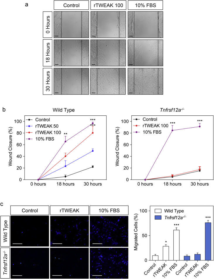
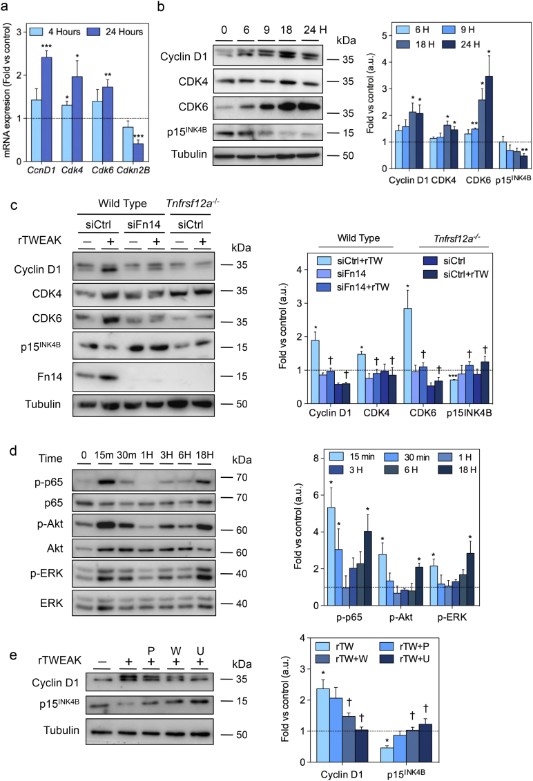
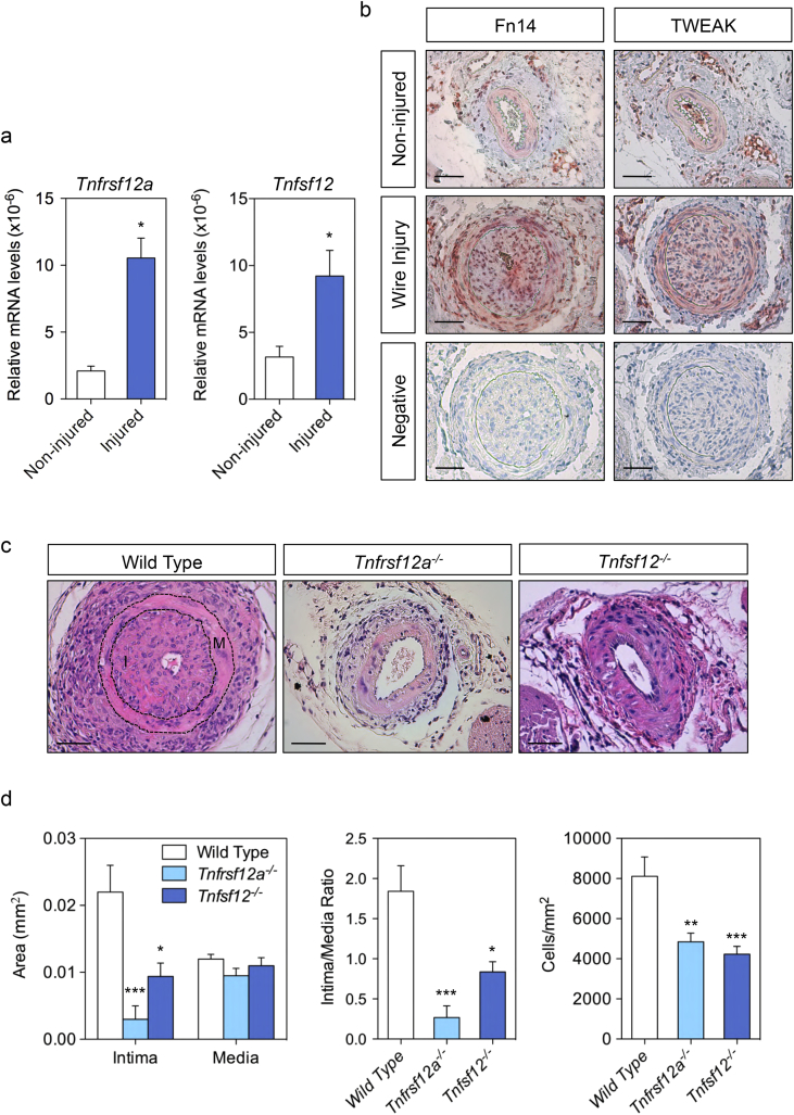
Findings: TWEAK up-regulated 1611 and down-regulated 1091 genes in VSMCs. Using a gene-set enrichment method, we found a functional module involved in cell proliferation defined as the minimal network connecting top TWEAK up-regulated genes. In vitro experiments in wild-type or Tnfrsf12a deficient VSMCs demonstrated that TWEAK increased cell proliferation, VSMCs motility and migration. Mechanistically, TWEAK increased cyclins (cyclinD1), cyclin-dependent kinases (CDK4, CDK6) and decreased cyclin-dependent kinase inhibitors (p15lNK4B) mRNA and protein expression. Downregulation of p15INK4B induced by TWEAK was mediated by mitogen-activated protein kinase ERK and Akt activation. Tnfrsf12a or Tnfsf12 genetic depletion and pharmacological intervention with TWEAK blocking antibody reduced neointimal formation, decreasing cell proliferation, cyclin D1 and CDK4/6 expression, and increasing p15INK4B expression compared with wild type or IgG-treated mice in wire-injured femoral arteries. Finally, immunohistochemistry in human coronary arteries with stenosis or in-stent restenosis revealed high levels of Fn14, TWEAK and PCNA in VSMCs enriched areas of the neointima as compared with healthy coronary arteries.

Interpretation: Our data define a major role of TWEAK/Fn14 in the control of VSMCs proliferation and migration during neointimal hyperplasia after wire injury in mice, and identify TWEAK/Fn14 as a potential target for treating in-stent restenosis. FUND: ISCiii-FEDER, CIBERCV and CIBERDEM.

Keywords: Cyclins; Fn14; Proliferation; Restenosis; TWEAK.


Conflict of interest statement

None.

Figures

Fig. 1
Fig. 1
RNA-Seq analysis of differentially expressed genes between TWEAK-treated and untreated VSMCs. A) Principal-component analysis (PCA) of VSMCs incubated 24 h in the presence or absence of rTWEAK (N = 3) based on RNA-sequencing gene expression levels. Color coded according the group of mice. B) Heat map representing differentially expressed genes between TWEAK-treated and untreated VSMCs (only genes with adjusted p-value by FDR < 0.05). C = control; TW = rTWEAK. C) Network linked to upregulated genes by rTWEAK in VSMCs. D) Top ten Gene Ontology (biological processes) terms significantly over-represented (adjusted p-value by FDR < 0.05) in the set of proteins from the network associated to TWEAK upregulated genes.
Fig. 2
Fig. 2
TWEAK increases VSMCs proliferation. A) Heat map shows cyclins (Ccn), cyclin-dependent kinases (Cdk) and cyclin-dependent kinase inhibitors (Cdkn) regulated by TWEAK in VSMCs (adjusted p < .05). B) Validation by RT-qPCR of select genes identified by RNASeq. RT-qPCR data represented as fold vs unstimulated cells of three biological replicates run in experimental duplicate and normalized to GADPH expression. Scatter plot showing the significant positive relationship between selected mRNA gene expression obtained by RNA-Seq and real-time PCR. Pearson correlation. r = 0.80; p = .006. C) Cell cycle analysis of VSMCs by propidium iodide staining and flow cytometry after 18 h of treatment. Control (0% FBS), rTWEAK (100 ng/mL rTWEAK) and 10% FBS (positive control). D) The percentage of VSMCs in G0/G1, S or G2/M phase of the cell cycle after 18 h of incubation with 0%FBS (Control), rTWEAK (50–100 ng/mL) or 10%FBS. Data represent mean ± SEM of 4 independent experiments (Student's t-test). **p < .01 vs Control and ***p < .001 vs Control). E) Proliferative curve of wild type or Tnfrsf12a−/− VSMCs cultured in the presence of 0%FBS (Control), rTWEAK (100 ng/mL) or 10%FBS from 0 to 72 h after serum starvation. Data represent mean ± SEM of 3 independent experiments (Student's t-test) *p < .05 vs Control and **p < .01 vs Control).
Fig. 3
Fig. 3
TWEAK increases migration of cultured VSMCs. A) Representative photograph of VSMC migration in the wound-healing assay. A single scrape wound was made on confluently plated wild type or Tnfrsf12a−/− VSMCs. Cells were then incubated for 18 to 30 h in the presence of 0.5% FBS (control), rTWEAK (50–100 ng/mL + 0.5% FBS) or 10% FBS. Wound closure images were captured and analyzed using an inverted microscope. Scale bars 200 μm. B) VSMCs migration was quantified by percentage of wound closure along time. Data represent mean ± SEM of 4 independent experiments (Student's t-test) **p < .01 vs Control and ***p < .001 vs Control. C) WT or Tnfrsf12a−/− cells were seeded in the upper surface of chemotaxis chambers and stimulated with 0.5% FBS (control), rTWEAK (100 ng/mL + 0.5% FBS) or 10% FBS. Quantification of migrated cells in ten fields per condition. Data represent the mean ± SEM of 4 independent experiments (Student's t-test) *p < .05 vs Control and ***p < .001 vs Control. Scale bars 20 μm.
Fig. 4
Fig. 4
TWEAK regulates cyclin D1, CDK4, CDK6 and p15INK4B expression in VSMCs. A) Quantitative real-time PCR analysis of CcnD1, Cdk4, Cdk6 and Cdkn2B mRNA expression in VSMCs upon rTWEAK (100 ng/mL) stimulation. Data represent the mean ± SEM of 4 independent experiments (Student's t-test) *p < .05 vs 0 h, **p < .01 vs 0 h and ***p < .001 vs 0 h). B) Western-blot analysis of cyclin D1, CDK4, CDK6 and p15INK4B in WT VSMCs. Data represent the mean ± SEM of 4 independent experiments (Student's t-test) *p < .05 vs 0 h, **p < .01 vs 0 h). C) Western-blot analysis of CcnD1, CDK4, CDK6, p15INK4B and Fn14 in WT or Tnfrsf12a−/− VSMCs transfected with negative control siRNA (siCtrl) or Fn14 siRNA after 18 h of rTWEAK (100 ng/mL) stimulation. Data represent the mean ± SEM of 4 independent experiments (Student's t-test) *p < .05 vs siControl, ***p < .001 vs siControl. p < .05 vs siControl + rTW). D) Western-blot analysis of ERK, Akt and p65 phosphorylation in WT VSMCs upon rTWEAK (100 ng/mL) stimulation. Data represent the mean ± SEM of 3 independent experiments (Student's t-test) *p < .05 vs 0 h. E) Western-blot analysis showing the effect of parthenolide (P), wormanin (W) and UO126 (U) on cyclin D1 and p15INK4B expression. Data represent the mean ± SEM of 3 independent experiments (Student's t-test) *p < .05 vs control; p < .05 vs rTW.
Fig. 5
Fig. 5
TWEAK participates in neointimal formation after wire injury. A) Relative Tnfrsf12a−/− or Tnfsf12−/− expression levels normalized to 18S rRNA of non-injured (N = 4) or injured (N = 5) femoral artery from wild type mice. Data represent the mean ± SEM (Student´s t test) *p < .05 vs non-injured artery. B) Representative images of non-injured and injured femoral artery cross-sections immunostained with anti-Fn14 or anti-TWEAK antibodies. Negative controls were incubated with non-specific IgG. Scale bars 50 μm. C) Representative images of injured femoral artery cross sections stained with hematoxylin and eosin from wild type, Tnfrsf12a−/− or Tnfsf12−/− mice. Scale bars 50 μm. M = media; I = intima. D) Cell number quantification per mm2, intima and media area, and intima/media ratio of cross-sections of injured femoral arteries from wild type (N = 11), Tnfrsf12a−/− (N = 11) or Tnfsf12−/− (N = 10) mice. Data represent the mean ± SEM (One-way ANOVA with Bonferroni's post-test) *p < .05 vs WT, **p < .01 vs WT and ***p < .001 vs WT.
Fig. 6
Fig. 6
TWEAK increases proliferation in vivo. A) Representative staining and quantification of positive PCNA cells in injured femoral artery cross-sections from Wild type (N = 9), Tnfrsf12a−/− (N = 11) or Tnfsf12−/− (N = 10) mice. Data represent the mean ± SEM (One-way ANOVA with Bonferroni's post-test) ***p < .001 vs WT. Scale bars 50 μm. B) Relative CcnD1, Cdk4, Cdk6 and Cdkn2B mRNA expression levels normalized to GAPDH mRNA expression of WT, Tnfrsf12a−/− or Tnfsf12−/− femoral arteries after wire injury. Data represent the mean ± SEM (N = 8 per group) (One-way ANOVA with Bonferroni's post-test) *p < .05 vs WT, **p < .01 vs WT and ***p < .001 vs WT. C) Representative images of anti-Cyclin D1, CDK6, CDK4 and p15INK4B staining of cross-sections of injured femoral arteries from WT, Tnfrsf12a−/− or Tnfsf12−/− mice. Quantification of intimal and medial percentage of Cyclin D1, CDK6, CDK4 and p15INK4B staining respectively, in injured femoral artery cross-sections of WT (N = 11), Tnfrsf12a−/− (N = 11) or Tnfsf12−/− (N = 8) Scale bars 50 μm. Data represent the mean ± SEM (One-way ANOVA with Bonferroni's post-test) *p < .05 vs WT, **p < .01 vs WT and ***p < .001 vs WT.
Fig. 7
Fig. 7
Anti-TWEAK treatment inhibits neointimal formation after wire injury. A) Experimental mouse design. Briefly, femoral arteries of 12-week-old male wild type mice were injured and treated with anti-TWEAK or IgG (10 mg/kg two times per week) for 14 days. B) Representative images of injured femoral artery cross sections from IgG or anti-TWEAK treated mice stained with H&E. Scale bars 50 μm. C) Quantification of number of cells per mm2, intima and media area, and intima/media ratio of cross-sections in injured conditions shown in B. IgG (N = 10), anti-TWEAK (N = 14). Data represent the mean ± SEM (Mann-Whitney U test) **p < .01 vs IgG. D) Representative images of anti-Cyclin D1, CDK6, CDK4 and p15INK4B staining of cross-sections of injured femoral arteries from IgG and anti-TWEAK treated mice. Quantification of intimal and medial percentage of Cyclin D1, CDK6, CDK4 and p15INK4B staining respectively, in the injured femoral artery of IgG (N = 10), anti-TWEAK (N = 11). Data represent the mean ± SEM (Mann-Whitney U test) **p < .01 vs IgG.
Fig. 8
Fig. 8
TWEAK and Fn14 are highly expressed in the neointima of human coronary arteries. Representative images of immunostaining for markers of VSMCs (α-SMA and calponin), TWEAK, Fn14 or PCNA in serial section from human coronary artery with or without stenosis. Negative controls were stained with non-specific IgG. Scale bars 100 μm.
Fig. 9
Fig. 9
Model illustrating the potential mechanism of TWEAK/Fn 14 axis in the development of neointimal formation after endovascular injury. Cartoon depicting TWEAK/Fn14 function in neointimal formation after wire injury. The interaction of TWEAK with its receptor Fn14 diminishes p15INK4B, increases cyclin D1, CDK4 and CDK6 expression and ERK1/2 and Akt activation in VSMCs, leading to an increase in VSMCs proliferation and migration. Therapeutic intervention with anti-TWEAK antibodies reduces neointimal formation.

References

    1. Dzau V.J., Braun-Dullaeus R.C., Sedding D.G. Vascular proliferation and atherosclerosis: new perspectives and therapeutic strategies. Nat Med. 2002;8:1249–1256. - PubMed
    1. Alexander M.R., Owens G.K. Epigenetic control of smooth muscle cell differentiation and phenotypic switching in vascular development and disease. Annu Rev Physiol. 2012;74:13–40. - PubMed
    1. Libby P., Hansson G.K. Inflammation and immunity in diseases of the arterial tree: players and layers. Circ Res. 2015;116:307–311. - PMC - PubMed
    1. Chaabane C., Otsuka F., Virmani R., Bochaton-Piallat M.-L. Biological responses in stented arteries. Cardiovasc Res. 2013;99:353–363. - PubMed
    1. Iakovou I., Schmidt T., Bonizzoni E., Ge L., Sangiorgi G.M., Stankovic G. Incidence, predictors, and outcome of thrombosis after successful implantation of drug-eluting stents. JAMA. 2005;293:2126–2130. - PubMed

MeSH terms