

doi: 10.3174/ajnr.A6145.
Epub 2019 Aug 8.
Optimal Detection of Subtle Gadolinium Leakage in CSF with Heavily T2-Weighted Fluid-Attenuated Inversion Recovery Imaging
Affiliations
- PMID: 31395665
- PMCID: PMC7048436
- DOI: 10.3174/ajnr.A6145
Item in Clipboard
Optimal Detection of Subtle Gadolinium Leakage in CSF with Heavily T2-Weighted Fluid-Attenuated Inversion Recovery Imaging
AJNR Am J Neuroradiol.
2019 Sep.
Abstract
Pericortical enhancement on postcontrast FLAIR images is a marker for subtle leptomeningeal blood-brain barrier leakage. We explored the optimal FLAIR sequence parameters for the detection of low gadolinium concentrations within the CSF. On the basis of phantom experiments and human in vivo data, we showed that detection of subtle pericortical enhancement can be facilitated by using a relatively long TE. Future studies should choose their FLAIR sequence parameters carefully when assessing pericortical enhancement due to subtle blood-brain barrier leakage.
© 2019 by American Journal of Neuroradiology.
Figures
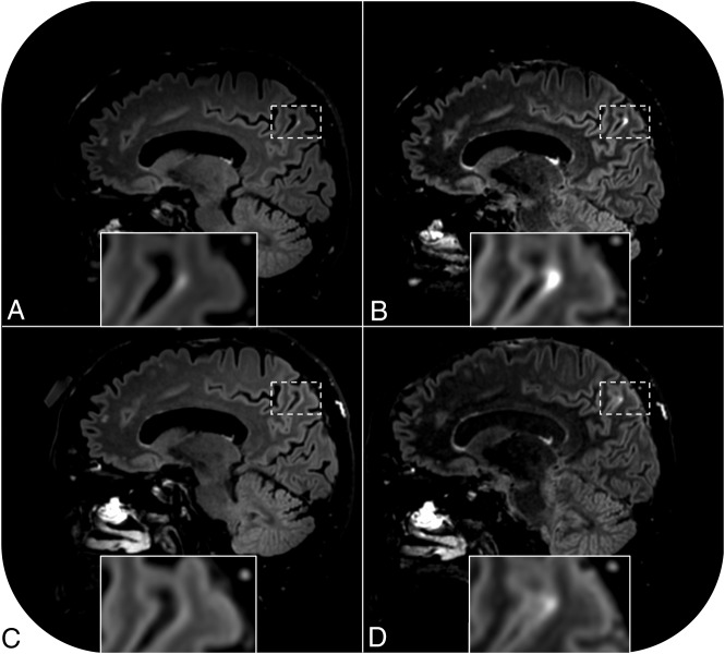
Pericortical enhancement is visible on postcontrast FLAIR images of a 68-year-old cognitively healthy female participant (case 1). Enhancement is apparent on the regular postcontrast FLAIR scan acquired 16 minutes after contrast administration (A) and even more conspicuous on the heavily T2-weighted FLAIR scan acquired immediately thereafter (B). The sequences were repeated 131 minutes after contrast administration. No signal enhancement is visible on the repeat regular postcontrast FLAIR scan (C), but enhancement is still visible on the repeat heavily T2-weighted FLAIR scan (D).
References
Publication types
MeSH terms
Substances
LinkOut - more resources
Full Text Sources
Medical