. 2019 Sep 1;33(17-18):1265-1279.
doi: 10.1101/gad.327593.119. Epub 2019 Aug 8.

Rationale for targeting BCL6 in MLL-rearranged acute lymphoblastic leukemia

Affiliations

Rationale for targeting BCL6 in MLL-rearranged acute lymphoblastic leukemia

Christian Hurtz et al. Genes Dev. .

Abstract

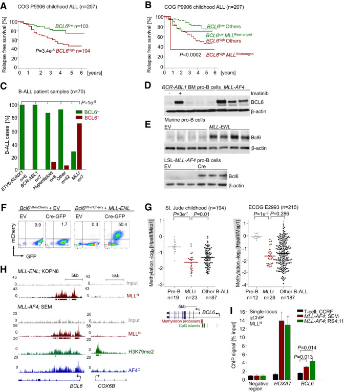
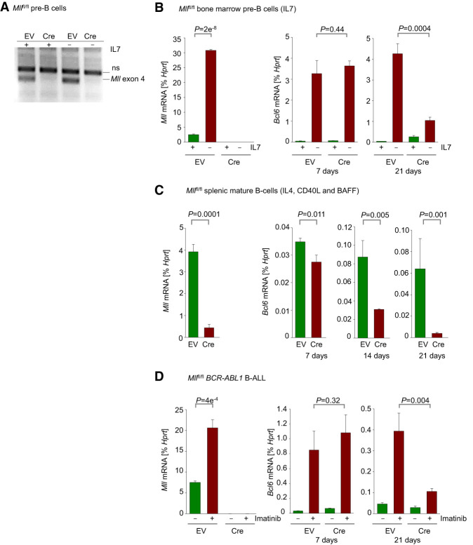
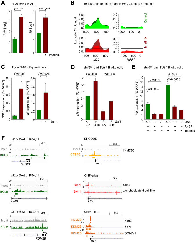
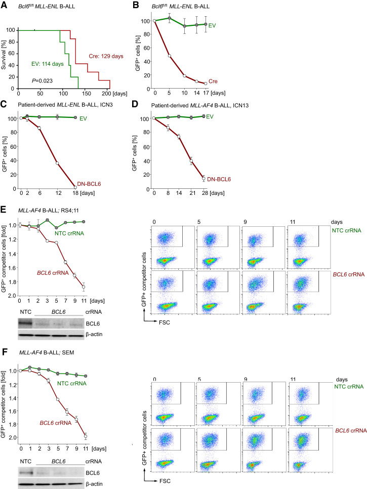
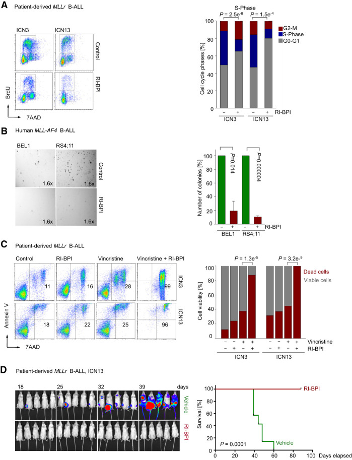
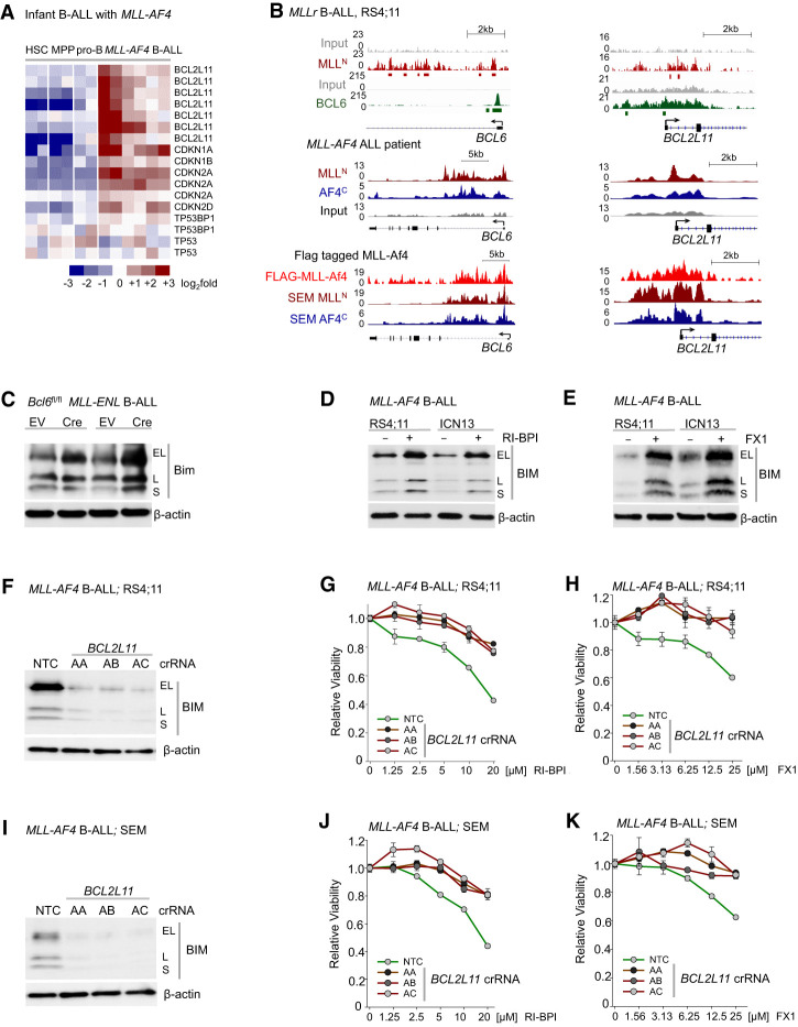
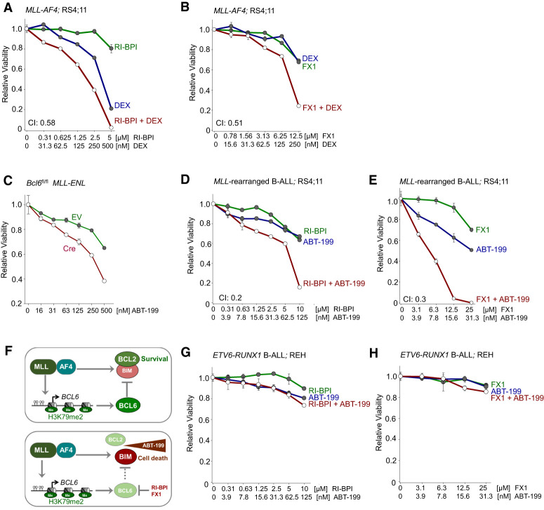
Chromosomal rearrangements of the mixed lineage leukemia (MLL) gene occur in ∼10% of B-cell acute lymphoblastic leukemia (B-ALL) and define a group of patients with dismal outcomes. Immunohistochemical staining of bone marrow biopsies from most of these patients revealed aberrant expression of BCL6, a transcription factor that promotes oncogenic B-cell transformation and drug resistance in B-ALL. Our genetic and ChIP-seq (chromatin immunoprecipitation [ChIP] combined with high-throughput sequencing) analyses showed that MLL-AF4 and MLL-ENL fusions directly bound to the BCL6 promoter and up-regulated BCL6 expression. While oncogenic MLL fusions strongly induced aberrant BCL6 expression in B-ALL cells, germline MLL was required to up-regulate Bcl6 in response to physiological stimuli during normal B-cell development. Inducible expression of Bcl6 increased MLL mRNA levels, which was reversed by genetic deletion and pharmacological inhibition of Bcl6, suggesting a positive feedback loop between MLL and BCL6. Highlighting the central role of BCL6 in MLL-rearranged B-ALL, conditional deletion and pharmacological inhibition of BCL6 compromised leukemogenesis in transplant recipient mice and restored sensitivity to vincristine chemotherapy in MLL-rearranged B-ALL patient samples. Oncogenic MLL fusions strongly induced transcriptional activation of the proapoptotic BH3-only molecule BIM, while BCL6 was required to curb MLL-induced expression of BIM. Notably, peptide (RI-BPI) and small molecule (FX1) BCL6 inhibitors derepressed BIM and synergized with the BH3-mimetic ABT-199 in eradicating MLL-rearranged B-ALL cells. These findings uncover MLL-dependent transcriptional activation of BCL6 as a previously unrecognized requirement of malignant transformation by oncogenic MLL fusions and identified BCL6 as a novel target for the treatment of MLL-rearranged B-ALL.

Keywords: B cells; BCL6; BIM; MLL.


Figures

Figure 1.
Figure 1.
BCL6 is up-regulated in MLL-rearranged ALL and correlates with poor clinical outcome. (A) Patients in a pediatric high-risk ALL trial (COG P9906; n = 207) were segregated into two groups based on whether BCL6 mRNA levels were higher (BCL6high) or lower (BCL6low) than the median expression value. RFS was assessed in the two groups by Kaplan-Meier analysis. Log-rank test, P = 3.39 × 10−05. (B) Multivariate analysis of RFS in pediatric B-ALL patients from the COG P9906 clinical trial. n = 207. Patients were segregated into four groups based on higher or lower than median expression levels of BCL6 and MLL status (rearranged or other). Log-rank test, P = 0.000208. (C) Immunohistochemical staining (Supplemental Fig. 2A) in bone marrow biopsies from B-ALL patients of different subtypes (n = 70) (Supplemental Table 2), including ETV6-RUNX1 (n = 6), BCR-ABL1 (n = 7), hyperdiploid (n = 8), MLL-rearranged (n = 7), and others (n = 42). Shown are percentages of different subtypes of B-ALLs that express (red) or do not express (green) BCL6. (D) BCL6 protein levels in CD19+ B cells from healthy donors (n = 3) and patient-derived MLL-rearranged B-ALL cells (n = 3). As a positive control for BCL6 expression, human Ph+ ALL (BV173) cells were treated with 10 µmol/L imatinib for 24 h. (E) Western blot analyses were performed to measure protein levels of Bcl6 upon overexpression of MLL-ENL (top panel) and Cre-mediated inducible activation of LSL-MLL-AF4 upon excision of a loxP-flanked Stop cassette (bottom panel) in murine pre-B cells. (F) A conditional Bcl6 knockout/mCherry reporter (Bcl6fl/fl-mCherry) mouse model in which exons 5–10 of Bcl6 are flanked by LoxP sites was developed (Geng et al. 2015). Cre-mediated deletion results in expression of a truncated Bcl6 protein fused to mCherry, allowing for simultaneous inducible ablation of Bcl6 and measurement of transcriptional activity of the Bcl6 promoter. Murine pre-B cells from Bcl6fl/fl-mCherry mice were transduced with MLL-ENL or an empty vector (EV) control, followed by transduction with a Cre-GFP expression vector or EV. Using the reporter capability, significantly higher transcriptional activation of Bcl6 in MLL-ENL transduced cells was observed, as reflected by increases in proportions of mCherry-positive cells. Transcriptional activation of Bcl6 was increased in concert with Cre-mediated deletion of Bcl6. (G) DNA methylation values of the BCL6 promoter region obtained from the HELP assays in pre-B cells from healthy donors, MLL-rearranged ALL patient samples, and other subtypes of B-ALL (St. Jude Childhood and Eastern Cooperative Oncology Group [ECOG] E2993). For DNA methylation at the BCL6 locus, methylation probe sets and CpG islands are shown. (H) ChIP-seq (chromatin immunoprecipitation [ChIP] combined with high-throughput sequencing) tracks on the BCL6 promoter region using an antibody specific for MLLN (red) in human B-ALL cell lines with MLL rearrangement: KOPN8 (MLL-ENL) and SEM (MLL-AF4). Gene models are shown in University of California at Santa Cruz Genome Browser view (hg18). ChIP-seq tracks for H3K79me2 (green) and AF4 C-terminal antibody (AF4C; blue) in the promoter region of BCL6 in SEM cells are also shown. The Y-axis represents the number of reads for peak summit normalized by the total number of reads per track (set to 1 Gb for each track). COX6B served as a negative control. (I) Quantitative single-locus ChIP validation of MLL binding to the promoter of BCL6 in SEM and RS4;11 cells was performed using HOXA7 (a known target of MLL fusion) as a positive control. ALL cells (CCRF-CEM) with no MLL rearrangement and an intergenic region with no binding enrichment were used as negative controls. Shown are mean values ± SD. n = 3.
Figure 2.
Figure 2.
Germline MLL is required for up-regulation of BCL6 expression during normal B-cell development. (A) Verification of deletion of Mll by genotyping. (B) Mllfl/fl pre-B cells expressing EV or Cre were cultured in the presence of IL-7. qRT-PCR was performed to measure levels of Mll (day 7) and Bcl6 (days 7 and 21) following Cre-mediated deletion of Mll with or without IL-7 withdrawal. Withdrawal of IL-7 was carried out 24 h prior to RNA extraction. (C) Mll (day 7) and Bcl6 (days 7, 14, and 21) mRNA levels were measured in Mllflfl spleen cells cultured in the presence of IL4, CD40L, and BAFF following Cre-mediated deletion of Mll. (D) qRT-PCR measurements of Mll (day 7) and Bcl6 (days 7 and 21) levels in Mllfl/fl BCR-ABL1 B-ALL cells following Cre-mediated deletion of Mll. Cells were treated with vehicle control or 1 µmol/L imatinib 24 h prior to RNA extraction. All qRT-PCR measurements were performed using Hprt as a reference. Shown are mean values ± SD. n = 3.
Figure 3.
Figure 3.
BCL6 positively regulates expression of MLL in pre-B and BCR-ABL1 pre-B-ALL cells. (A) Microarray analysis of Bcl6 and Mll expression in murine BCR-ABL1-driven B-ALL cells treated with either vehicle control or 10 µmol/L imatinib for 16 h. n = 3; GSE20987. (B) Human Ph+ ALL cells (Tom1) were treated with vehicle control or 10 µmol/L imatinib for 16 h and then subjected to ChIP-on-chip analysis using a BCL6 antibody (GSE24426). The Y-axis indicates enrichment versus input, while the X-axis indicates the location of probes within the respective locus relative to the transcriptional start site. The dark-green and light-green (vehicle control-treated) or red (imatinib-treated) tracings depict two replicates. Recruitment to MLL and HPRT (negative control) is shown. (C) Pre-B cells from a Tg(tetO-BCL6) mouse were cultured in the presence of IL-7 and treated with either vehicle control or 1 µg/mL Dox for 24 h to induce BCL6 expression. qRT-PCR was performed to measure mRNA levels of BCL6 and Mll relative to Hprt. (D) Bcl6+/+ and Bcl6−/− BCR-ABL1 B-ALL cells transduced with either a control or a BCL6-ER-overexpressing vector. qRT-PCR was performed to measure mRNA levels of Bcl6 and Mll relative to Hprt. (E) Bcl6+/+ and Bcl6−/− BCR-ABL1 B-ALL cells were treated with 10 µmol/L RI-BPI for 4 h, 1 µmol/L imatinib for 4 h, or a combination of both. Cells were then subjected to qRT-PCR to measure mRNA levels of Mll relative to Hprt. (F) ChIP-seq analyses of human MLL-rearranged B-ALL (RS4;11) cells revealed binding of BCL6 to the loci of CTBP2, BMI1, and KDM2B (GSE38403). ChIP-seq tracks showing binding of CTBP2 to the MLL promoter in human H1-hESC cells (ENCODE), binding of BMI1 to the MLL promoter in K562 (GSM937872) and a lymphoblastoid cell line (GSM3384454), and binding of KDM2B to the MLL promoter in K562 (GSM1812033), SEM (GSM2212235), and OCI-LY1 (GSM2171650) cell lines.
Figure 4.
Figure 4.
BCL6 is required for initiation and progression of MLL-rearranged B-ALL. (A) One million Bcl6fl/fl MLL-ENL B-ALL cells expressing EV or Cre were injected (intravenously) into sublethally irradiated NSG recipient mice. Cells were induced with 4-OHT for 24 h prior to injection. Overall survival of mice was compared by Kaplan-Meier analysis. Log-rank test, P = 0.023. Median survival for each group is indicated. (B) Bcl6fl/fl pre-B cells expressing MLL-ENL were transduced with a GFP-tagged 4-OHT-inducible Cre or EV control. Following 4-OHT induction, enrichment or depletion of GFP+ cells was monitored by flow cytometry. n = 3. (C,D) Patient-derived MLL-rearranged B-ALL cells (ICN3 [C] and ICN13 [D]) were transduced with a GFP-tagged 4-OHT-inducible dominant-negative form of BCL6 (DNBCL6-ERT2-GFP) or EV control. (BD) Relative changes of GFP+ cells were monitored over time following induction. n = 3. GFP+ cells (percentage) were normalized to EV of each day. (E,F) Electroporation of Cas9 ribonucleoproteins (RNPs), complexes of recombinant Cas9 with nontargeting (NT) crRNAs or crRNAs targeting BCL6, and tracrRNA was performed to transfect MLL-rearranged B-ALL cells (RS4;11 and SEM). The efficiency of CRISPR/Cas9-mediated deletion of BCL6 was assessed by Western blot analyses. RS4;11 (E) and SEM (F) cells transfected with Cas9/RNPs carrying NT or BCL6 crRNAs were mixed with GFP+ RS4;11 and GFP+ SEM competitor cells, respectively. Enrichment or depletion of GFP+ competitor cells was monitored by flow cytometry. n = 3.
Figure 5.
Figure 5.
Inhibition of BCL6 compromises proliferation, colony formation, and leukemia initiation of human MLL-rearranged B-ALL cells. (A) Patient-derived MLL-rearranged B-ALL cells (ICN3 and ICN13) were treated with 5 µmol/L RI-BPI or vehicle control for 2 h and then subjected to cell cycle analysis by measuring BrdU incorporation in combination with 7AAD staining. Representative FACS plots from three independent experiments are shown. Percentages of cells in the G0/G1, S, and G2/M phases are indicated. (B) Ten-thousand human MLL-rearranged B-ALL cells (BEL1 and RS4;11) were treated with 5 µmol/L RI-BPI or vehicle control and plated on semisolid methylcellulose. Colonies were counted after 14 d. The bar graphs show the mean values of the number of colonies ± SD. n = 3. (C) ICN3 and ICN13 cells were treated with vehicle control, 1 nmol/L vincristine, 5 µmol/L RI-BPI, or a combination of both for 5 d. Viability was measured by Annexin V/7AAD staining. Shown are the mean values from three independent experiments. (D) ICN13 cells (5 × 10⁵) labeled with firefly luciferase were injected (intrafemorally) into sublethally irradiated (2.5 Gy) NOD/SCID mice that were treated with intraperitoneal injections of vehicle or 25 mg/kg RI-BPI five times. Leukemia engraftment and progression were monitored by luciferase bioimaging at the times indicated. Kaplan-Meier analysis showing overall survival for each group (n = 7) of the recipient mice. Log-rank test, P = 0.0001.
Figure 6.
Figure 6.
BCL6-mediated transcriptional repression of BIM represents a vulnerability in MLL-rearranged B-ALL. (A) Gene expression analysis of proapoptotic and cell cycle checkpoint molecules in hematopoietic stem cells, myeloid hematopoietic progenitor cells, pre-B cells, and MLL-AF4+ blasts from infant ALL (GSE79450). (B, top) ChIP-seq analyses of human MLL-rearranged ALL (RS4;11) cells showed that MLL and BCL6 bind to the loci of BCL6 and BCL2L11 (BIM) (GSE38403). (Middle) ChIP-seq tracks showing binding of MLLN and AF4C to the loci of BCL6 and BCL2L11 in a MLL-AF4 ALL patient (GSE83671). (Bottom) MLL-Af4-Flag ChIP-seq peaks in MLL-Af4 leukemia cells and MLLN and AF4C ChIP-seq peaks in MLL-rearranged ALL (SEM) cells (GSE84116). (C) Protein levels of Bim upon 4-OHT-inducible Cre-mediated deletion of Bcl6 in pre-B cells expressing MLL-ENL. (D) Effects of vehicle control or 10 µmol/L RI-BPI for 48 h on BIM protein levels in human MLL-rearranged ALL cells. (E) Protein levels of BIM upon treatment with 50 µmol/L FX1 in human MLL-rearranged ALL cells. (FK) Electroporation of Cas9 RNPs, complexes of recombinant Cas9 with nontargeting (NT) crRNAs or crRNAs targeting BIM (BCL2L11), and tracrRNA was performed to transfect MLL-rearranged B-ALL cells (RS4;11 [FH] and SEM [IK]). (F,I) The efficiency of CRISPR/Cas9-mediated deletion of BIM was assessed by Western blot analysis. RS4;11 (G,H) and SEM (J,K) cells transfected with Cas9/RNPs carrying NT or BIM crRNAs were treated with increasing concentrations of RI-BPI (G,J) or FX1 (H,K). Shown is average relative viability. n = 3.
Figure 7.
Figure 7.
Dual targeting of BCL2 and BCL6 in MLL-rearranged B-ALL. (A) Human MLL-rearranged B-ALL cells were treated with Dex, RI-BPI, or a combination of both. CI values for ED50 are shown. Relative viability was assessed. n = 3. (B) Human MLL-rearranged B-ALL cells were treated with Dex, FX1, or a combination of both. Relative viability and CI were assessed. n = 3. (C) Relative viability (n = 3) was assessed upon treatment with increasing concentrations of ABT-199 in pre-B cells expressing MLL-ENL following 4-OHT Cre-mediated deletion of Bcl6. (D) Human MLL-rearranged B-ALL cells were treated with ABT-199, RI-BPI, or ABT-199 in combination with RI-BPI. Relative viability and CI were assessed. (E) Relative viability and CI were examined following treatment of human MLL-rearranged B-ALL cells with ABT-199, FX1, or a combination of both. (F) MLL-rearranged fusion proteins induce aberrant expression of BCL6, which contributes to MLL-rearranged-driven leukemogenesis by restricting expression of BIM, a proapoptotic molecule. The BCL2 inhibitor ABT-199 disrupts the interaction between BCL2 and BIM. Pharmacological inhibition of BCL6 (RI-BPI or FX1) synergizes with ABT-199 in eradicating MLL-rearranged B-ALL cells. (G,H) Relative viability was determined upon treatment with increasing concentrations of ABT-199, RI-BPI (G), FX1 (H), or a combination of ABT-199 with RI-BPI (G) or FX1 (H) in ETV6-RUNX1 B-ALL cells.

References

    1. Andersson AK, Ma J, Wang J, Chen X, Gedman AL, Dang J, Nakitandwe J, Holmfeldt L, Parker M, Easton J, et al. 2015. The landscape of somatic mutations in infant MLL-rearranged acute lymphoblastic leukemias. Nat Genet 47: 330–337. 10.1038/ng.3230 - DOI - PMC - PubMed
    1. Ayton PM, Cleary ML. 2001. Molecular mechanisms of leukemogenesis mediated by MLL fusion proteins. Oncogene 20: 5695–5707. 10.1038/sj.onc.1204639 - DOI - PubMed
    1. Baron BW, Nucifora G, McCabe N, Espinosa R III, Le Beau MM, McKeithan TW. 1993. Identification of the gene associated with the recurring chromosomal translocations t(3;14)(q27;q32) and t(3;22)(q27;q11) in B-cell lymphomas. Proc Natl Acad Sci 90: 5262–5266. 10.1073/pnas.90.11.5262 - DOI - PMC - PubMed
    1. Baron BW, Anatasi J, Montag A, Huo D, Baron RM, Karrison T. 2004. The human BCL6 transgene promotes the development of lymphomas in the mouse. Proc Natl Acad Sci 101: 14198–14203. 10.1073/pnas.0406138101 - DOI - PMC - PubMed
    1. Baron BW, Anatasi J, Hyjek EM, Bies J, Reddy PL, Dong J, Joseph L, Thirman MJ, Wroblewski K, Wolff L, et al. 2012. PIM1 gene cooperates with human BCL6 gene to promote the development of lymphomas. Proc Natl Acad Sci 109: 5735–5739. 10.1073/pnas.1201168109 - DOI - PMC - PubMed

Publication types

MeSH terms

Substances