

Functional Brain Changes During Mindfulness-Based Cognitive Therapy Associated With Tinnitus Severity
- PMID: 31396035
- PMCID: PMC6667657
- DOI: 10.3389/fnins.2019.00747
Functional Brain Changes During Mindfulness-Based Cognitive Therapy Associated With Tinnitus Severity
Abstract
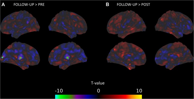
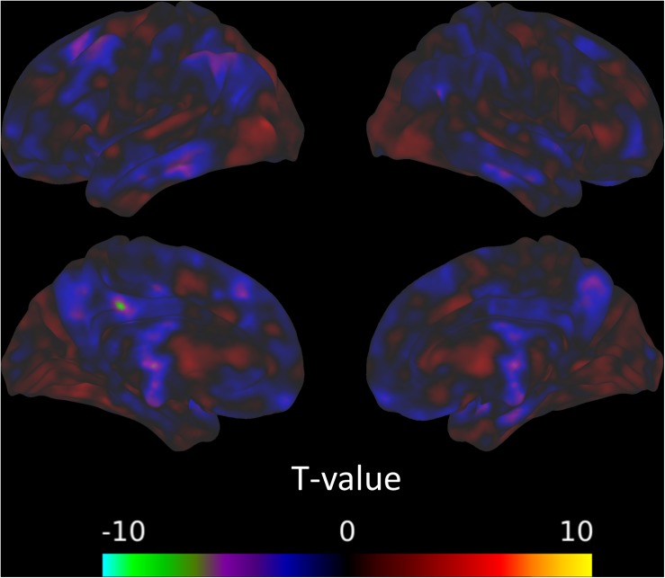
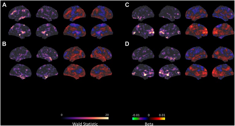
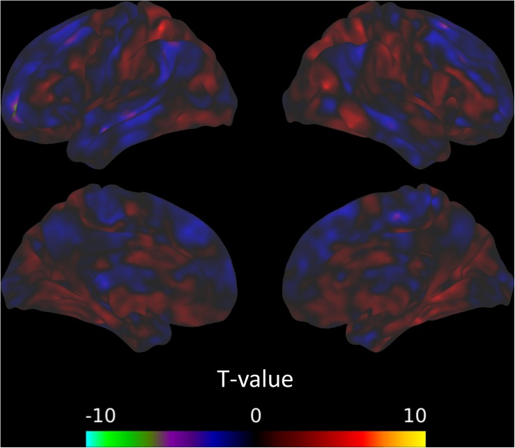
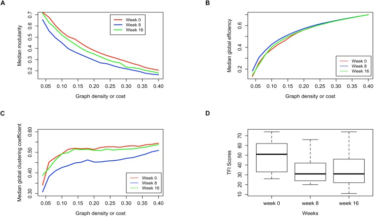
Mindfulness-based therapies have been introduced as a treatment option to reduce the psychological severity of tinnitus, a currently incurable chronic condition. This pilot study of twelve subjects with chronic tinnitus investigates the relationship between measures of both task-based and resting state functional magnetic resonance imaging (fMRI) and measures of tinnitus severity, assessed with the Tinnitus Functional Index (TFI). MRI was measured at three time points: before, after, and at follow-up of an 8-week long mindfulness-based cognitive therapy intervention. During the task-based fMRI with affective sounds, no significant changes were observed between sessions, nor was the activation to emotionally salient compared to neutral stimuli significantly predictive of TFI. Significant results were found using resting state fMRI. There were significant decreases in functional connectivity among the default mode network, cingulo-opercular network, and amygdala across the intervention, but no differences were seen in connectivity with seeds in the dorsal attention network (DAN) or fronto-parietal network and the rest of the brain. Further, only resting state connectivity between the brain and the amygdala, DAN, and fronto-parietal network significantly predicted TFI. These results point to a mostly differentiated landscape of functional brain measures related to tinnitus severity on one hand and mindfulness-based therapy on the other. However, overlapping results of decreased amygdala connectivity with parietal areas and the negative correlation between amygdala-parietal connectivity and TFI is suggestive of a brain imaging marker of successful treatment.
Keywords: functional MRI; graph connectivity analysis; mindfulness-based cognitive therapy; resting state MRI; tinnitus.
Figures
Similar articles
-
Effects of mindfulness based stress reduction therapy on subjective bother and neural connectivity in chronic tinnitus.Otolaryngol Head Neck Surg. 2015 May;152(5):919-26. doi: 10.1177/0194599815571556. Epub 2015 Feb 24. Otolaryngol Head Neck Surg. 2015. PMID: 25715350 Free PMC article.
-
Network-level functional topological changes after mindfulness-based cognitive therapy in mood dysregulated adolescents at familial risk for bipolar disorder: a pilot study.BMC Psychiatry. 2021 Apr 28;21(1):213. doi: 10.1186/s12888-021-03211-4. BMC Psychiatry. 2021. PMID: 33910549 Free PMC article.
-
Connectivity of precuneus to the default mode and dorsal attention networks: A possible invariant marker of long-term tinnitus.Neuroimage Clin. 2017 Jul 22;16:196-204. doi: 10.1016/j.nicl.2017.07.015. eCollection 2017. Neuroimage Clin. 2017. PMID: 28794980 Free PMC article.
-
Using resting state functional connectivity to unravel networks of tinnitus.Hear Res. 2014 Jan;307:153-62. doi: 10.1016/j.heares.2013.07.010. Epub 2013 Jul 26. Hear Res. 2014. PMID: 23895873 Review.
-
The Neural Mechanisms of Tinnitus: A Perspective From Functional Magnetic Resonance Imaging.Front Neurosci. 2021 Feb 11;15:621145. doi: 10.3389/fnins.2021.621145. eCollection 2021. Front Neurosci. 2021. PMID: 33642982 Free PMC article. Review.
Cited by
-
Mindfulness-Based Cognitive Therapy in Recurrent MDD Patients With Residual Symptoms: Alterations in Resting-State Theta Oscillation Dynamics Associated With Changes in Depression and Rumination.Front Psychiatry. 2022 Mar 7;13:818298. doi: 10.3389/fpsyt.2022.818298. eCollection 2022. Front Psychiatry. 2022. PMID: 35321228 Free PMC article.
-
The Impact of Mindfulness on Functional Brain Connectivity and Peripheral Inflammation in Breast Cancer Survivors with Cognitive Complaints.Cancers (Basel). 2023 Jul 15;15(14):3632. doi: 10.3390/cancers15143632. Cancers (Basel). 2023. PMID: 37509292 Free PMC article.
-
Mindfulness-based cognitive therapy on bereavement grief: Alterations of resting-state network connectivity associate with changes of anxiety and mindfulness.Hum Brain Mapp. 2021 Feb 1;42(2):510-520. doi: 10.1002/hbm.25240. Epub 2020 Oct 17. Hum Brain Mapp. 2021. PMID: 33068043 Free PMC article.
-
A Scoping Review of the Role of Attention in Tinnitus Management.Semin Hear. 2025 Mar 6;45(3-04):317-330. doi: 10.1055/s-0045-1804903. eCollection 2024 Aug. Semin Hear. 2025. PMID: 40256370 Review.
-
An examination of the reliability of seed-to-seed resting state functional connectivity in tinnitus patients.Neuroimage Rep. 2023 Feb 15;3(1):100158. doi: 10.1016/j.ynirp.2023.100158. eCollection 2023 Mar. Neuroimage Rep. 2023. PMID: 40568052 Free PMC article.
References
-
- Bartels H., Middel B. L., van der Laan B. F. A. M., Staal M. J., Albers F. W. J. (2008). The additive effect of co-occurring anxiety and depression on health status, quality of life and coping strategies in help-seeking tinnitus sufferers. Ear Hear. 29 947–956. 10.1097/AUD.0b013e3181888f83 - DOI - PubMed
LinkOut - more resources
Full Text Sources
Miscellaneous