. 2019 Jul 24:13:338.
doi: 10.3389/fncel.2019.00338. eCollection 2019.

Bone Marrow Stromal Cells Alleviate Secondary Damage in the Substantia Nigra After Focal Cerebral Infarction in Rats

Affiliations

Bone Marrow Stromal Cells Alleviate Secondary Damage in the Substantia Nigra After Focal Cerebral Infarction in Rats

Jizi Jin et al. Front Cell Neurosci. .

Abstract

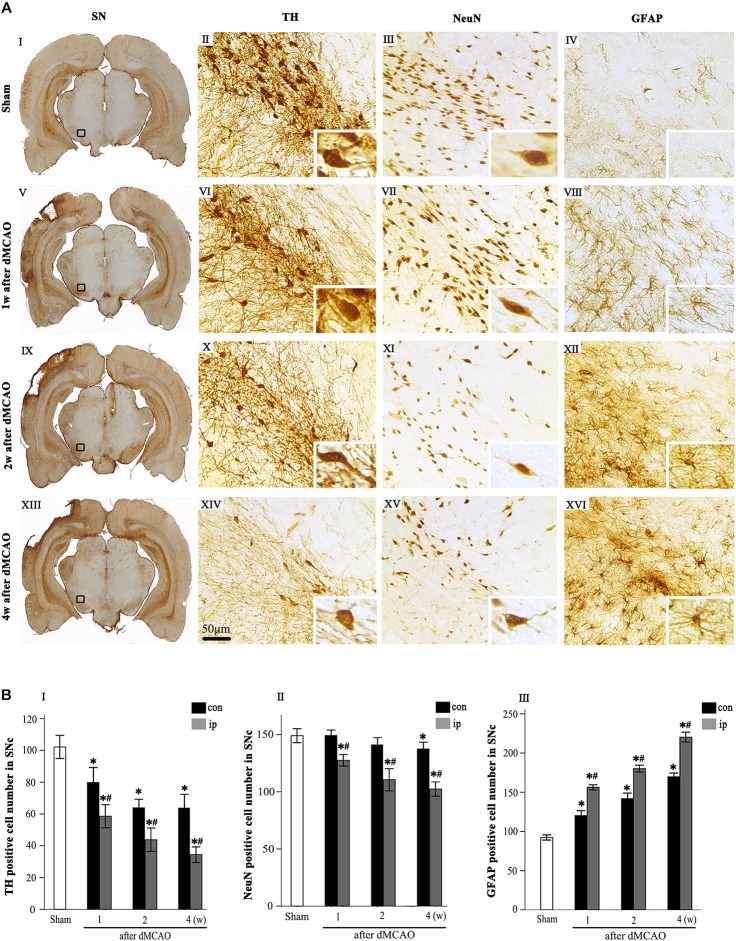
Transplantation of bone marrow stromal cells (BMSCs) is a promising therapy for ischemic stroke. Previously, we had reported that the secondary degeneration occurred in the ipsilateral substantia nigra (SN) after permanent distal branch of middle cerebral artery occlusion (dMCAO) in Sprague-Dawley rats. However, whether BMSCs have neurorestorative effects on the secondary damage in the SN after focal cerebral infarction has not known. In this study, rats were subjected to dMCAO followed by intravenous administration of BMSCs 1 day later. We found that transplanted BMSCs survived and migrated to cortical infarct areas and ipsilateral SN. Furthermore, BMSCs promoted neurogenesis through proliferation and differentiation in the SN after dMCAO. Rats implanted with BMSCs showed significant improvement in their performance of modified neurological severity scores and adhesive-removal test. Engrafted BMSCs enhanced survival of dopaminergic neuron, reduced gliosis in the ipsilateral SN, and increased contents of dopamine (DA) and its metabolites in the ipsilateral striatum after dMCAO. With pseudorabies virus-152 as a retrograde tracer, we also demonstrated that BMSCs could effectively enhance the cortico-striatum-nigral connections. These results suggest that BMSCs transplantation exerts neurorestorative effects after cortical infarction through promoting endogenous neurogenesis, increasing contents of DA and its metabolites, alleviating the secondary neuronal damage in the SN, enhancing the cortico-striatum-nigral projections pathway, and finally improving the neurological functional outcome.

Keywords: bone marrow stromal cells; cerebral infarction; neurorestoration; secondary degeneration; substantia nigra.


Figures

FIGURE 1
FIGURE 1
Secondary degeneration in the ipsilateral SN after dMCAO in rats. (A) Representative microphotographs of immunohistochemistry for TH (II,VI,X,XIV), NeuN (III,VII,XI,XV) and GFAP (IV,VIII,XII,XVI) in SNc at 1 week (V–VIII), 2 weeks (IX–XII), and 4 weeks (XIII–XVI) after dMCAO and sham-operated groups (I–IV). The pictures on the right are magnified from the square area on the left. Scale bar, 50 μm. (B) Quantitative analyses of TH+ (I), NeuN+ (II), and GFAP+ (III) cells in the SNc at 1, 2, and 4 weeks after dMCAO. Quantitative analyses showed that dMCAO decreased the number of TH+ cells in the ipsilateral [F(3, 101) = 94.59, p < 0.01] and contralateral SNc [F(3, 101) = 25.78, p < 0.01], and NeuN+ cells in the ipsilateral SNc [F(3, 83) = 38.19, p < 0.01], and increased the number of GFAP+ cells in the ipsilateral SNc [F(3, 122) = 638.85, p < 0.01] at 1, 2, and 4 weeks. Each bar represents the mean ± SD (n = 6 in each group). *p < 0.05 vs. sham-operated group; #p < 0.05 vs. contralateral groups at the same time point. SN, substantia nigra; SNc, substantia nigra compact part; TH, tyrosine hydroxylase; NeuN, neuron-specific nuclear-binding protein; GFAP, glial fibrillary acidic protein; Sham, sham-operated; w, week; dMCAO, distal middle cerebral artery occlusion.
FIGURE 2
FIGURE 2
Transplanted BMSCs migrate to the ipsilateral SN after dMCAO in rats. (A) The in vivo fluorescence images of migrated BMSCs-DiR+ in the brain at 2 h and 1, 7, 14 days post-intravenous transplantation following focal cortical infarction. (B) The ex vivo fluorescence images of migrated BMSCs-DiR+ in the cortex (I–IV) and SN (V–VIII) at 2 h and 1, 7, 14 days post-intravenous transplantation following focal cortical infarction. (C) Quantitative analyses (in vivo and ex vivo) of migrated BMSCs-DiR+ in the brain (I), cortex (II), and SN (III) at 2 h and 1, 7, 14 days post-intravenous transplantation following focal cortical infarction. Compared with the sham-operated group, the fluorescence intensities of BMSCs-DiR+ groups both in vivo [F(4, 25) = 7.67, p < 0.01] and ex vivo [cortex: F(4, 25) = 6.58, p < 0.01, SN: F(4, 25) = 4.09, p < 0.05] were significantly increased. *p < 0.05 vs. BMSCs-DiR group (n = 7 in each group). Sham, sham-operated; h, hour; d, day; w, week; L, left; R, right; SN, substantia nigra; dMCAO, distal middle cerebral artery occlusion; BMSCs, bone marrow stromal cells; DiR (1,1-dioctadecyltetramethyl indotricarbocyanine iodide); DiR+, DiR-labeled; DiR, DiR-nonlabeled.
FIGURE 3
FIGURE 3
The proliferation and differentiation of BMSCs in SNc after dMCAO. (A) Representative microphotographs of immunohistochemistry for Ki-67 and DCX in SN after dMCAO with or without BMSCs transplantation. (I–III) Sham group; (IV–VI) vehicle group at 2 days after dMCAO; (VII–IX) BMSCs group at 2 days after dMCAO; (B,C) Quantitative analyses of Ki-67+ and DCX+ cells in SNc at 2 days, 1, 2, and 4 weeks after dMCAO. BMSCs transplantation increased the numbers of Ki-67+ cell [F(4, 106) = 90.12, p < 0.01] and DCX+ cell [F(4, 106) = 154.17, p < 0.01] in the ipsilateral SNc after dMCAO). (D) Quantitative analyses of DiR+ cells in SNc at 2 days, 1 and 2 weeks after dMCAO. The number of ipsilateral BMSCs-DiR+ cells was significantly increased after dMCAO [F(2, 57) = 13.93, p < 0.01]. (E) Representative images of fluorescent staining of BMSCs-DiR+ (I,V,IX, green) and Ki-67 (II, red), DCX (VI, red), TH (X, red) and DAPI (III,VII,XI, blue) in SNc with BMSCs transplantation at 1 week after dMCAO. The overlapped images showed that DiR+ BMSCs were colocalized with Ki-67+, DCX+, and TH+ cells, respectively at 1 week after dMCAO (IV,VIII,XII). Scale bar: A, I, IV, VII, 250 μm; II, III, V, VI, VIII, IX, 50 μm; (E), 25 μm. Data are represented with mean ± S.D. *p < 0.05 vs. sham-operated group, #p < 0.05 vs. contralateral groups at the same time point and &p < 0.05 vs. ipsilateral vehicle groups (n = 6 in each group). SN, substantia nigra; SNc, substantia nigra compact part; DCX, doublecortin; Sham, sham-operated; BMSCs, bone marrow stromal cells; con, contralateral; ip, ipsilateral; d, day; w, week; dMCAO, distal middle cerebral artery occlusion, DiR (1,1-dioctadecyltetramethyl indotricarbocyanine iodide), Ki-67, nuclear-associated antigen Ki-67; DAPI, 4’,6- diamidino-2-phenylindole; TH, tyrosine hydroxylase.
FIGURE 4
FIGURE 4
Relative infarct volume and neurological functional evaluations with or without BMSCs transplantation after dMCAO in rats. (A) Schematic diagram of cortical infarction (marked with arrowhead) and ipsilateral SN (marked with asterisk). (B) Nissl-stained coronal brain sections of focal cortical infarction at 4 weeks after dMCAO. The infarcted tissue appeared white, while the intact tissue is colored. The ipsilateral SN was marked with quadrangle. (C) Quantitative analyses of relative infarct volume at 4 weeks after dMCAO. There was no significant difference in the relative infarct volume among dMCAO, vehicle and BMSCs groups [F(2, 18) = 0.478, p > 0.05]. (D) Adhesive removal test at 1–4 weeks after cortical infarction. Quantitative analyses showed that transplantation of BMSCs reduced the mean time to remove the adhesive from the forepaws after dMCAO [F(2, 19) 16 d = 16.87, p < 0.01; F(2, 19) 20 d = 19.76, p < 0.01; F(2, 19) 24 d = 30.14, p < 0.01; F(2, 22) 28 d = 52.75, p < 0.01]. (E) NSS at 1–4 weeks after dMCAO. Quantitative analyses showed that transplantation of BMSCs raised NSS after dMCAO [F(2, 19) 16 d = 22.08, p < 0.01; F(2, 19) 20 d = 54.54, p < 0.01; F(2, 19) 24 d = 60.91, p < 0.01; F(2, 18) 28 d = 79.67, p < 0.01]. *p < 0.05 vs. dMCAO groups, #p < 0.05 vs. vehicle groups (n = 7 in each group). dMCAO, distal middle cerebral artery occlusion; BMSCs, bone marrow stromal cells; NSS, neurological severity score; d, day.
FIGURE 5
FIGURE 5
Effects of BMSCs treatment on the number of TH, NeuN, and GFAP positive cells in SN, and DA and its metabolites in striatum at 4 weeks after dMCAO. (A) Representative microphotographs of immunohistochemistry for TH (II,VI,X), NeuN (III,VII,XI), and GFAP (IV,VIII,XII) in SN. The pictures on the right are magnified from the square area on the left. Scale bar: I, V, IX, 250 μm; II–IV, VI–VIII, X–XII, 50 μm. (B) Quantitative analyses of TH, NeuN, GFAP-positive cells in the SNc at 1 and 4 weeks after dMCAO. Transplantation of BMSCs increased the numbers of TH+ [F(2, 60) = 31.51, p < 0.01] and NeuN+ cells [F(2, 78) = 17.20, p < 0.01], and decreased the number of GFAP+ cells [F(2, 78) = 1848.10, p < 0.01] in the ipsilateral SNc after dMCAO. Each bar represents the mean ± S.D. *p < 0.05 vs. sham-operated group, #p < 0.05 vs. contralateral groups at the same time point and &p < 0.05 vs. ipsilateral vehicle groups (n = 6 in each group). (C) The DA (I), DOPAC (II), and HVA (III) concentrations in striata at 1 and 4 weeks after dMCAO with or without BMSCs transplantation. Transplantation with BMSCs increased the concentration of DA [F(2, 8) = 6.33, p < 0.05] in the ipsilateral striatum after dMCAO, maintained the concentration of DOPAC [F(2, 11) = 0.53, p > 0.05] and HVA [F(2, 11) = 0.67, p > 0.05] in the ipsilateral striatum after dMCAO. Each bar represents the mean ± SD *p < 0.05 vs. sham-operated group and #p < 0.05 vs. contralateral groups at the same time point and &p < 0.05 vs. ipsilateral vehicle groups (n = 4 in each group). SN, substantia nigra; SNc, substantia nigra compact part; TH, tyrosine hydroxylase; NeuN, neuron-specific nuclear-binding protein; GFAP, Glial fibrillary acidic protein; Sham, sham-operated; BMSCs, bone marrow stromal cells; DA, dopamine; DOPAC, 3,4-dihydroxyphenylacetic acid; HVA, homovanillic acid; con, contralateral; ip, ipsilateral; w, week; dMCAO, distal middle cerebral artery occlusion.
FIGURE 6
FIGURE 6
Cortico-striatum-nigral tract retrograde tracing with PRV-152. (A) PRV-152 was injected into the ipsilateral SNr. Regions of interest (ROI) of PRV-152 positive cell counting were shown. Dark shaded area represents ischemic region after dMCAO; lighter shaded area represents ROI. (B) Schematic illustration of the fluorescent signal of PRV-152 in sham-operated and dMCAO groups with or without BMSCs transplantation after PRV-152 was injected into the ipsilateral SNr. (C) Representative photographs of fluorescent double staining of PRV-152 (green) and DAPI (blue) in the ipsilateral cortex (I–IV), striatum (V–VIII) and SNr (IX-XII) at 4 days after PRV-152 injection in sham-operated group. (D) Representative photographs of fluorescent double staining of PRV-152 (green) and DAPI (blue) in the ipsilateral cortex (I–IV), striatum (V–VIII), and SNr (IX–XII) at 4 weeks after PRV-152 injection in vehicle group. (E) Representative photographs of fluorescent double staining of PRV-152 (green) and DAPI (blue) in the ipsilateral cortex (I–IV), striatum (V–VIII) and SNr (IX–XII) at 4 weeks after PRV-152 injection in BMSCs group. Scale bar, 50 μm. (F) Quantitative analyses of PRV-152+ cell number in the ipsilateral cortex, striatum and SNr after dMCAO. Transplantation with BMSCs increased PRV-152+ cells in the ipsilateral cortex [F(2, 19) = 9.85, p < 0.01], striatum [F(2, 41) = 10.67, p < 0.01] and SNr [F(2, 19) = 6.05, p < 0.01] after dMCAO. Each bar represents the mean ± SD *p < 0.05 vs. sham-operated group and #p < 0.05 vs. vehicle group (n = 7 in each group). SNr, substantia nigra pars reticulata; Cor, cortex; Str, striatum; PRV, pseudorabies virus; Sham, sham-operated; w, week; dMCAO, distal middle cerebral artery occlusion; BMSCs, bone marrow stromal cells; DAPI, 4’, 6- diamidino-2-phenylindole.

Similar articles

Cited by

References

    1. Bosnakovski D., Mizuno M., Kim G., Takagi S., Okumura M., Fujinaga T. (2005). Isolation and multilineage differentiation of bovine bone marrow mesenchymal stem cells. Cell Tissue Res. 319 243–253. 10.1007/s00441-004-1012-5 - DOI - PubMed
    1. Bouet V., Boulouard M., Toutain J., Divoux D., Bernaudin M., Schumann-Bard P., et al. (2009). The adhesive removal test: a sensitive method to assess sensorimotor deficits in mice. Nat. Protoc. 4 1560–1564. 10.1038/nprot.2009.12 - DOI - PubMed
    1. Campbell B. C. V., Mitchell P. J., Churilov L., Yassi N., Kleinig T. J., Dowling R. J., et al. (2018). Tenecteplase versus alteplase before thrombectomy for ischemic stroke. N. Engl. J. Med. 378 1573–1582. 10.1056/NEJMoa1716405 - DOI - PubMed
    1. Chen J., Li Y., Wang L., Zhang Z., Lu D., Lu M., et al. (2001). Therapeutic benefit of intravenous administration of bone marrow stromal cells after cerebral ischemia in rats. Stroke 32 1005–1011. 10.1161/01.STR.32.4.1005 - DOI - PubMed
    1. Chen X., Dang G., Dang C., Liu G., Xing S., Chen Y., et al. (2015). An ischemic stroke model of nonhuman primates for remote lesion studies: a behavioral and neuroimaging investigation. Restor. Neurol. Neurosci. 33 131–142. 10.3233/RNN-140440 - DOI - PubMed

LinkOut - more resources