

Reirradiation of recurrent breast cancer with proton beam therapy: A case report and literature review
- PMID: 31396475
- PMCID: PMC6682500
- DOI: 10.5306/wjco.v10.i7.256
Reirradiation of recurrent breast cancer with proton beam therapy: A case report and literature review
Abstract
Background: Locoregional recurrence of breast cancer is challenging for clinicians, due to the various former treatments patients have undergone. However, treatment of the recurrence with systemic therapy and subsequent reirradiation of chest wall is accompanied by increased toxicities, particularly radiation-induced cardiovascular disease. Reirradiation by proton beam therapy (PBT) enables superior preservation of adjacent organs at risk as well as concurrent dose escalation for delivery to the gross tumor. This technology is expected to improve the overall outcome of recurrent breast cancer.
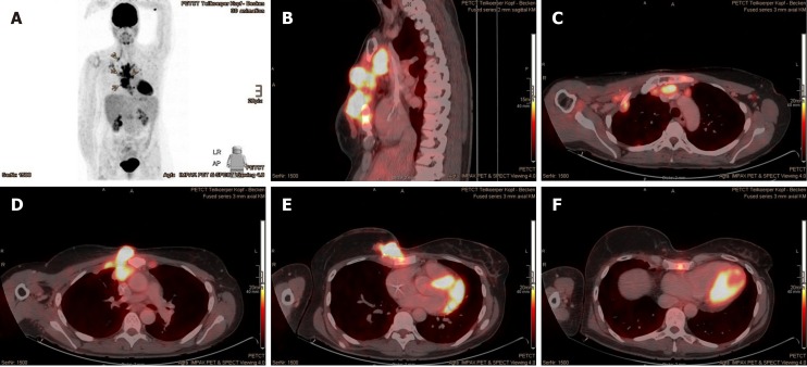
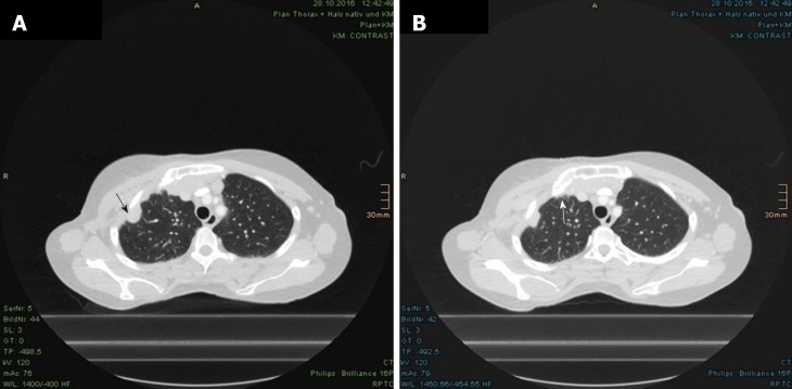
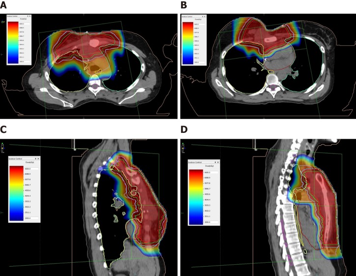
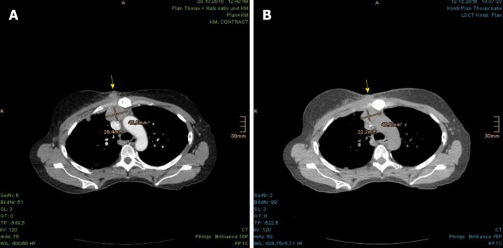
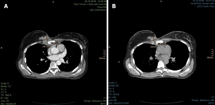
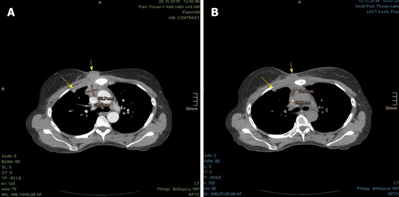
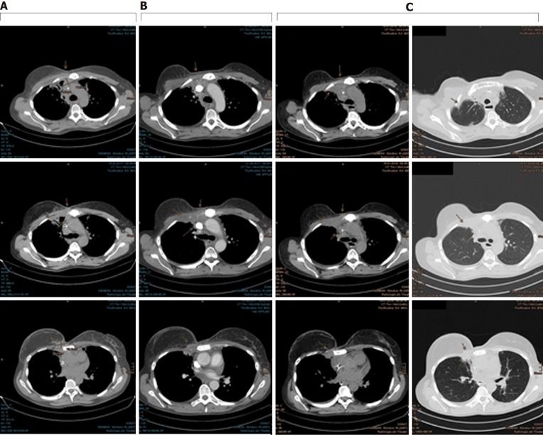
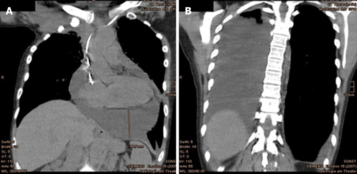
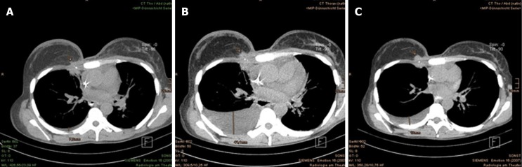
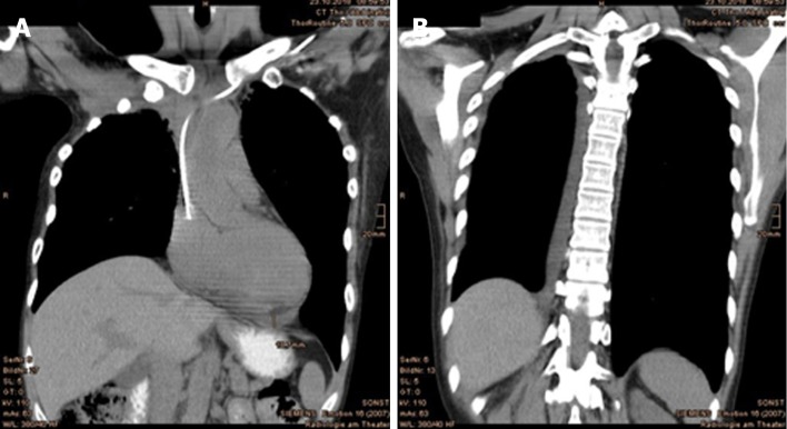
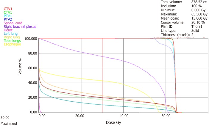
Case summary: A 47-year-old female presented with an extensive locoregional recurrence at 10 yr after primary treatment of a luminal A breast cancer. Because of tumor progression despite having undergone bilateral ovarectomy and systemic therapy, the patient was treated with PBT total dose of 64.40 Gy to each gross tumor and 56.00 Gy to the upper mediastinal and retrosternal lymphatics including the entire sternum in 28 fractions. Follow-up computed tomography showed a partial remission, without evidence of newly emerging metastasis. At 19 mo after the PBT, the patient developed a radiation-induced pericardial disease and pleural effusions with clinical burden of dyspnea, which were successfully treated by drainage and corticosteroid. Cytological analysis of the puncture fluid showed no malignancy, and the subsequent computed tomography scan indicated stable disease as well as significantly decreased pericardial and pleural effusions. The patient remains free of progression to date.
Conclusion: PBT was a safe and effective method of reirradiation for locoregionally recurrent breast cancer in our patient.
Keywords: Case report; Chest wall recurrence; Pericarditis; Proton beam therapy; Radiation-induced cardiovascular disease; Recurrent breast cancer; Reirradiation.
Conflict of interest statement
Conflict-of-interest statement: The author states there are no potential conflicts of interest relevant to this publication.
Figures
Similar articles
-
Clinical Outcomes in Patients with Recurrent Glioblastoma Treated with Proton Beam Therapy Reirradiation: Analysis of the Multi-Institutional Proton Collaborative Group Registry.Adv Radiat Oncol. 2020 Apr 22;5(5):978-983. doi: 10.1016/j.adro.2020.03.022. eCollection 2020 Sep-Oct. Adv Radiat Oncol. 2020. PMID: 33083661 Free PMC article.
-
Reirradiation With Proton Therapy for Recurrent Malignancies of the Esophagus and Gastroesophageal Junction: Results of the Proton Collaborative Group Multi-Institutional Prospective Registry Trial.Adv Radiat Oncol. 2024 Feb 6;9(5):101459. doi: 10.1016/j.adro.2024.101459. eCollection 2024 May. Adv Radiat Oncol. 2024. PMID: 38596455 Free PMC article.
-
Proton Reirradiation for Locoregionally Recurrent Breast Cancer.Adv Radiat Oncol. 2021 May 9;6(4):100710. doi: 10.1016/j.adro.2021.100710. eCollection 2021 Jul-Aug. Adv Radiat Oncol. 2021. PMID: 34409209 Free PMC article.
-
Reirradiation for locoregionally recurrent non-small cell lung cancer.J Thorac Dis. 2018 Aug;10(Suppl 21):S2522-S2536. doi: 10.21037/jtd.2017.12.50. J Thorac Dis. 2018. PMID: 30206496 Free PMC article. Review.
-
How successful is high-dose (> or = 60 Gy) reirradiation using mainly external beams in salvaging local failures of nasopharyngeal carcinoma?Int J Radiat Oncol Biol Phys. 1998 Mar 1;40(4):897-913. doi: 10.1016/s0360-3016(97)00854-7. Int J Radiat Oncol Biol Phys. 1998. PMID: 9531376 Review.
Cited by
-
Proton Radiation Therapy: A Systematic Review of Treatment-Related Side Effects and Toxicities.Int J Mol Sci. 2024 Oct 11;25(20):10969. doi: 10.3390/ijms252010969. Int J Mol Sci. 2024. PMID: 39456752 Free PMC article.
-
Concomitant fulvestrant with reirradiation for unresectable locoregional recurrent estrogen receptor positive (ER+) breast cancer: A case report and narrative review.Medicine (Baltimore). 2020 Jul 24;99(30):e21344. doi: 10.1097/MD.0000000000021344. Medicine (Baltimore). 2020. PMID: 32791733 Free PMC article.
-
Proton beam therapy causing pericarditis - a rare case of radiation induced cardiotoxicity.Cardiooncology. 2022 Apr 18;8(1):9. doi: 10.1186/s40959-022-00135-0. Cardiooncology. 2022. PMID: 35436973 Free PMC article.
-
Target motion management in breast cancer radiation therapy.Radiol Oncol. 2021 Oct 8;55(4):393-408. doi: 10.2478/raon-2021-0040. Radiol Oncol. 2021. PMID: 34626533 Free PMC article. Review.
References
-
- Voinea SC, Sandru A, Blidaru A. Management of Breast Cancer Locoregional Recurrence. Chirurgia (Bucur) 2017;112:429–435. - PubMed
-
- Cadoo KA, Fornier MN, Morris PG. Biological subtypes of breast cancer: current concepts and implications for recurrence patterns. Q J Nucl Med Mol Imaging. 2013;57:312–321. - PubMed
-
- Anderson WF, Chen BE, Jatoi I, Rosenberg PS. Effects of estrogen receptor expression and histopathology on annual hazard rates of death from breast cancer. Breast Cancer Res Treat. 2006;100:121–126. - PubMed
-
- Hess KR, Pusztai L, Buzdar AU, Hortobagyi GN. Estrogen receptors and distinct patterns of breast cancer relapse. Breast Cancer Res Treat. 2003;78:105–118. - PubMed
Publication types
LinkOut - more resources
Full Text Sources
