. 2019 Oct 10;47(18):9829-9841.
doi: 10.1093/nar/gkz693.

Remodeling of the Caenorhabditis elegans non-coding RNA transcriptome by heat shock

Affiliations

Remodeling of the Caenorhabditis elegans non-coding RNA transcriptome by heat shock

William P Schreiner et al. Nucleic Acids Res. .

Abstract

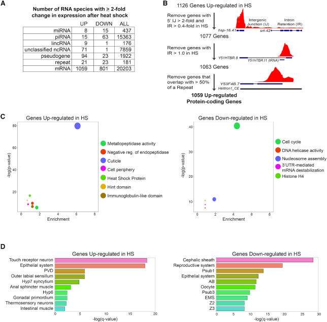
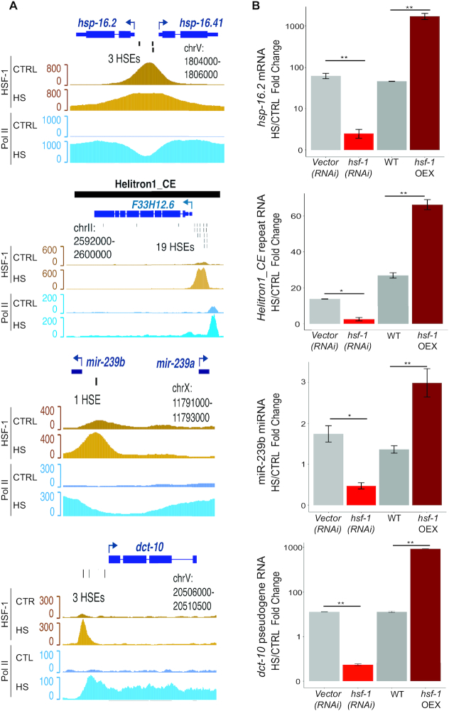
Elevated temperatures activate a heat shock response (HSR) to protect cells from the pathological effects of protein mis-folding, cellular mis-organization, organelle dysfunction and altered membrane fluidity. This response includes activation of the conserved transcription factor heat shock factor 1 (HSF-1), which binds heat shock elements (HSEs) in the promoters of genes induced by heat shock (HS). The upregulation of protein-coding genes (PCGs), such as heat shock proteins and cytoskeletal regulators, is critical for cellular survival during elevated temperatures. While the transcriptional response of PCGs to HS has been comprehensively analyzed in a variety of organisms, the effect of this stress on the expression of non-coding RNAs (ncRNAs) has not been systematically examined. Here we show that in Caenorhabditis elegans HS induces up- and downregulation of specific ncRNAs from multiple classes, including miRNA, piRNA, lincRNA, pseudogene and repeat elements. Moreover, some ncRNA genes appear to be direct targets of the HSR, as they contain HSF-1 bound HSEs in their promoters and their expression is regulated by this factor during HS. These results demonstrate that multiple ncRNA genes respond to HS, some as direct HSF-1 targets, providing new candidates that may contribute to organismal survival during this stress.


Figures

Figure 1.
Figure 1.
HS alters the expression of coding and non-coding RNAs. (A) Small RNA-seq and stranded paired-end RNA-seq were used to analyze changes in RNA expression in Caenorhabditis elegans shifted from 20°C (control, CTRL) to 35°C (HS) for 4 (RNA-seq) or 6 h (smRNA-seq). The numbers of differentially expressed genes in each RNA category are indicated. ‘ALL’ refers to the total number of annotated genes in each class. (B) Strategy used to filter out false positive upregulated mRNAs. See ‘Materials and Methods’ section for further details; codes used for filtering are available at https://github.com/wschrein. (C) DAVID Functional Annotation Clustering of Genes up- and downregulated in response to HS (41). Representative members of each cluster with an enrichment score > 2 are shown. Size of dot corresponds to number of genes in each cluster. (D) TEA for genes up- and downregulated in response to HS. TEA was performed using the Wormbase TEA tool (45). Abbreviations: PVD—Sensory neuron (polymodal nociceptive for mechanosensation and thermosensation), Hyp7—entire syncytium of hyp7, Hyp6—Cylindrical hypodermal syncytium in head, Psub1—Embryonic founder cell, AB—Embryonic founder cell, Psub3—Embryonic founder cell, EMS—Embryonic Cell, Z2—Germ line precursor cell, Z3—Germ line precursor cell.
Figure 2.
Figure 2.
The expression of specific miRNAs is regulated by HS. (A) Expression of miRNAs in CTRL versus HS detected by small RNA-sequencing. Results represent the average of two independent biological replicates and miRNAs reproducibly up- (red) and down- (blue) regulated by ≥ 2-fold are indicated. (B) List of guide strand miRNAs within the top 100 expressed miRNAs that are reproducibly up- or downregulated in response to HS. The final column shows the most highly enriched biological process GO term of potential targets for each miRNA (see Supplementary Table S2). GO analysis was performed using DAVID (41). (C) Analysis of miR-4936 expression in CTRL versus HS conditions by northern blotting. 5S rRNA serves as a loading control. (D and E) Network analysis of differentially regulated mRNAs targeted by at least three up- or downregulated miRNAs. Cytoscape was used to draw networks (https://cytoscape.org) (25).
Figure 3.
Figure 3.
HS alters the expression of long non-coding RNAs. (A) Expression of long intergenic non-coding RNAs (lincRNAs) in CTRL versus HS detected by stranded paired-end sequencing. Significantly up- (red) and downregulated (blue) lincRNAs (≥2-fold change with baseMean ≥ 50 and padj < 0.01 from three independent replicates) are indicated. (B) qRT-PCR analysis of hsp-70 and linc-7 RNA levels after 15, 30 and 180 min of HS versus CTRL conditions. Mean fold changes and SEM from three independent replicates are shown. *P <0.05, **P <0.01, ***P <0.001 (t-test, two-sided). (C) Expression of ncRNAs in CTRL versus HS detected by stranded paired-end sequencing. Significantly up- (red) and downregulated (blue) ncRNAs (≥2-fold change with baseMean ≥ 50 and padj < 0.01 from three independent replicates) are indicated. (D) Expression of pseudogene RNAs in CTRL versus HS detected by stranded paired-end sequencing. Significantly up- (red) and downregulated (blue) pseudogene RNAs (≥2-fold change with baseMean ≥ 50 and padj < 0.05 from three independent replicates) are indicated.
Figure 4.
Figure 4.
Upregulation of multiple repetitive element RNAs during HS. (A) Expression of repetitive element RNAs in CTRL versus HS detected by stranded paired-end sequencing and mapped to a database of repetitive elements (24). Significantly up- (red) and downregulated (blue) repeat RNAs (≥2-fold change with baseMean ≥ 100 and padj < 0.05 from three independent replicates) are indicated. (B) List of repetitive element RNAs upregulated at least 10-fold in HS. (C) Semi-quantitative RT-PCR detection of the indicated RNAs in CTRL and after 15 or 30 min of HS. (D) Quantitative RT-PCR analysis of hsp-16.2 and Helitron1_CE RNA expression during a time course of HS. Mean fold changes and SEM from three independent replicates are shown. *P < 0.05, **P < 0.01, ***P < 0.001 (t-test, two-sided).
Figure 5.
Figure 5.
NcRNAs are regulated by HSF-1 during HS. (A) Genome browser screenshots of HSF-1 (yellow) and Pol II (blue) ChIP-seq data from control (CTRL) and HS conditions (data from (26)) for representative genes (hsp-16.2 and hsp-16.41, mRNA; Helitron1_CE, repeat RNA, miR-239a and miR-239b, miRNA; dct-10, pseudogene). Individual HSEs identified using FIMO (P < 1e-04) are indicated (30). (B) Fold change in RNA levels of hsp16.2, Helitron1_CE, miR-239b and dct-10 after 30 min of HS in animals subjected to empty vector or hsf-1 RNAi, and WT versus a strain overexpressing HSF-1 (hsf-1 OEX) determined by qRT-PCR analyses. The mean fold changes and SEM from three independent replicates are graphed. *P < 0.05, **P < 0.01 (t-test, two-sided).

References

    1. Richter K., Haslbeck M., Buchner J.. The heat shock response: life on the verge of death. Mol. Cell. 2010; 40:253–266. - PubMed
    1. Åkerfelt M., Morimoto R.I., Sistonen L.. Heat shock factors: integrators of cell stress, development and lifespan. Nat. Rev. Mol. Cell Biol. 2010; 11:545–555. - PMC - PubMed
    1. Duarte F.M., Fuda N.J., Mahat D.B., Core L.J., Guertin M.J., Lis J.T.. Transcription factors GAF and HSF act at distinct regulatory steps to modulate stress-induced gene activation. Genes Dev. 2016; 30:1731–1746. - PMC - PubMed
    1. Mahat D.B., Salamanca H.H., Duarte F.M., Danko C.G., Lis J.T.. Mammalian heat shock response and mechanisms underlying its Genome-wide transcriptional regulation. Mol. Cell. 2016; 62:63–78. - PMC - PubMed
    1. Solís E.J., Pandey J.P., Zheng X., Jin D.X., Gupta P.B., Airoldi E.M., Pincus D., Denic V.. Defining the essential function of Yeast Hsf1 reveals a compact transcriptional program for maintaining eukaryotic proteostasis. Mol. Cell. 2016; 63:60–71. - PMC - PubMed

Publication types

MeSH terms

Substances