

Advanced Imaging Modalities to Monitor for Cardiotoxicity
- PMID: 31396720
- PMCID: PMC6687672
- DOI: 10.1007/s11864-019-0672-z
Advanced Imaging Modalities to Monitor for Cardiotoxicity
Abstract
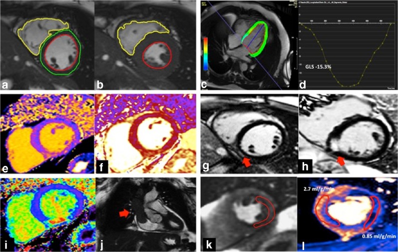
Early detection and treatment of cardiotoxicity from cancer therapies is key to preventing a rise in adverse cardiovascular outcomes in cancer patients. Over-diagnosis of cardiotoxicity in this context is however equally hazardous, leading to patients receiving suboptimal cancer treatment, thereby impacting cancer outcomes. Accurate screening therefore depends on the widespread availability of sensitive and reproducible biomarkers of cardiotoxicity, which can clearly discriminate early disease. Blood biomarkers are limited in cardiovascular disease and clinicians generally still use generic screening with ejection fraction, based on historical local expertise and resources. Recently, however, there has been growing recognition that simple measurement of left ventricular ejection fraction using 2D echocardiography may not be optimal for screening: diagnostic accuracy, reproducibility and feasibility are limited. Modern cancer therapies affect many myocardial pathways: inflammatory, fibrotic, metabolic, vascular and myocyte function, meaning that multiple biomarkers may be needed to track myocardial cardiotoxicity. Advanced imaging modalities including cardiovascular magnetic resonance (CMR), computed tomography (CT) and positron emission tomography (PET) add improved sensitivity and insights into the underlying pathophysiology, as well as the ability to screen for other cardiotoxicities including coronary artery, valve and pericardial diseases resulting from cancer treatment. Delivering screening for cardiotoxicity using advanced imaging modalities will however require a significant change in current clinical pathways, with incorporation of machine learning algorithms into imaging analysis fundamental to improving efficiency and precision. In the future, we should aspire to personalized rather than generic screening, based on a patient's individual risk factors and the pathophysiological mechanisms of the cancer treatment they are receiving. We should aspire that progress in cardiooncology is able to track progress in oncology, and to ensure that the current 'one size fits all' approach to screening be obsolete in the very near future.
Keywords: Cancer; Cancer treatment; Cardiac MRI; Cardiac imaging; Cardiotoxicity; Chemotherapy; Echocardiography; Nuclear imaging.
Conflict of interest statement
The authors declare that they have no conflict of interest.
Figures
References
References and Recommended Reading
Papers of particular interest, published recently, have been highlighted as: • Of importance •• Of major importance
-
- Heymach J, Krilov L, Alberg A, Baxter N, Chang SM, Corcoran RB, et al. Clinical Cancer Advances 2018: annual report on progress against cancer from the American Society of Clinical Oncology. J Clin Oncol. 2018;36(10):1020–1044. - PubMed
-
- Cardinale D, Colombo A, Sandri MT, Lamantia G, Colombo N, Civelli M, et al. Prevention of high-dose chemotherapy-induced cardiotoxicity in high-risk patients by angiotensin-converting enzyme inhibition. Circulation. 2006;114(23):2474–2481. - PubMed
Publication types
MeSH terms
Substances
Grants and funding
LinkOut - more resources
Full Text Sources
Medical
