. 2019 Oct 1;317(4):H732-H742.
doi: 10.1152/ajpheart.00132.2019. Epub 2019 Aug 9.

Role of B1 and B2 lymphocytes in placental ischemia-induced hypertension

Affiliations

Role of B1 and B2 lymphocytes in placental ischemia-induced hypertension

Connor F Laule et al. Am J Physiol Heart Circ Physiol. .

Abstract

Preeclampsia is a prevalent pregnancy complication characterized by new-onset maternal hypertension and inflammation, with placental ischemia as the initiating event. Studies of others have provided evidence for the importance of lymphocytes in placental ischemia-induced hypertension; however, the contributions of B1 versus B2 lymphocytes are unknown. We hypothesized that peritoneal B1 lymphocytes are important for placental ischemia-induced hypertension. As an initial test of this hypothesis, the effect of anti-CD20 depletion on both B-cell populations was determined in a reduced utero-placental perfusion pressure (RUPP) model of preeclampsia. Anti-murine CD20 monoclonal antibody (5 mg/kg, Clone 5D2) or corresponding mu IgG2a isotype control was administered intraperitoneally to timed pregnant Sprague-Dawley rats on gestation day (GD)10 and 13. RUPP or sham control surgeries were performed on GD14, and mean arterial pressure (MAP) was measured on GD19 from a carotid catheter. As anticipated, RUPP surgery increased MAP and heart rate and decreased mean fetal and placental weight. However, anti-CD20 treatment did not affect these responses. On GD19, B-cell populations were enumerated in the blood, peritoneal cavity, spleen, and placenta with flow cytometry. B1 and B2 cells were not significantly increased following RUPP. Anti-CD20 depleted B1 and B2 cells in peritoneum and circulation but depleted only B2 lymphocytes in spleen and placenta, with no effect on circulating or peritoneal IgM. Overall, these data do not exclude a role for antibodies produced by B cells before depletion but indicate the presence of B lymphocytes in the last trimester of pregnancy is not critical for placental ischemia-induced hypertension.NEW & NOTEWORTHY The adaptive and innate immune systems are implicated in hypertension, including the pregnancy-specific hypertensive condition preeclampsia. However, the mechanism of immune system dysfunction leading to pregnancy-induced hypertension is unresolved. In contrast to previous reports, this study reveals that the presence of classic B2 lymphocytes and peritoneal and circulating B1 lymphocytes is not required for development of hypertension following third trimester placental ischemia in a rat model of pregnancy-induced hypertension.


Conflict of interest statement

No conflicts of interest, financial or otherwise, are declared by the authors.

Figures

Fig. 1.
Fig. 1.
Anti-CD20 does not favorably affect placental ischemia-induced changes in mean arterial pressure (MAP), heart rate, or mean fetal and placental weight at gestation day (GD)19. Animals were treated with 5 mg/kg anti-CD20 or isotype control by intraperitoneal injection on GD10 and 13. Values represent means ± SE; n, values for each end point ranged from 8 to 14 with the actual number for each end point reported in Supplementary Table S1 (https://doi.org/10.17605/OSF.IO/NTMHJ). *P < 0.05 for indicated comparisons; #P < 0.05, reduced utero-placental perfusion pressure (RUPP) compared with sham surgery as a main effect by ANOVA. A: RUPP surgery increased MAP (main effect P = 0.003) when RUPP isotype was compared with sham isotype (P = 0.025) and RUPP anti-CD20 to sham anti-CD20 (P = 0.03). Anti-CD20 did not inhibit the RUPP increase in MAP. B: by ANOVA, RUPP surgery increased heart rate (main effect P = 0.049) with no significant main anti-CD20 treatment effect; bpm. beats/min. In anti-CD20-treated animals, heart rate in RUPP vs. sham was significantly different (P = 0.034). C: average fetal weight was significantly reduced (main effect, P = 0.0004) in RUPP isotype vs. sham isotype (P = 0.007), and this difference persisted with anti-CD20 treatment when RUPP was compared with sham (P = 0.01). D: average placenta weight was significantly decreased with RUPP surgery (main effect P = 0.042), and anti-CD20 had no effect.
Fig. 2.
Fig. 2.
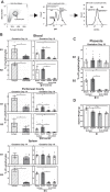
Anti-CD20 significantly depletes B1 lymphocytes in the blood and peritoneal cavity and B2 cells in all measured tissues and compartments on gestation day (GD)19. Values represent means ± SE. Data were logit transformed for analysis; n values for each end point ranged from 4 to 14 with the actual number for each end point reported in Supplementary Table S1 (https://doi.org/10.17605/OSF.IO/NTMHJ). *P < 0.05 for indicated contrast; ‡P < 0.0001, indicating significant effect by ANOVA for anti-CD20 compared with isotype treatment at GD19; †P < 0.05, GD14 anti-CD20 treatment compared with GD19 anti-CD20 sham and GD19 anti-CD20 reduced utero-placental perfusion pressure (RUPP). A: representative rat spleen cell population showing lymphocytes gated based on characteristic forward and side scatter profiles. Gated IgM+ lymphocytes were characterized as mature B cells and further classified as CD11b B2 or CD11b+ B1 cells. B-lymphocyte populations are reported as a percentage of total lymphocytes. B: RUPP surgery did not significantly change peritoneal B1 and B2 cells when RUPP isotype was compared with sham isotype at GD19. Anti-CD20 depleted both B-cell lineages in the blood and peritoneal cavity with ANOVA analysis when RUPP and sham isotype groups were compared with RUPP anti-CD20 and sham anti-CD20 at GD19 and when isotype was compared with anti-CD20 at GD14. However, peritoneal B1 (P < 0.0003) and B2 (P < 0.0001) populations were significantly less at GD19 compared with GD14. Anti-CD20 depleted splenic B2, but not B1, cells at GD14 and 19. However, splenic B1 populations were significantly increased at GD19 compared with GD14 in the anti-CD20-treated animals. Blood: at GD19, P < 0.0003; at GD14, significant change in B1 cells (P = 0.04) and B2 cells (P < 0.0001). Peritoneal cavity: B1 and B2 cells decreased in sham isotype vs. sham anti-CD20 (P = 0.047 and 0.0008, respectively) and RUPP isotype vs. RUPP anti-CD20 (P < 0.0001 for both); at GD14, significant change in B1 cells (0.04) and B2 cells (0.049). Spleen: B2 cells decreased in sham isotype vs. sham anti-CD20 and RUPP isotype vs. RUPP anti-CD20 (P < 0.0001 for both); B1 cells in anti-CD20-treated animals were greater at GD19 than GD14 (†P < 0.02). C: anti-CD20 depleted B2 cells, but not B1 cells, in the placenta at GD19 when sham isotype was compared with sham anti-CD20 (P < 0.0001) and RUPP isotype was compared with RUPP anti-CD20 (P < 0.0001). D: spleen weight was decreased when RUPP isotype was compared with sham isotype (P = 0.038) or sham isotype was compared with sham anti-CD20 (P = 0.004). ANOVA analysis indicated that anti-CD20 treatment significantly reduced spleen weight when compared with isotype animals (‡P = 0.002).
Fig. 3.
Fig. 3.
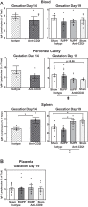
Anti-CD20 effect on IgM-negative lymphocytes in all measured tissues and compartments on gestation day (GD)14 and GD19. Values represent means ± SE of the IgM-negative lymphocytes as percentage of total cells. Data were logit transformed for analysis; n values for each end point ranged from 4 to 14 with the actual number for each end point reported in Supplementary Table S1 (https://doi.org/10.17605/OSF.IO/NTMHJ). *P < 0.05 for indicated contrast; ‡P < 0.05, significant effect by ANOVA for anti-CD20 compared with isotype treatment at GD19; †P < 0.05, GD14 anti-CD20 treatment compared with GD19 anti-CD20 sham and GD19 anti-CD20 reduced utero-placental perfusion pressure (RUPP). A: anti-CD20 did not significantly decrease IgM-negative lymphocytes at GD14 or 19 in blood. In the peritoneal cavity, an overall anti-CD20 affect (P = 0.015) suggests a reduction in the IgM-negative lymphocytes at GD19. In the spleen, anti-CD20 treatment significantly increased IgM-negative lymphocytes at both GD14 (P = 0.002) and 19 (P = 0.004, sham isotype compared with sham anti-CD20; P < 0.0001, RUPP isotype compared with RUPP anti-CD20; main treatment effect P < 0.0001). B: in placenta, anti-CD20 treatment had no effect.
Fig. 4.
Fig. 4.
Effect of reduced utero-placental perfusion pressure (RUPP) surgery and anti-CD20 on IgM in serum and peritoneal cavity. Values represent means ± SE expressed as μg/ml or percentage of total protein. Data were log transformed for analysis; n values for each end point ranged from 5 to 14 with the actual number for each end point reported in Supplementary Table S1 (https://doi.org/10.17605/OSF.IO/NTMHJ). *P < 0.05 for indicated contrast. A: RUPP surgery decreased serum IgM when RUPP isotype was compared with sham isotype (P = 0.007, μg/ml; P = 0.026, %total protein). Anti-CD20 treatment affected circulating IgM concentrations in RUPP when expressed as μg/ml (P = 0.02) but not as percentage of total serum protein. A significant interaction between surgery and treatment was detected (P = 0.019) in the analysis of μg/ml IgM, with no significant main effects. B: peritoneal IgM levels were not altered by RUPP surgery or anti-CD20 treatment. ANOVA showed no significant overall anti-CD20 or surgery effect, nor any significant interaction, in either serum or peritoneal cavity.
Fig. 5.
Fig. 5.
Anti-CD20 increases preproendothelin (PPE) message in placenta. Values represent means ± SE; n values ranged from 8 to 14 with the actual number for each end point reported in Supplementary Table S1 (https://doi.org/10.17605/OSF.IO/NTMHJ). Reduced utero-placental perfusion pressure (RUPP) surgery did not significantly change placental message for PPE when comparing RUPP was compared with sham isotype (P = 0.09), but a significant difference was detected when sham isotype was compared with sham anti-CD20 (P = 0.006). ANOVA indicated that anti-CD20 significantly increased placenta PPE (‡P = 0.012). *P < 0.05 for indicated comparisons; ‡P < 0.05, significant treatment effect by ANOVA for anti-CD20 compared with isotype.

Comment in

References

    1. Abais-Battad JM, Lund H, Fehrenbach DJ, Dasinger JH, Mattson DL. Rag1-null Dahl SS rats reveal that adaptive immune mechanisms exacerbate high protein-induced hypertension and renal injury. Am J Physiol Regul Integr Comp Physiol 315: R28–R35, 2018. doi:10.1152/ajpregu.00201.2017. - DOI - PMC - PubMed
    1. Ahuja A, Anderson SM, Khalil A, Shlomchik MJ. Maintenance of the plasma cell pool is independent of memory B cells. Proc Natl Acad Sci USA 105: 4802–4807, 2008. doi:10.1073/pnas.0800555105. - DOI - PMC - PubMed
    1. Alexander BT, Kassab SE, Miller MT, Abram SR, Reckelhoff JF, Bennett WA, Granger JP. Reduced uterine perfusion pressure during pregnancy in the rat is associated with increases in arterial pressure and changes in renal nitric oxide. Hypertension 37: 1191–1195, 2001. doi:10.1161/01.HYP.37.4.1191. - DOI - PubMed
    1. Alexander BT, Rinewalt AN, Cockrell KL, Massey MB, Bennett WA, Granger JP. Endothelin type a receptor blockade attenuates the hypertension in response to chronic reductions in uterine perfusion pressure. Hypertension 37: 485–489, 2001. doi:10.1161/01.HYP.37.2.485. - DOI - PubMed
    1. Amaral LM, Faulkner JL, Elfarra J, Cornelius DC, Cunningham MW, Ibrahim T, Vaka VR, McKenzie J, LaMarca B. Continued investigation into 17-OHPC: results from the preclinical rupp rat model of preeclampsia. Hypertension 70: 1250–1255, 2017. doi:10.1161/HYPERTENSIONAHA.117.09969. - DOI - PMC - PubMed

Publication types

MeSH terms