Review
. 2019 Dec;14(12):2054-2062.
doi: 10.4103/1673-5374.262572.

From cortex to cord: motor circuit plasticity after spinal cord injury

Affiliations
Review

From cortex to cord: motor circuit plasticity after spinal cord injury

Andrew R Brown et al. Neural Regen Res. 2019 Dec.

Abstract

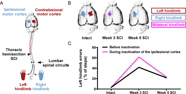
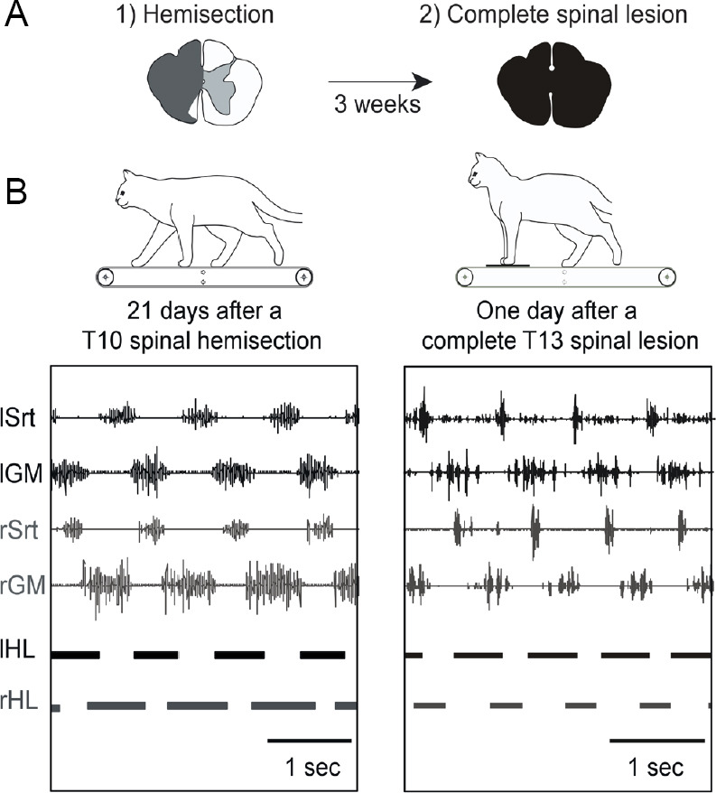
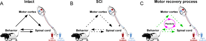
Spinal cord injury is associated with chronic sensorimotor deficits due to the interruption of ascending and descending tracts between the brain and spinal cord. Functional recovery after anatomically complete spinal cord injury is limited due to the lack of long-distance axonal regeneration of severed fibers in the adult central nervous system. Most spinal cord injuries in humans, however, are anatomically incomplete. Although restorative treatment options for spinal cord injury remain currently limited, research from experimental models of spinal cord injury have revealed a tremendous capability for both spontaneous and treatment-induced plasticity of the corticospinal system that supports functional recovery. We review recent advances in the understanding of corticospinal circuit plasticity after spinal cord injury and concentrate mainly on the hindlimb motor cortex, its corticospinal projections, and the role of spinal mechanisms that support locomotor recovery. First, we discuss plasticity that occurs at the level of motor cortex and the reorganization of cortical movement representations. Next, we explore downstream plasticity in corticospinal projections. We then review the role of spinal mechanisms in locomotor recovery. We conclude with a perspective on harnessing neuroplasticity with therapeutic interventions to promote functional recovery.

Keywords: animal models; corticospinal tract; forelimb; functional recovery; hindlimb; locomotion; motor cortex; motor map; neuroplasticity; spinal cord injury.


Conflict of interest statement

None

Figures

Figure 1
Figure 1
Motor cortex plasticity is involved in spontaneous locomotor recovery after spinal cord injury (SCI). (A) Thoracic hemisection SCI on the left side in the rat severs the crossed corticospinal tract from the contralesional motor cortex to the left hindlimb, but spares the crossed corticospinal projection from the ipsilesional motor cortex to the right hindlimb. The lumbosacral spinal circuits are located below the lesion. (B) In the intact state, intracortical microstimulation of either motor cortex elicits movement in the contralateral hindlimb. After SCI, stimulation of the contralesional motor cortex no longer elicits hindlimb movement for up to 5 weeks. Three weeks after SCI, stimulation of the ipsilesional motor cortex, that retains access to lumbosacral spinal circuits, elicits movement in both hindlimbs. This time point coincides with significant locomotor recovery of the affected (left) hindlimb (Brown and Martinez, 2018). Five weeks after SCI, stimulation of the ipsilesional motor cortex no longer elicits bilateral hindlimb movements. (C) Reversible inactivation of the ipsilesional motor cortex during skilled locomotion on a horizontal rung-ladder 3 weeks after SCI reinstated deficits in the affected hindlimb, but not in the intact state or 5 weeks after SCI. These findings indicate that after SCI, the ipsilesional motor cortex spontaneously gains a novel functional access to the affected hindlimb during the motor recovery process. Data presented are from a representational rat (Brown and Martinez, 2018).
Figure 2
Figure 2
Episodes of treadmill locomotion at 0.4 m/s displayed by a cat 3 weeks after a spinal hemisection at T10 and 24 hours after a second complete spinal lesion at T13. (A) Three weeks after hemisection, hindlimb locomotion was already re-expressed and well-organized. (B) After the subsequent complete spinal section isolating spinal circuits from all supraspinal inputs, the cat could express hindlimb locomotion as soon as tested (24 hours). The locomotor pattern was asymmetrical and consisted mainly in a better capacity to walk with the hindlimb previously impacted by the hemisection. For instance, the phase of support on the side of the previous hemisection was longer than on the other side (horizontal bars below the EMG traces). Schematic drawings above each panel represent the extent of the hemisection (in grey) and of the subsequent complete spinalization (in black). Top traces of EMGs recordings are shown. Duty cycles (horizontal bars) below the EMGs illustrate the support periods. Srt: Sartorius; GM: median gastrocnemius; HL: hindlimb; sec: second; EMG: electromyography; l: left; r: right.
Figure 3
Figure 3
Schematic representation of the interactions between the motor cortex, spinal cord, and behavior during the motor recovery process after an incomplete spinal cord injury (SCI). (A) In the intact state, behavioral output (movement) is generated by the spinal cord which, in turn, is regulated by the motor cortex. Feedback (sensory afferents) is provided to the spinal cord and motor cortex to adjust the desired output response of the system. (B) After SCI, behavioral output is impaired due to disruptions in both its generation and feedback control. (C) During the recovery process, spontaneous plasticity can occur at multiple levels of the neuraxis, and be further promoted by therapeutic interventions to re-establish connectivity within the system. Circuit interactions are depicted indicating the directionality of communication (solid black arrows). Disruptions in communicative flow in the system occur after SCI (dashed black arrows). Strengthening of systems interactions (green dashed arrows) can be mediated through plasticity (purple arrows).

References

    1. Anderson KD, Sharp KG, Steward O. Bilateral cervical contusion spinal cord injury in rats. Exp Neurol. 2009;220:9–22. - PMC - PubMed
    1. Antri M, Barthe JY, Mouffle C, Orsal D. Long-lasting recovery of locomotor function in chronic spinal rat following chronic combined pharmacological stimulation of serotonergic receptors with 8-OHDPAT and quipazine. Neurosci Lett. 2005;384:162–167. - PubMed
    1. Armand J. The origin course and terminations of corticospinal fibers in various mammals. Prog Brain Res. 1982;57:329–360. - PubMed
    1. Asanuma H, Sakata H. Functional organization of a cortical efferent system examined with focal depth stimulation in cats. J Neurophysiol. 1967;30:35–54.
    1. Asboth L, Friedli L, Beauparlant J, Martinez-Gonzalez C, Anil S, Rey E, Baud L, Pidpruzhnykova G, Anderson MA, Shkorbatova P, Batti L, Pages S, Kreider J, Schneider BL, Barraud Q, Courtine G. Cortico-reticulo-spinal circuit reorganization enables functional recovery after severe spinal cord contusion. Nat Neurosci. 2018;21:576–588. - PubMed