. 2019;8(4):625-642.
doi: 10.1016/j.jcmgh.2019.07.005. Epub 2019 Aug 6.

TRPV6 and Cav1.3 Mediate Distal Small Intestine Calcium Absorption Before Weaning

Affiliations

TRPV6 and Cav1.3 Mediate Distal Small Intestine Calcium Absorption Before Weaning

Megan R Beggs et al. Cell Mol Gastroenterol Hepatol. 2019.

Abstract

Background & aims: Intestinal Ca2+ absorption early in life is vital to achieving optimal bone mineralization. The molecular details of intestinal Ca2+ absorption have been defined in adults after peak bone mass is obtained, but they are largely unexplored during development. We sought to delineate the molecular details of transcellular Ca2+ absorption during this critical period.

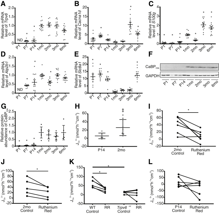
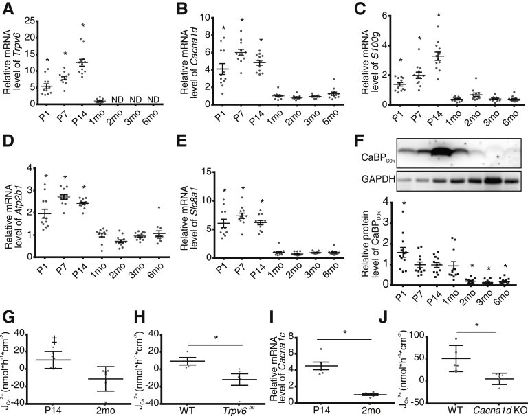
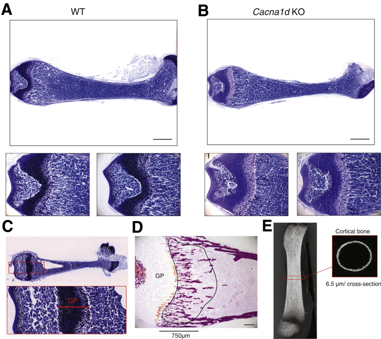
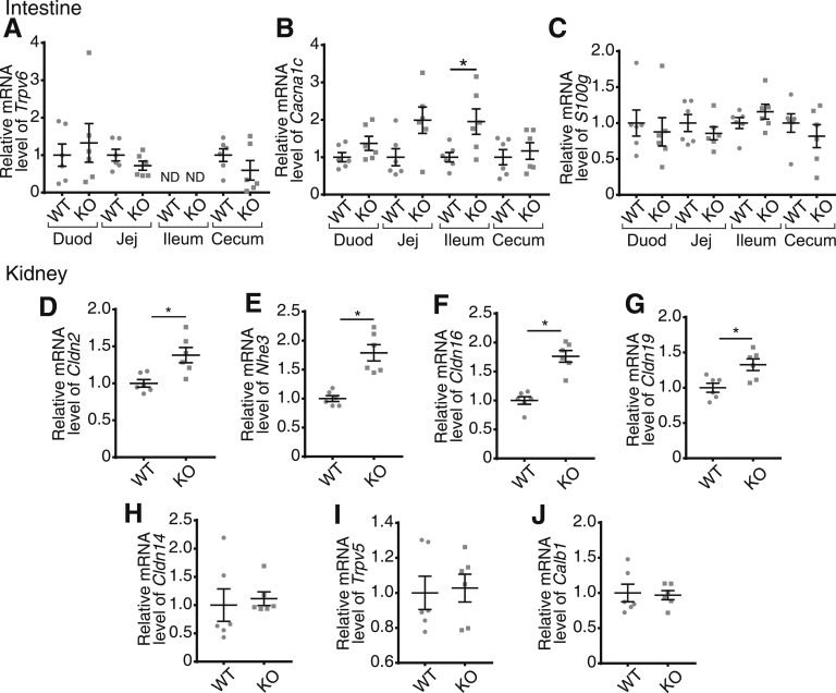
Methods: Expression of small intestinal and renal calcium transport genes was assessed by using quantitative polymerase chain reaction. Net calcium flux across small intestinal segments was measured in Ussing chambers, including after pharmacologic inhibition or genetic manipulation of TRPV6 or Cav1.3 calcium channels. Femurs were analyzed by using micro-computed tomography and histology.

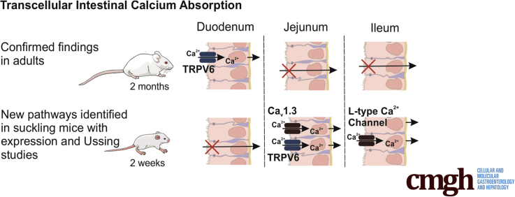
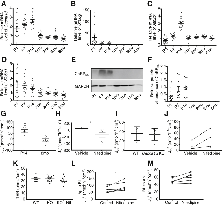
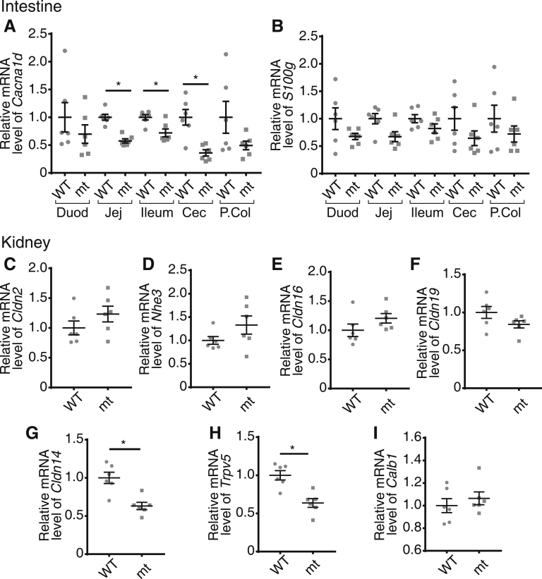
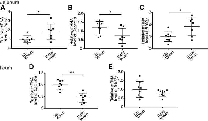
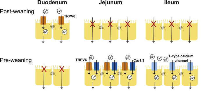
Results: Net TRPV6-mediated Ca2+ flux across the duodenum was absent in pre-weaned (P14) mice but present after weaning. In contrast, we found significant transcellular Ca2+ absorption in the jejunum at 2 weeks but not 2 months of age. Net jejunal Ca2+ absorption observed at P14 was not present in either Trpv6 mutant (D541A) mice or Cav1.3 knockout mice. We observed significant nifedipine-sensitive transcellular absorption across the ileum at P14 but not 2 months. Cav1.3 knockout pups exhibited delayed bone mineral accrual, compensatory nifedipine-insensitive Ca2+ absorption in the ileum, and increased expression of renal Ca2+ reabsorption mediators at P14. Moreover, weaning pups at 2 weeks reduced jejunal and ileal Cav1.3 expression.

Conclusions: We have detailed novel pathways contributing to transcellular Ca2+ transport across the distal small intestine of mice during development, highlighting the complexity of the multiple mechanisms involved in achieving a positive Ca2+ balance early in life.

Keywords: Bone; Calcium Channel; Development; Pediatric.


Figures

None
Graphical abstract
Figure 1
Figure 1
Transcellular JCa2+flux across the duodenum is not detectable at P14 but mediated by TRPV6 in 2-month-old mice. Relative expression of (A) Trpv6, (B) Cacna1d, (C) S100g, (D) Atb2b1, and (E) Slc8a1 in the duodenum across ages (n = 12/group). Expression is normalized to Gapdh and relative to 1 month. (F) Representative immunoblot from 3 replicates and (G) semi-quantification of calbinidin-D9k. Protein abundance is normalized to GAPDH and presented relative to the 1-month group (n = 6/group). Groups compared by 1-way analysis of variance with Dunnett multiple comparisons test. *P < .05 compared with 1-month group. (H) Net JCa2+ across ex vivo sections of mouse duodenum is not different from 0 in P14 mice (n = 6, P = .095) but significantly greater than 0, consistent with absorption, in 2-month-old mice (n = 7, P = .013) (two-tailed, one-sample t test). Net JCa2+ is significantly reduced in 2-month-old mice after addition of 100 μmol/L ruthenium red apically under (I) apical hyperosmolar (n = 6; two-tailed, paired t test; *P = .006) and (J) iso-osmolar conditions (n = 5; two-tailed, paired t test; *P = .02). (K) Net JCa2+ is significantly lower in 2-month-old Trpv6mt mice compared with WT littermates (n = 6/group; two-tailed, unpaired t test; *P < .001). One hundred μmol/L apical ruthenium red significantly decreases net JCa2+ in WT (*P = .002) but not Trpv6mt mice (P = .474) (two-tailed, paired t test). Both paired experiments were performed under apical hyperosmolar conditions. (L) One hundred μmol/L ruthenium red did not decrease net JCa2+ in P14 mice (n = 6; two-tailed, paired t test; P = .2). Data are presented as mean ± standard error of the mean. CaBPD9k, calbindin-D9k; ND, not detected; RR, ruthenium red; Trpv6mt, Trpv6 mutant.
Figure 2
Figure 2
Net Ca2+absorption across the jejunum of P14 mice is mediated by TRPV6 and Cav1.3 and is not present at 2 months. Relative expression of (A) Trpv6, (B) Cacna1d, (C) S100g, (D) Atp2b1, and (E) Slc8a1 by age (n = 12/group). Expression is normalized to Gapdh and relative to 1 month. (F) Representative calbindin-D9k (CaBPD9k) immunoblot of 12 replicates and quantification by age (n = 12/group). Groups compared by 1-way analysis of variance with Dunnett multiple comparisons test. *P < .05 compared with 1-month group. (G) Net JCa2+ across ex vivo sections of mouse jejunum are greater than 0, indicating absorption at P14 (n = 7; P = .03) but not 2-month-old mice (n = 6; P = .11; two-tailed, one-sample t test). (H) Net JCa2+ is significantly reduced across the jejunum of P14 Trpv6mt mice compared with WT littermates (n = 4 WT and 5 mt; two-tailed unpaired t test; *P = .04). (I) Greater expression of Cacna1c, encoding Cav1.2, at P14 (n = 6/group; two-tailed unpaired t test; *P < .0001) normalized to Gapdh. (J) Significantly reduced net JCa2+ across the jejunum of P14 Cacna1d KO mice compared with WT mice (n = 5/group; Mann-Whitney test; *P = .008). Data are presented as mean ± standard error of the mean. CaBPD9k, calbindin-D9k; ND, not detected; Trpv6mt, Trpv6 mutant.
Figure 3
Figure 3
Cav1.3 expression in jejunum of P14 mice. (A and B) Immunoreactivity (red) of HA reveals apical localization of HA-tagged Cav1.3 on sections (7 μm) of jejunum from P14 mice expressing HA-tagged Cacna1d. (C) Section of jejunum from WT mice (control) shows no HA immunoreactivity. Cell nuclei were stained with bisbenzimide (Hoechst 33342) (blue). (D) Hematoxylin-eosin staining of sections of jejunum of Cav1.3-HA mice. Scale bars: 50 μm.
Figure 4
Figure 4
P14 but not 2-month-old mice display net apical to basolateral calcium flux across the ileum, mediated by L-type Ca2+channel. Expression of (A) Cacna1d, (B) S100g, (C) Atp2b1, and (D) Slc8a1 by age (n = 12/group). Expression is normalized to Gapdh and relative to 1 month. (E) Representative immunoblot of 12 repeats and (F) semi-quantification of calbindin-D9k (CaBPD9k) demonstrates expression in ileum only in younger mice. Results are normalized to GAPDH and displayed relative to P14 age (n = 12/group). Groups are compared by 1-way analysis of variance with Dunnett multiple comparisons test; *P < .05. (G) Net JCa2+ across ex vivo sections of mouse ileum are greater than 0 at P14 (n = 6; P = .001) but not 2 months (n = 6; P = .359) (two-tailed, one-sample t tests). (H) Ten μmol/L apical nifedipine decreases net JCa2+ in P14 mice compared with vehicle (n = 5 vehicle and 9 nifedipine; Mann-Whitney test; *P = .001). (I) No difference in net JCa2+ across ileum between WT and Cacna1d KO mice at P14 (n = 5 WT and 6 KO; Mann-Whitney test; P = .54). (J) Ten μmol/L apical nifedipine does not decrease net JCa2+ in Cacna1d KO mice at P14 mice (n = 4; Wilcoxon matched-pairs signed rank test; P = .25). (K) Transepithelial resistance (TER) across the ileum of P14 WT, Cacna1d KO, or Cacna1d KO with nifedipine (1-way analysis of variance; P = .5) (n = 7–10/group). Unidirectional apical to basolateral (L) and basolateral to apical (M) JCa2+ across ileum of P14 Cacna1d KO mice before (control) and after apical addition of 10 μmol/L nifedipine (paired t test; *P < .05) (n = 6). Data are presented as mean ± standard error of the mean.
Figure 5
Figure 5
Bone phenotype of Cacna1d KO pups. Representative toluidine blue–stained sections from fixed non-decalcified femurs of (A) WT and (B) Cacna1d KO mice at P14 (P13-P15). The growth plate thickness was measured in middle of the section as indicated below. Scale bar = 1 mm (upper panels) and 0.2 mm (lower panels). (C) Representative toluidine blue–stained sections obtained from non-decalcified femur (top) and enlarged region covering the growth plate (GP) used to determine thickness of growth plate shown in Tables 1 and 2. (D) Representative alizarin red stain used to visualize calcified bone (red) and to calculate trabecular parameters shown in Tables 1 and 2. Region of interest (ROI) starting at growth plate (GP) and covering primary spongiosa over 750 μm is indicated. (E) Lateral scout view of femur indicating midshaft section used to analyze cortical bone.
Figure 6
Figure 6
Renal compensation in Cacna1d KO mice at P14. Quantitative real-time PCR results of (A) Trpv6, (B) Cacna1c encoding Cav1.2, and (C) S100g along the intestine. Renal expression of (D) Cldn2, (E) Nhe3, (F) Cldn16, (G) Cldn19, (H) Cldn14, (I) Trpv5, and (J) Calb1 encoding calbindin-D28k reveals compensatory increases in Cacna1d KO pups. Small intestine and kidney results are normalized to Gapdh; cecum results are normalized to β-actin. All expression results are displayed relative to WT group for each tissue. *P < .05 vs WT by Mann-Whitney test. (n = 6/group). Data are presented as mean ± standard error of the mean. Duod, duodenum; Jej, jejunum.
Figure 7
Figure 7
Compensatory expression changes in Trpv6mtpups. Quantitative real-time PCR results of (A) Cacna1d and (B) S100g encoding calbindin-D9k along the intestine from Trpv6mt pups relative to WT expression in each tissue. Quantitative real-time PCR expression of mediators of renal Ca2+ reabsorption, (C) Cldn2, (D) Nhe3, (E) Cldn16, (F) Cldn19, (G) Cldn14, (H) Trpv5, and (I) Calb1 encoding calbindin-D28k in Trpv6mt pups relative to WT. Small intestine and kidney results are normalized to Gapdh; cecum and proximal colon (P.Col) results are normalized to β-actin. All expression results are relative to WT group for each tissue. *P < .05 vs WT by Mann-Whitney test. (n = 6/group). Data presented as mean ± standard error of the mean. Duod, duodenum; Jej, jejunum; mt, mutant.
Figure 8
Figure 8
Early weaning to rodent chow alters Trpv6, Cacna1d, and S100g expression in jejunum and ileum at P14. Quantitative real-time PCR results of (A) Trpv6, (B) Cacna1d, and (C) S100g in jejunum and (D) Cacna1d and (E) S100g in ileum. Tissue was taken from mice at P14 after either early weaning to rodent chow at P12 or not. Results are normalized to β-actin. *P < .05, ***P < .0001 vs P14 mice not weaned by Mann-Whitney or unpaired t test. (n = 7–8/group). Data are presented as mean ± standard error of the mean.
Figure 9
Figure 9
Summary of apical entry mechanisms contributing transcellular Ca2+absorption across the small intestine before and after weaning. Significant net transcellular Ca2+ absorption across the duodenum is mediated by apical TRPV6 and is present only after weaning. In the jejunum, significant net transcellular Ca2+ absorption is present only before weaning and is mediated by apical TRPV6 and Cav1.3. Similarly, significant net transcellular Ca2+ absorption occurs only before weaning across the ileum and is mediated by an L-type calcium channel.

Comment in

References

    1. Matkovic V. Calcium metabolism and calcium requirements during skeletal modeling and consolidation of bone mass. Am J Clin Nutr. 1991;54:245S–260S. - PubMed
    1. Bronner F. Recent developments in intestinal calcium absorption. Nutr Rev. 2009;67:109–113. - PubMed
    1. Bachrach L.K., Levine M.A., Cowell C.T., Shaw N.J. Clinical indications for the use of DXA in pediatrics. In: Sawyer A.J., editor. Bone densitometry in growing patients. Humana Press; New York, NY: 2007. pp. 59–72.
    1. Cauley J., Wampler N., Barnhart J., Wu L., Allison M., Chen Z., Hendrix S., Robbins J., Jackson R. Incidence of fractures compared to cardiovascular disease and breast cancer: the Women’s Health Initiative Observational Study. Osteoporos Int. 2008;19:1717. - PMC - PubMed
    1. Tarride J.E., Guo N., Hopkins R., Leslie W.D., Morin S., Adachi J.D., Papaioannou A., Bessette L., Brown J.P., Goeree R. The burden of illness of osteoporosis in Canadian men. J Bone Miner Res. 2012;27:1830–1838. - PMC - PubMed

Publication types

MeSH terms