. 2019 Dec:58:101540.
doi: 10.1016/j.media.2019.101540. Epub 2019 Aug 1.

Surface-constrained volumetric registration for the early developing brain

Affiliations

Surface-constrained volumetric registration for the early developing brain

Sahar Ahmad et al. Med Image Anal. 2019 Dec.

Abstract

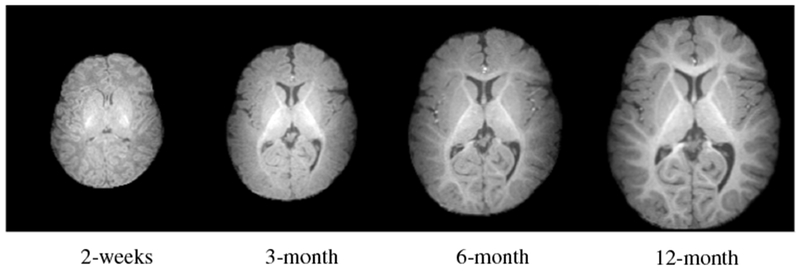
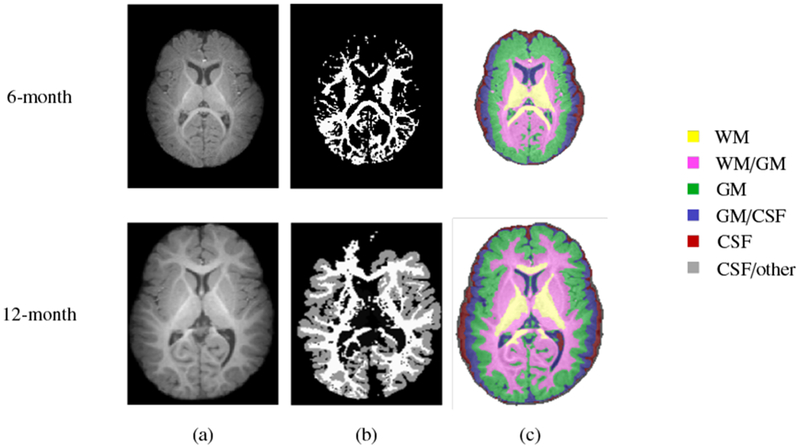
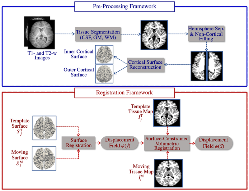
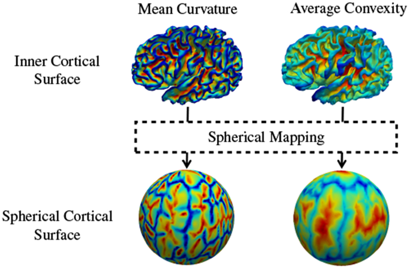
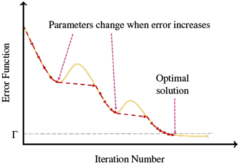
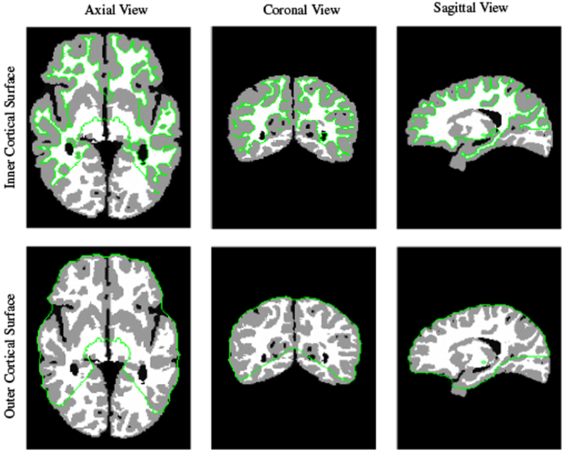
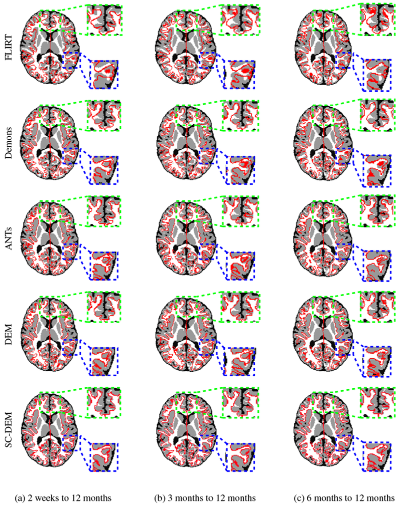
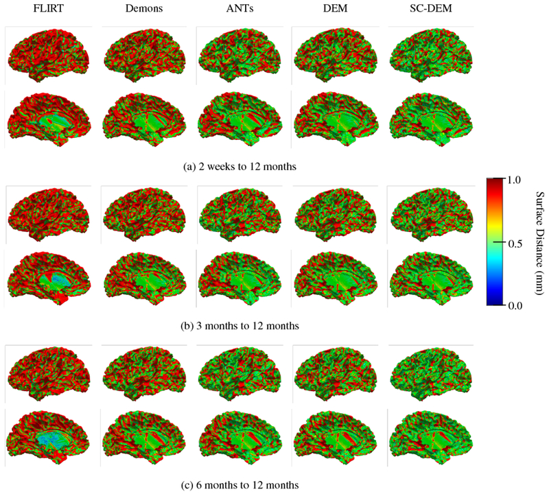
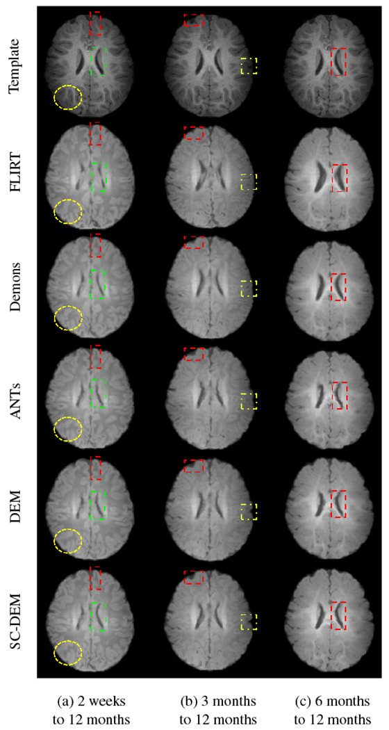
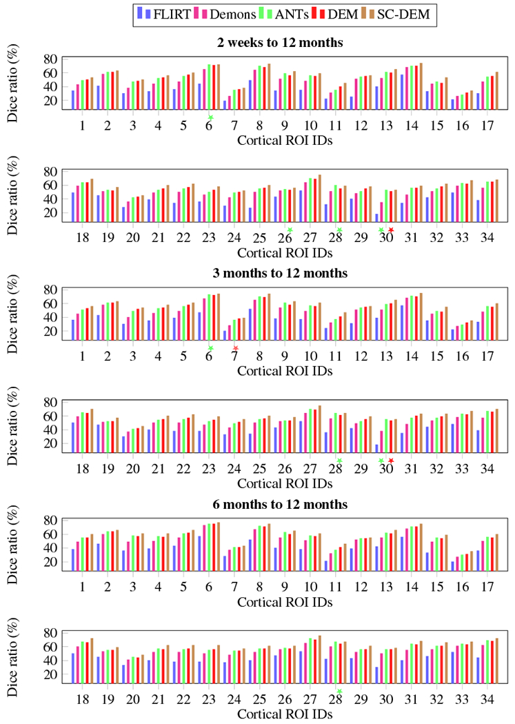
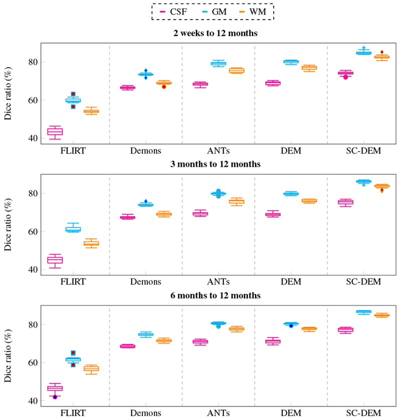
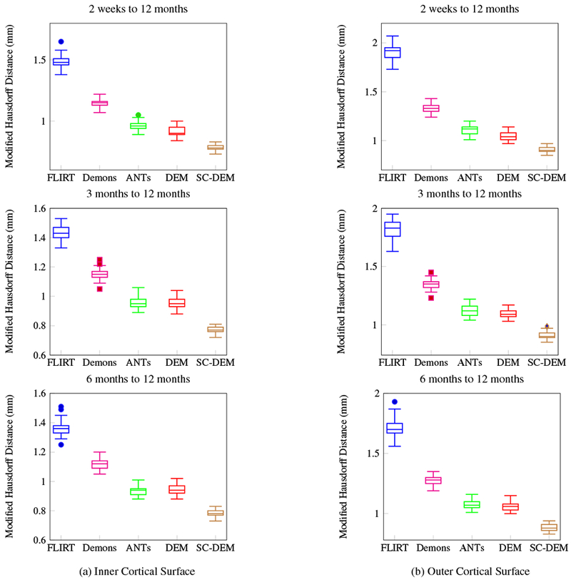
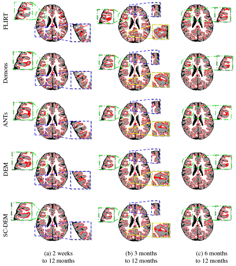
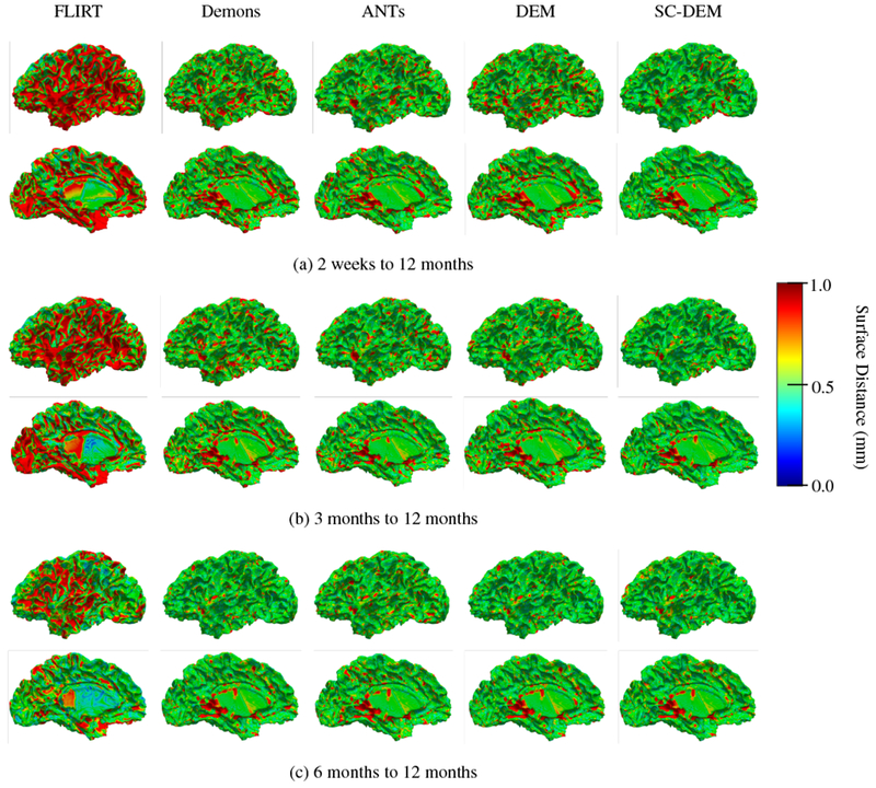
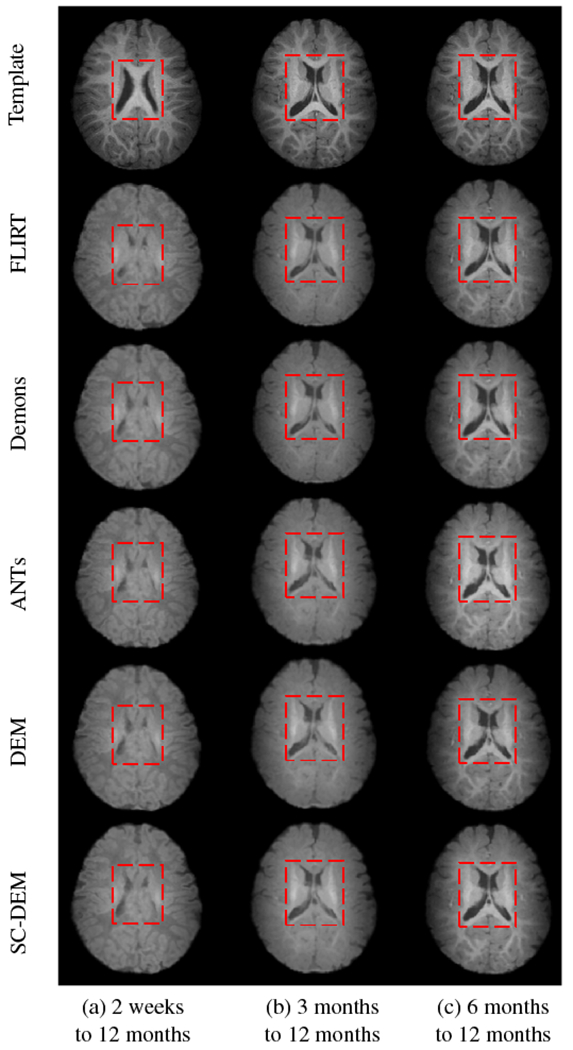
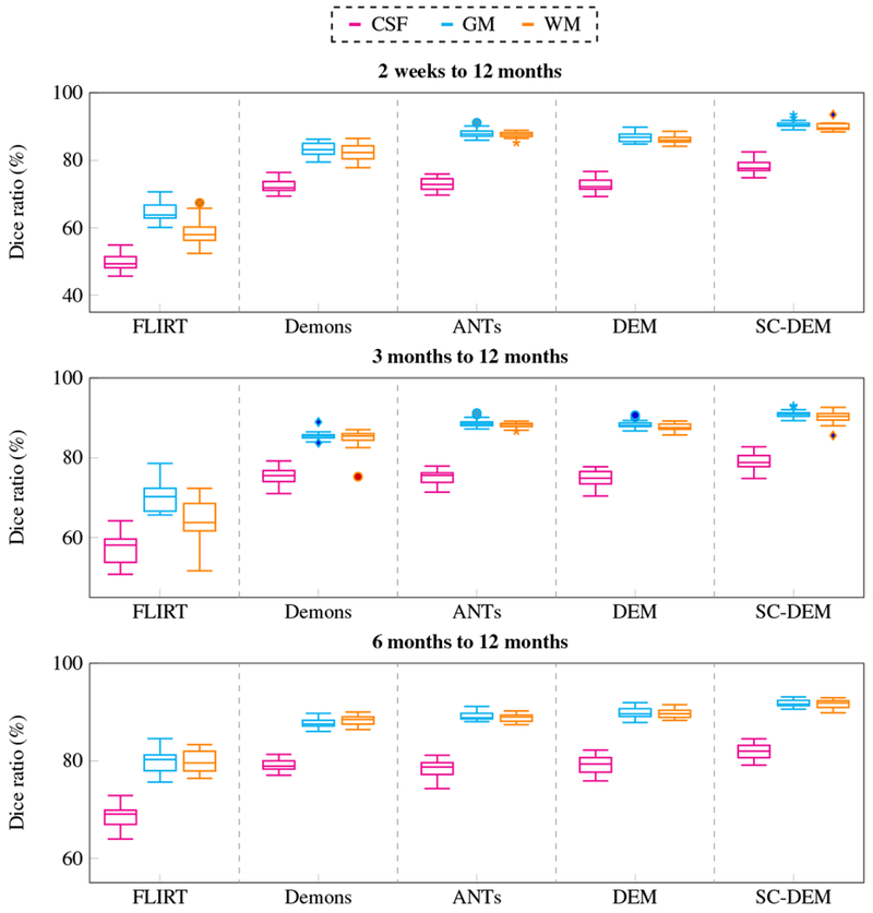
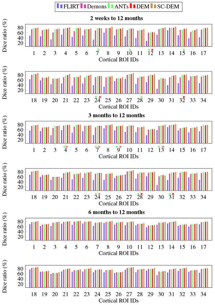
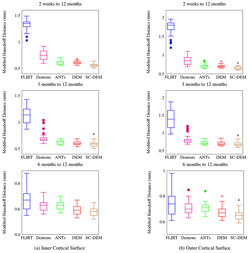
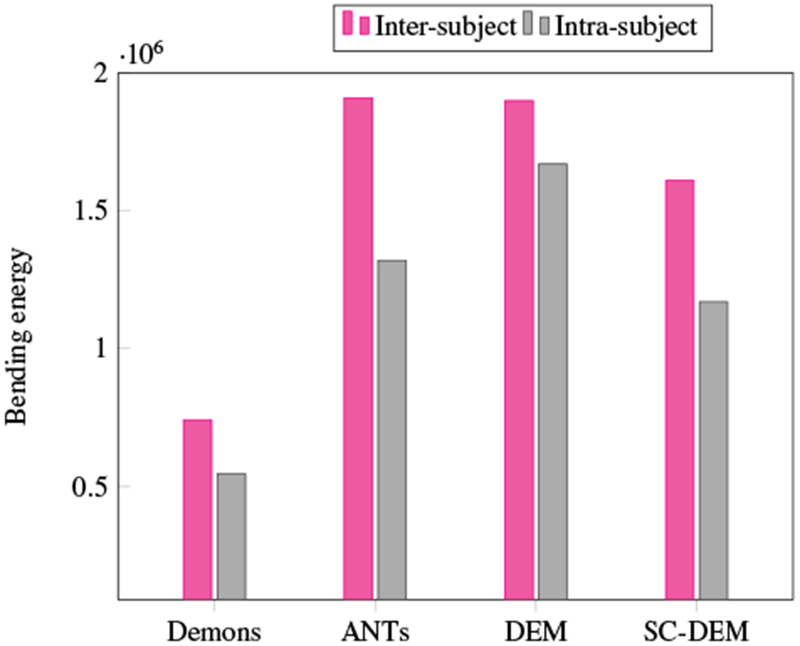
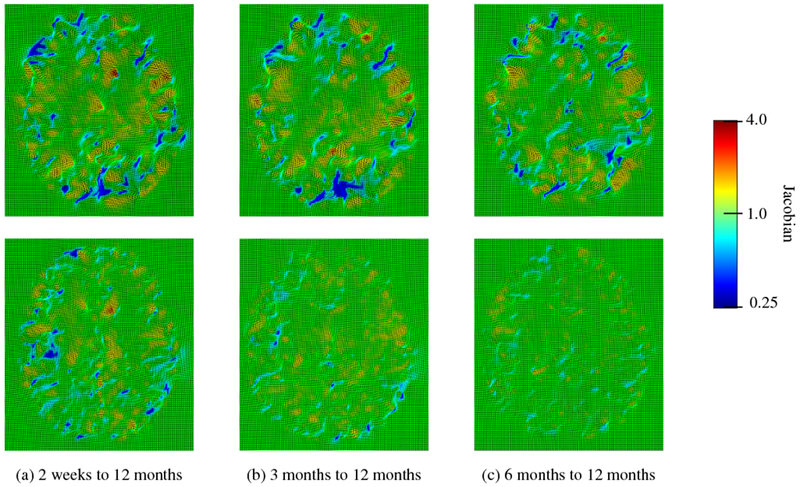
The T1-weighted and T2-weighted MRI contrasts of the infant brain evolve drastically during the first year of life. This poses significant challenges to inter- and intra-subject registration, which is key to subsequent statistical analyses. Existing registration methods that do not consider temporal contrast changes are ineffective for infant brain MRI data. To address this problem, we present in this paper a method for deformable registration of infant brain MRI. The key advantage of our method is threefold: (i) To deal with appearance changes, registration is performed based on segmented tissue maps instead of image intensity. Segmentation is performed by using an infant-centric algorithm previously developed by our group. (ii) Registration is carried out with respect to both cortical surfaces and volumetric tissue maps, thus allowing precise alignment of both cortical and subcortical structures. (iii) A dynamic elasticity model is utilized to allow large non-linear deformation. Experimental results in comparison with well-established registration methods indicate that our method yields superior accuracy in both cortical and subcortical alignment.

Keywords: Infant magnetic resonance imaging; Joint cortical surface and volumetric registration.


Conflict of interest statement

Conflict of Interest

None declared.

Figures

Fig. 1:
Fig. 1:
Dynamic morphological and appearance changes in a typical infant brain from 2 weeks to 12 months of age demonstrated using T1-weighted MR images.
Fig. 2:
Fig. 2:
Tissue segmentation of a typical 6-month-old (first row) and 12-month-old (second row). (a) T1-weighted MR brain image and segmentation results using (b) FreeSurfer and (c) BrainSuite.
Fig. 3:
Fig. 3:
Pipeline for surface-constrained volumetric registration.
Fig. 4:
Fig. 4:
Spherical mapping of the inner cortical surface.
Fig. 5:
Fig. 5:
Illustration of optimization procedure.
Fig. 6:
Fig. 6:
Inner and outer cortical surfaces generated by BrainSuite.
Fig. 7:
Fig. 7:
Inner cortical surface of the moving image warped using FLIRT (first row), diffeomorphic Demons (second row), ANTs (third row), DEM (fourth row) and SC-DEM (fifth row) overlaid on the template tissue map used in inter-subject registration.
Fig. 8:
Fig. 8:
Cortical surface distance between the template cortical surface and the moving cortical surface warped using FLIRT (first column), diffeomorphic Demons (second column), ANTs (third column), DEM (fourth column) and SC-DEM (fifth column) for inter-subject registration.
Fig. 9:
Fig. 9:
Moving T1-weighted image warped using FLIRT (second row), diffeomorphic Demons (third row), ANTs (fourth row), DEM (fifth row) and SC-DEM (sixth row), in comparison with the template image (first row) used in inter-subject registration.
Fig. 10:
Fig. 10:
Inter-subject registration results in terms of Dice ratio for cortical ROIs. The colored stars mark differences that are not statistically significant (p > 0.01) with reference to SC-DEM.
Fig. 11:
Fig. 11:
Inter-subject registration results in terms of Dice ratio for different tissue types (CSF, GM, WM).
Fig. 12:
Fig. 12:
Inter-subject registration results in terms of modified Hausdorff distance for inner and outer cortical surfaces.
Fig. 13:
Fig. 13:
Inner cortical surface of the moving image warped using FLIRT (first row), diffeomorphic Demons (second row), ANTs (third row), DEM (fourth row) and SC-DEM (fifth row) overlaid on the template tissue map for intra-subject registration.
Fig. 14:
Fig. 14:
Cortical surface distance between the template cortical surface and the moving cortical surface warped using FLIRT (first column), diffeomorphic Demons (second column), ANTs (third column), DEM (fourth column) and SC-DEM (fifth column) for intra-subject registration.
Fig. 15:
Fig. 15:
Moving image warped using FLIRT (second row), diffeomorphic Demons (third row), ANTs (fourth row), DEM (fifth row) and SC-DEM (sixth row), compared to the template image (first row) for intra-subject registration.
Fig. 16:
Fig. 16:
Intra-subject registration results in terms of Dice ratio for different tissue types (CSF, GM, WM).
Fig. 17:
Fig. 17:
Intra-subject registration results in terms of Dice ratio for cortical ROIs. The colored stars mark differences that are not statistically significant (p > 0.01) with reference to SC-DEM.
Fig. 18:
Fig. 18:
Intra-subject registration results in terms of modified Hausdorff distance for inner and outer cortical surfaces.
Fig. 19:
Fig. 19:
Bending energy of the deformation fields estimated using different registration methods.
Fig. 20:
Fig. 20:
Deformation Jacobian maps of SC-DEM for inter-subject (first row) and intra-subject (second row) registration.
Fig. 21:
Fig. 21:
Parameter sensitivity analysis in terms of Dice ratio and modified Hausdorff distance.

References

    1. Acosta O, Fripp J, Rueda A, Xiao D, Bonner E, Bourgeat P, Salvado O, 2010. 3D shape context surface registration for cortical mapping, in: IEEE International Symposium on Biomedical Imaging: From Nano to Macro (ISBI’10), Rotterdam, Netherlands pp. 1021–1024. doi: 10.1109/ISBI.2010.5490163. - DOI
    1. Ahmad S, Khan MF, 2017. Dynamic elasticity model for inter-subject non-rigid registration of 3D MRI brain scans. Biomedical Signal Processing and Control 33, 346–357. doi: 10.1016/j.bspc.2016.12.016. - DOI
    1. Ahmad S, Wu Z, Li G, Wang L, Lin W, Yap PT, Shen D, 2019. Surface-volume consistent construction of longitudinal atlases for the early developing brains, in: Shen D, Liu T (Eds.), Medical Image Computing and Computer Assisted Intervention - MICCAI 2019, Springer. - PMC - PubMed
    1. Avants B, Epstein C, Grossman M, Gee J, 2008. Symmetric diffeomorphic image registration with cross-correlation: Evaluating automated labeling of elderly and neurodegenerative brain. Medical Image Analysis 12, 26–41. doi: 10.1016/j.media.2007.06.004. - DOI - PMC - PubMed
    1. Budday S, Nay R, de Rooij R, Steinmann P, Wyrobek T, Ovaert TC, Kuhl E, 2015. Mechanical properties of gray and white matter brain tissue by indentation. Journal of the Mechanical Behavior of Biomedical Materials 46, 318–330. doi: 10.1016/j.jmbbm.2015.02.024. - DOI - PMC - PubMed

Publication types