Review
. 2020 Jan 24:15:23-50.
doi: 10.1146/annurev-pathmechdis-012419-032824. Epub 2019 Aug 9.

Liver Progenitors and Adult Cell Plasticity in Hepatic Injury and Repair: Knowns and Unknowns

Affiliations
Review

Liver Progenitors and Adult Cell Plasticity in Hepatic Injury and Repair: Knowns and Unknowns

Sungjin Ko et al. Annu Rev Pathol. .

Abstract

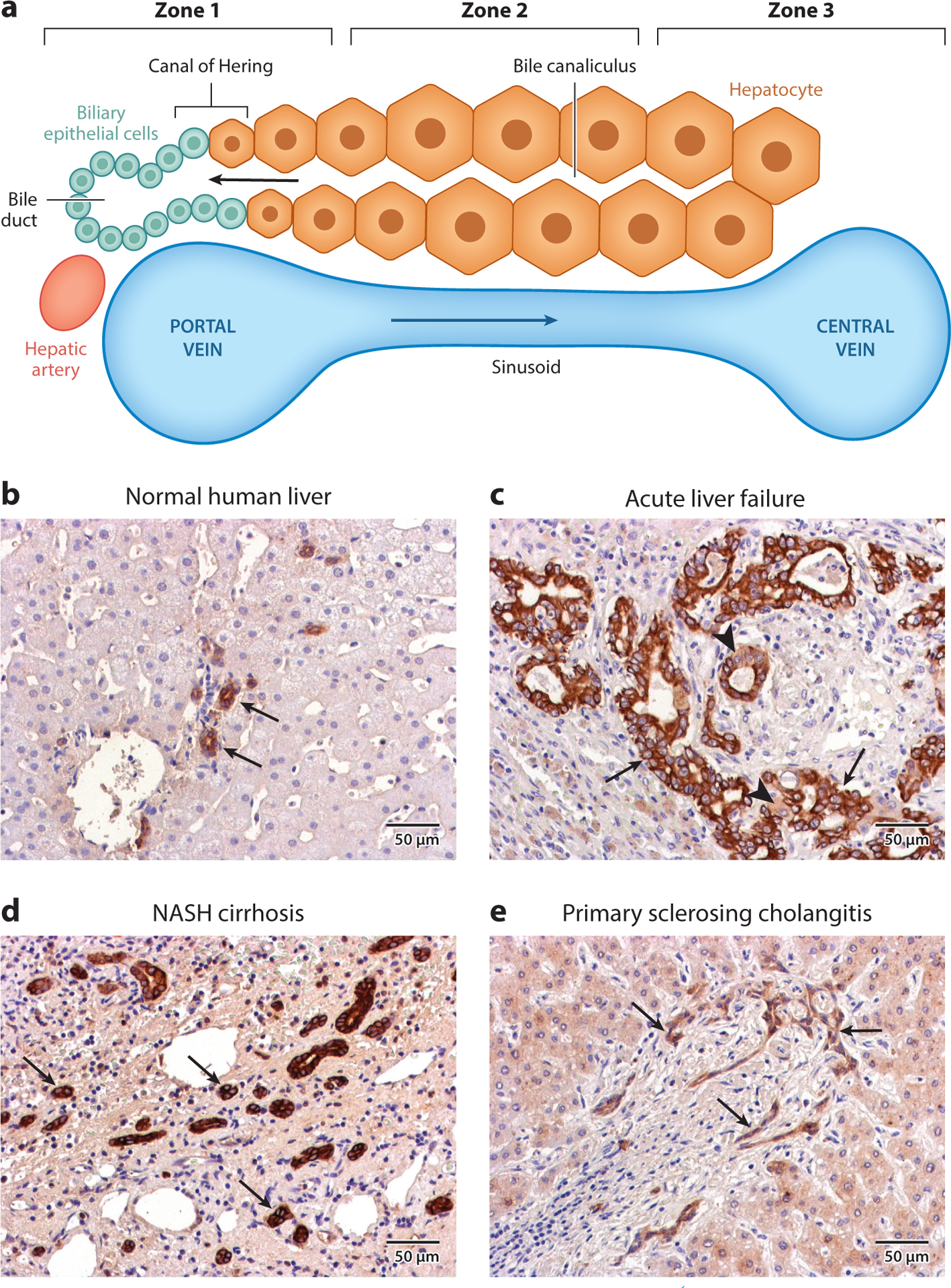
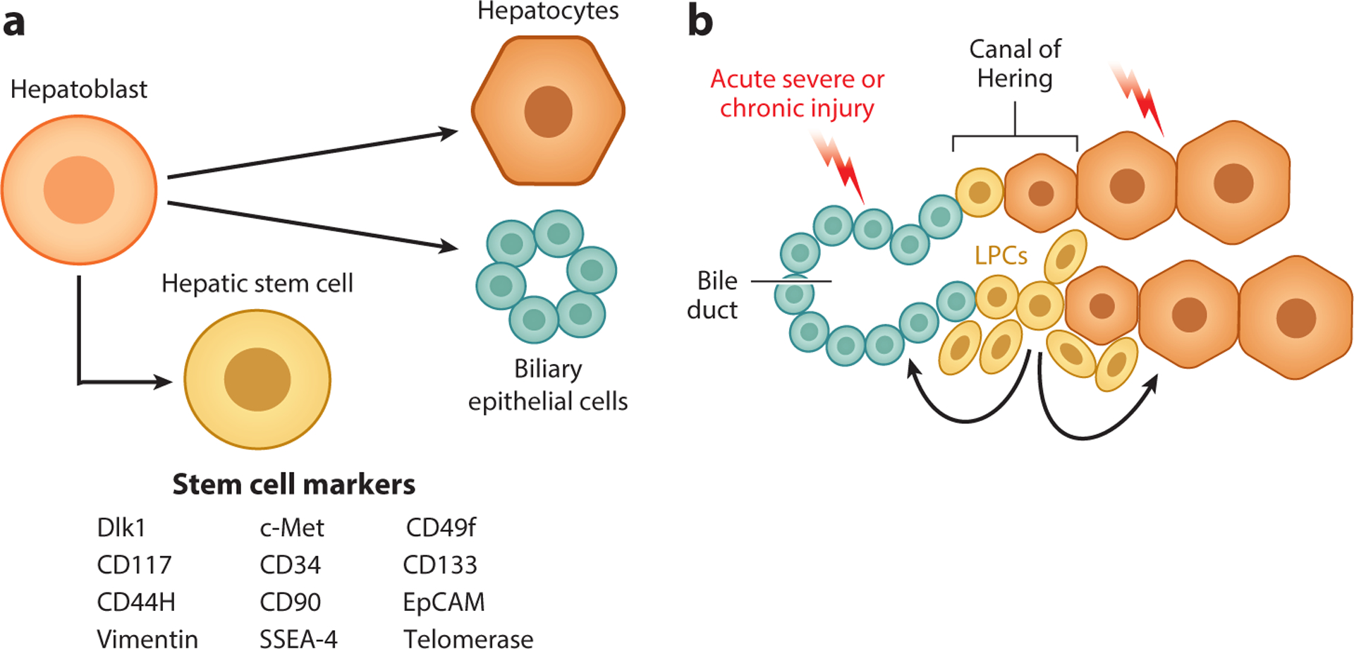
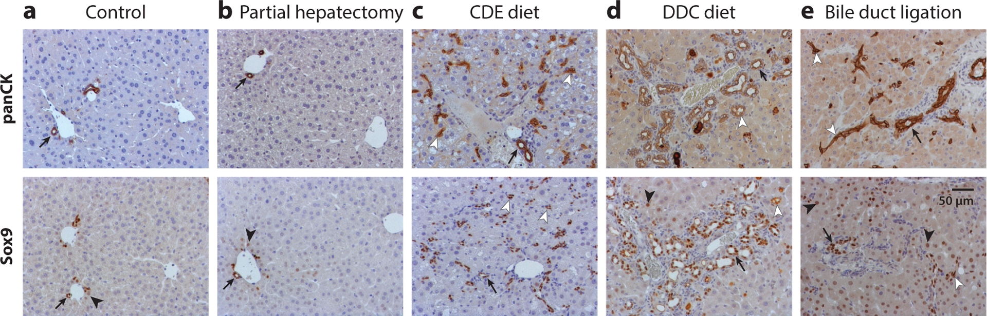
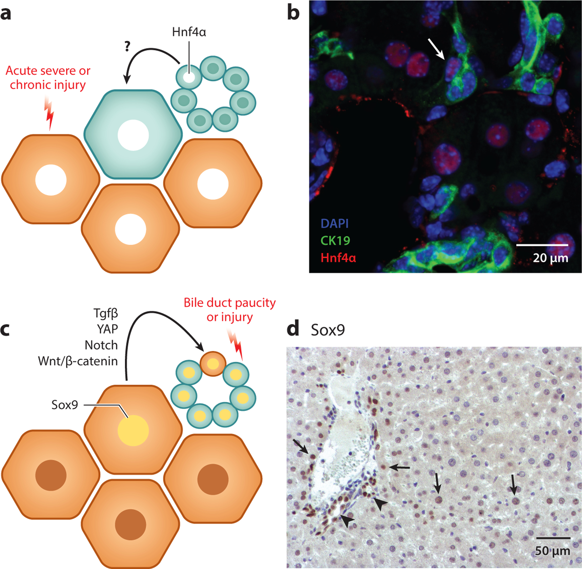
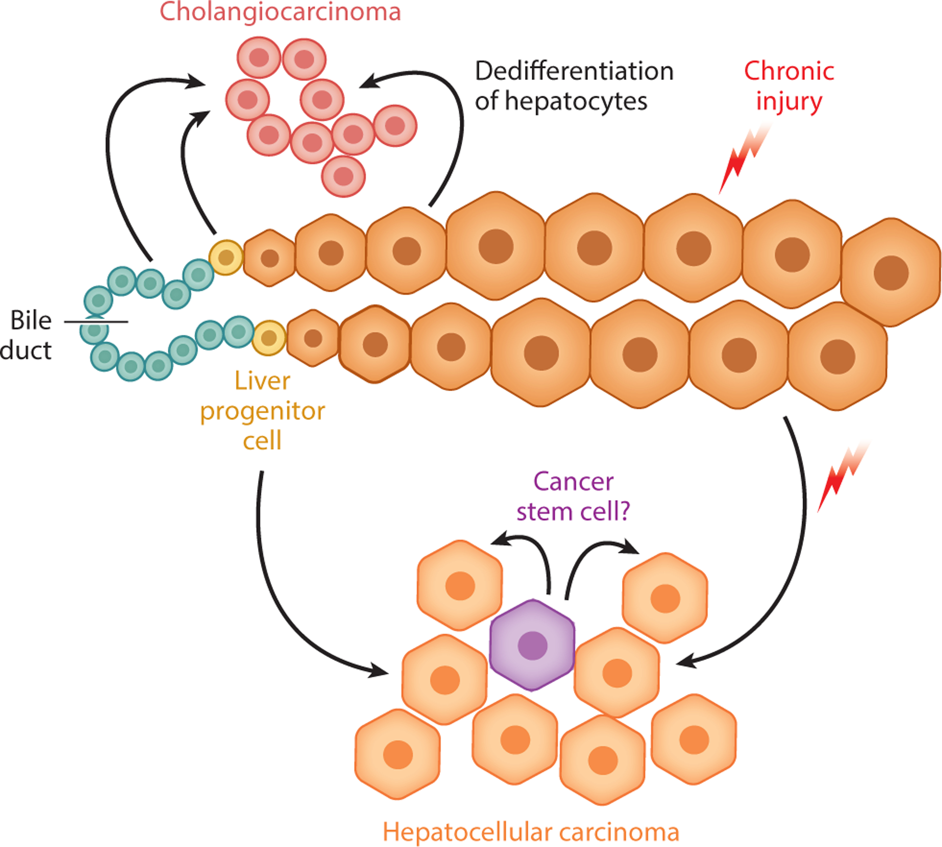
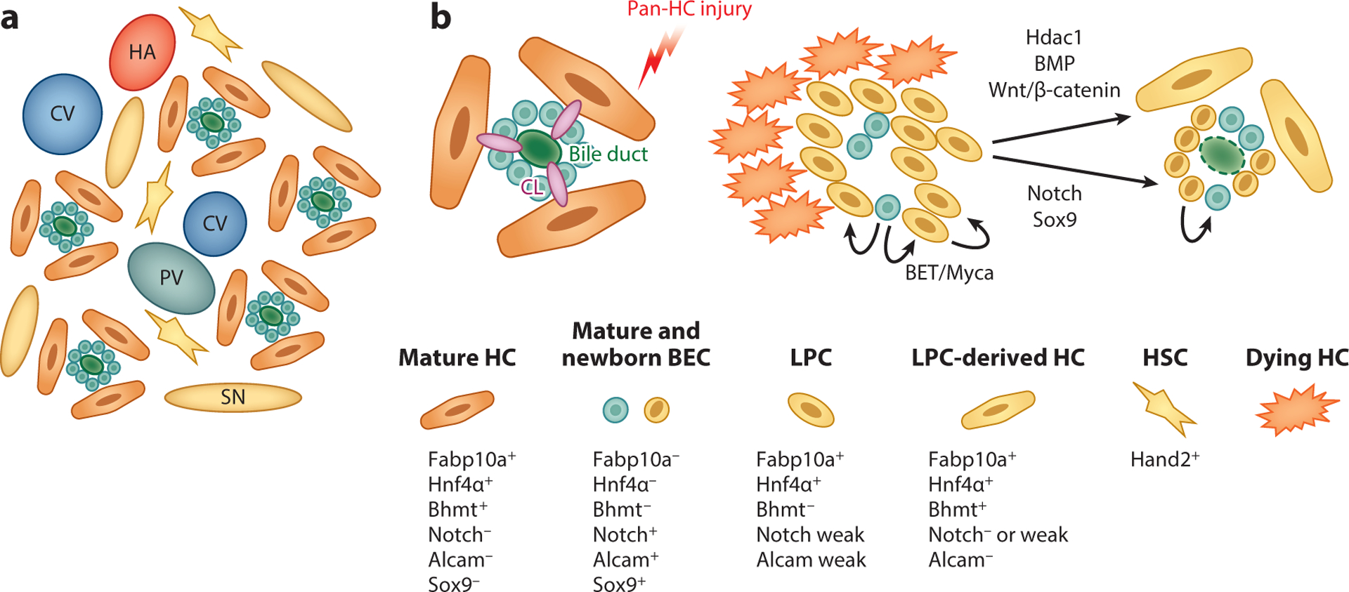
The liver is a complex organ performing numerous vital physiological functions. For that reason, it possesses immense regenerative potential. The capacity for repair is largely attributable to the ability of its differentiated epithelial cells, hepatocytes and biliary epithelial cells, to proliferate after injury. However, in cases of extreme acute injury or prolonged chronic insult, the liver may fail to regenerate or do so suboptimally. This often results in life-threatening end-stage liver disease for which liver transplantation is the only effective treatment. In many forms of liver injury, bipotent liver progenitor cells are theorized to be activated as an additional tier of liver repair. However, the existence, origin, fate, activation, and contribution to regeneration of liver progenitor cells is hotly debated, especially since hepatocytes and biliary epithelial cells themselves may serve as facultative stem cells for one another during severe liver injury. Here, we discuss the evidence both supporting and refuting the existence of liver progenitor cells in a variety of experimental models. We also debate the validity of developing therapies harnessing the capabilities of these cells as potential treatments for patients with severe and chronic liver diseases.

Keywords: ductular reaction; liver cancer; liver regeneration; liver stem cell; oval cell.


Figures

Figure 1
Figure 1
Architecture of normal and diseased liver. (a) The structure of a liver lobule consists of three zones. Zone 1 consists of the portal vein, bile ducts, and hepatic artery, which together form the portal triad. Oxygenated blood from the hepatic artery mixes with blood from the portal vein and flows through the hepatic sinusoids toward the central vein, which constitutes zone 3. The interface between the end of a bile canaliculus and the start of a bile duct is known as the canal of Hering. Cords of hepatocytes (HCs) form the bile canaliculi, which transport HC-derived bile to the bile ducts in the opposite direction of blood flow. The HCs found between zones 1 and 3 constitute zone 2, and these are also known as midzonal HCs. (b) Normal human liver stained with a pan-cytokeratin (panCK) antibody. The arrows denote bile ducts, structures with obvious lumina lined by panCK-positive cells. (c) Liver from a patient with acute liver failure stained with a panCK antibody. A ductular reaction (DR) is evidenced by a large increase in the number of panCK-positive cells (arrows). Intermediate HCs, or cells that are panCK-positive but exhibit HC morphology (arrowhead), can be observed adjacent to the cells of the DR. (d) Liver from a patient with nonalcoholic steatohepatitis (NASH)– induced cirrhosis stained with a panCK antibody. A DR is evident in the increase in the number of panCK-positive cells, which form bile duct–like structures without obvious lumina (arrows). (e) Liver from a patient with primary sclerosing cholangitis stained with a panCK antibody. A DR is evident by the increase in the number of panCK-positive cells (arrows), which have a distinctly different morphology from normal bile ducts.
Figure 2
Figure 2
The liver progenitor cell theory. (a) During fetal liver development, hepatoblasts give rise to the two primary epithelial cell types of the mature liver: hepatocytes and biliary epithelial cells. Studies have identified potential markers of hepatoblasts with characteristics of pluripotent stem cells (shown in the figure). (b) It is theorized that quiescent liver progenitor cells (LPCs), potentially residing in the canals of Hering, are activated in the context of severe acute injury or chronic liver injury. These LPCs proliferate and are capable of giving rise to both hepatocytes and cholangiocytes, thus contributing to liver repair.
Figure 3
Figure 3
Images depict (a) normal mouse liver, and liver from a mouse (b) 1 day after two-thirds partial hepatectomy, (c) after 2 weeks of a choline-deficient, ethionine-supplemented (CDE) diet, (d) after 5 weeks of a 3,5-diethoxycarbonyl-1,4-dihydrocollidine (DDC) diet, and (e) 1 week after bile duct ligation. Liver tissues were stained with pan-cytokeratin (panCK) (top row) or Sox9 antibody (bottom row). Arrows denote bile ducts with luminal structures, lined by panCK- or Sox9-positive cells, while arrowheads denote Sox9-positive hepatocytes. (c–e) Ductular reactions are evinced by a large increase in the number of panCK- or Sox9-positive cells (white arrowheads). Dilation of luminal structures is observed after (d) the DDC diet, and (e) preexisting bile ducts are thickened, with increased numbers of panCK-positive cells, after bile duct ligation. A few Sox9-positive hepatocytes are observed adjacent to the portal vein in (a) normal liver, while this population is (d) modestly increased after administration of the DDC diet, and it is (e) hugely increased throughout the whole liver following bile duct ligation.
Figure 4
Figure 4
The transdifferentiation theory. (a) During acute severe or chronic liver injury in which hepatocyte (HC) function is significantly impaired, biliary epithelial cells (BECs) can directly transdifferentiate into HCs to mediate liver repair. During this process, BECs express HC markers such as Hnf4α. (b) The liver from a mouse fed a choline-deficient, ethionine-supplemented diet for 2 weeks, followed by 3 days of recovery on a normal diet, stained for DAPI (blue), cytokeratin (CK) 19 (green) and Hnf4α (red). A transdifferentiating cell that expressed both CK19 and Hnf4α is evident (white arrow). (c) Following bile duct paucity or severe injury to BECs, HCs can transdifferentiate into BECs, and during this process, HCs will express BEC markers, such as Sox9. Signaling pathways, including transforming growth factor-β (Tgfβ), yes-associated protein (YAP), Notch, and Wnt/β-catenin, have been identified as playing roles in promoting this process. (d) Liver from a mouse stained for Sox9 2 days after bile duct ligation reveals HCs that express Sox9 (arrows) in comparison to the presence of Sox9 in the BECs lining the bile ducts (arrowheads).
Figure 5
Figure 5
Liver progenitor cells (LPCs) in liver cancer. Generally, the two major forms of adult liver cancer are thought to arise from their corresponding epithelial cell counterparts, often in the setting of chronic injury and inflammation: hepatocellular carcinoma from hepatocytes and cholangiocarcinoma from biliary epithelial cells. At the same time, the question remains whether LPCs serve as another source of both hepatocellular carcinoma and cholangiocarcinoma due to their stem cell–like properties and their long-term activation in the setting of liver injury. In addition, studies have identified potential cancer stem cells in liver cancer, which may or may not be related in origin to LPCs.
Figure 6
Figure 6
Schematic of zebrafish liver anatomy and liver progenitor cell (LPC)–driven liver regeneration. (a) Schematic of zebrafish liver architecture. Unlike the hepatic zonation found in the mammalian liver, portal veins (PVs), central veins (CVs), and hepatic arteries (HAs) are randomly distributed in the zebrafish liver. Hepatocytes (HCs) are arranged in tubules around bile ducts interspersed with hepatic stellate cells (HSCs) and surrounded by fenestrated sinusoidal endothelium (SN). (b) During near-total HC ablation, bile canaliculi (CL) and biliary networks completely collapse, and remnant biliary epithelial cells (BECs) are packed without a central lumen. Subsequently, BECs rapidly dedifferentiate into LPCs, which coexpress Notch signaling targets, Alcam, Fabp10a, and Hnf4α. This is followed by redifferentiation of LPCs into functional HCs and BECs. Using this model, signaling pathways, including BET/Myca, Hdac1/Sox9, BMP, and Wnt/β-catenin have been identified as regulators of LPC-mediated liver regeneration.

References

    1. Stoick-Cooper CL, Moon RT, Weidinger G. 2007. Advances in signaling in vertebrate regeneration as a prelude to regenerative medicine. Genes Dev. 21:1292–315 - PubMed
    1. Cordero-Espinoza L, Huch M. 2018. The balancing act of the liver: tissue regeneration versus fibrosis. J. Clin. Investig 128:85–96 - PMC - PubMed
    1. Vivien CJ, Hudson JE, Porrello ER. 2016. Evolution, comparative biology and ontogeny of vertebrate heart regeneration. NPJ Regen. Med 1:16012. - PMC - PubMed
    1. Ferretti P, Zhang F, O’Neill P. 2003. Changes in spinal cord regenerative ability through phylogenesis and development: lessons to be learnt. Dev. Dyn 226:245–56 - PubMed
    1. Wynn TA. 2008. Cellular and molecular mechanisms of fibrosis. J. Pathol 214:199–210 - PMC - PubMed

Publication types