. 2019 Aug 9;20(1):181.
doi: 10.1186/s12931-019-1129-4.

Role of KRAS in regulating normal human airway basal cell differentiation

Affiliations

Role of KRAS in regulating normal human airway basal cell differentiation

Fumihiro Ogawa et al. Respir Res. .

Abstract

Background: KRAS is a GTPase that activates pathways involved in cell growth, differentiation and survival. In normal cells, KRAS-activity is tightly controlled, but with specific mutations, the KRAS protein is persistently activated, giving cells a growth advantage resulting in cancer. While a great deal of attention has been focused on the role of mutated KRAS as a common driver mutation for lung adenocarcinoma, little is known about the role of KRAS in regulating normal human airway differentiation.

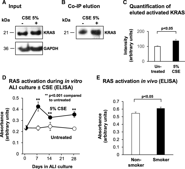
Methods: To assess the role of KRAS signaling in regulating differentiation of the human airway epithelium, primary human airway basal stem/progenitor cells (BC) from nonsmokers were cultured on air-liquid interface (ALI) cultures to mimic the airway epithelium in vitro. Modulation of KRAS signaling was achieved using siRNA-mediated knockdown of KRAS or lentivirus-mediated over-expression of wild-type KRAS or the constitutively active G12 V mutant. The impact on differentiation was quantified using TaqMan quantitative PCR, immunofluorescent and immunohistochemical staining analysis for cell type specific markers. Finally, the impact of cigarette smoke exposure on KRAS and RAS protein family activity in the airway epithelium was assessed in vitro and in vivo.

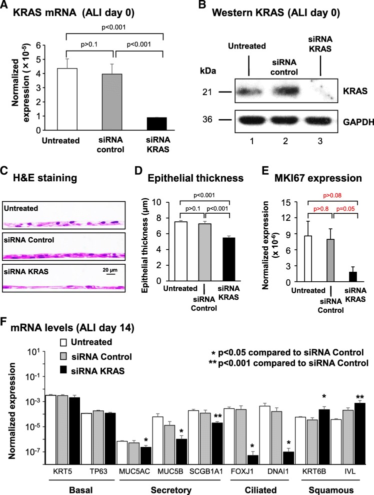
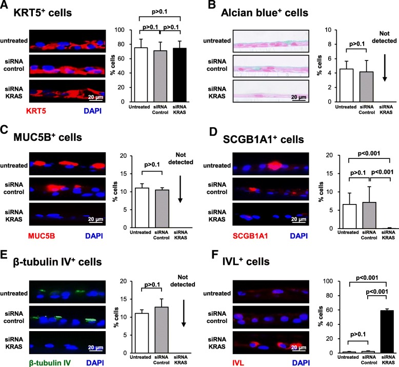
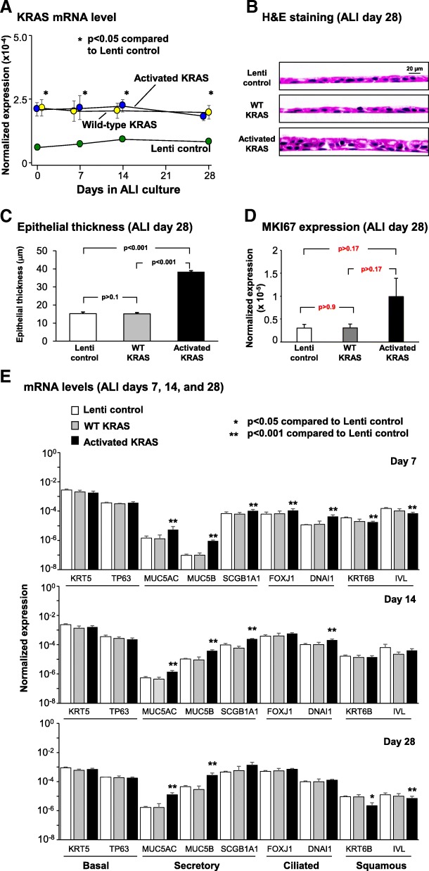
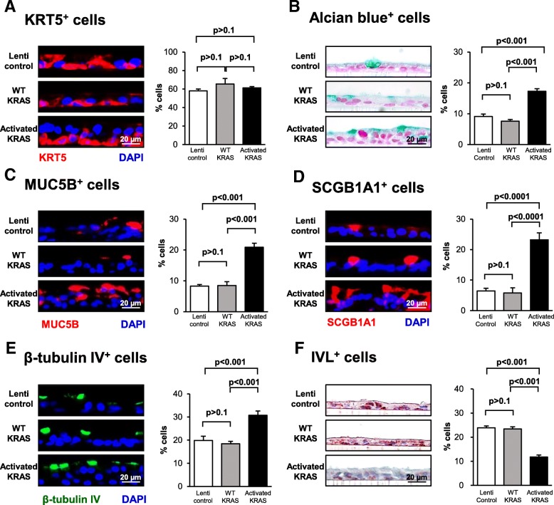
Results: siRNA-mediated knockdown of KRAS decreased differentiation of BC into secretory and ciliated cells with a corresponding shift toward squamous cell differentiation. Conversely, activation of KRAS signaling via lentivirus mediated over-expression of the constitutively active G12 V KRAS mutant had the opposite effect, resulting in increased secretory and ciliated cell differentiation and decreased squamous cell differentiation. Exposure of BC to cigarette smoke extract increased KRAS and RAS protein family activation in vitro. Consistent with these observations, airway epithelium brushed from healthy smokers had elevated RAS activation compared to nonsmokers.

Conclusions: Together, these data suggest that KRAS-dependent signaling plays an important role in regulating the balance of secretory, ciliated and squamous cell differentiation of the human airway epithelium and that cigarette smoking-induced airway epithelial remodeling is mediated in part by abnormal activation of KRAS-dependent signaling mechanisms.

Keywords: Airway; Basal cell; Cigarette smoking; Differentiation; KRAS; Stem/progenitor.


Conflict of interest statement

The authors declare that they have no competing interests.

Figures

Fig. 1
Fig. 1
KRAS expression in the normal human airway epithelium in vivo and in vitro a Immunohistochemical staining analysis of KRAS in the airway epithelium. Normal nonsmoker human airway sections were analyzed for expression of KRAS. Isotype specific antibody was used as negative control. Scale bar 20 μm. b Expression of KRAS during differentiation of human airway basal cells (BC) on air-liquid interface (ALI) culture. KRAS mRNA expression was assessed by qPCR. Data points indicate the mean of n = 3 independent experiments, each performed in triplicate with independent donors of BC. Error bars indicate standard error of the mean. c Immunohistochemical staining analysis of KRAS during ALI culture. ALI day 0 and day 28 sections were analyzed for expression of KRAS. Isotype specific antibody was used as negative control. Scale bar 20 μm
Fig. 2
Fig. 2
Effect of silencing of KRAS expression on regulation of basal cell (BC) differentiation into a mucociliated epithelium. Primary human airway BC were untreated or transfected with either control, or KRAS specific siRNA and cultured on ALI for 14 days to assess the impact of KRAS on BC differentiation. a qPCR analysis to assess mRNA expression of KRAS to confirm efficacy of siRNA-mediated knockdown at ALI day 0. Bars indicate the normalized gene expression. Error bars indicate standard error of the mean. Data from n = 3 independent experiments, each performed in triplicate with independent donors of BC. b Western analysis of KRAS expression following siRNA mediated knockdown at ALI day 0. Lane 1- untreated; lane 2- siRNA control; and lane 3- siRNA KRAS. GAPDH was used as a loading control. c Histology of untreated, siRNA control and siRNA KRAS cells at ALI day 14. d Quantification of epithelial thickness of ALI day 14. Bars indicate the epithelial thickness. Error bars indicate standard error of the mean. Data from n = 3 independent experiments, each performed with an independent donor of BC. e qPCR analysis to assess mRNA expression of the proliferation marker MKI67 at ALI day 14. Bars indicate the normalized mRNA expression. Error bars indicate standard error. Data from n = 3 independent experiments, each performed in triplicate with independent donors of BC. f qPCR analysis to assess mRNA expression of BC markers (KRT5, TP63), secretory cell markers (MUC5AC, MUC5B, SCGB1A1), ciliated cell markers (FOXJ1, DNAI1) and squamous cell markers (KRT6B, IVL) at ALI day 14. Bars indicate the normalized mRNA expression. Error bars indicate standard error. Asterisks indicate p < 0.05 (*) or p < 0.001 (**). ANOVA was used to determine the statistical significance among groups as described in the methods section. Data from n = 3 independent experiments, each performed in triplicate with independent donors of BC
Fig. 3
Fig. 3
Effect of silencing KRAS expression on regulation of basal cell (BC) differentiation into a mucociliated epithelium. Primary human airway BC were untreated or transfected with either control, or KRAS specific siRNA and cultured on ALI for 14 days to assess the impact of KRAS on BC differentiation into a mucociliated epithelium. a Immunofluorescence staining of KRT5+ BC. Sections of cells on ALI day 14 stained for KRT5 (red) and DAPI (nuclei, blue). b Alcian blue staining of secretory cells. Sections of cells on ALI day 14 stained with Alcian blue (blue). c Immunofluorescence staining of MUC5B+ secretory cells. Sections of cells on ALI day 14 stained for MUC5B (red) and DAPI (nuclei, blue). d Immunofluorescence staining of SCGB1A1+ secretory cells. Sections of cells on ALI day 14 stained for SCGB1A1 (red) and DAPI (nuclei, blue). e Immunofluorescence staining of β-tubulin IV+ ciliated cells. Sections of cells on ALI day 14 stained for β-tubulin IV (green) and DAPI (nuclei, blue). f Immunofluorescence staining of IVL+ squamous cells. Sections of cells on ALI day 14 stained for IVL (red). The data for a-f are the mean percentage of positively stained cells for n = 3 independent experiments performed with independent donors of BC. Error error bars indicate standard error of the mean. ANOVA was used to determine the statistical significance among groups as described in the methods section. Scale bar 20 μm
Fig. 4
Fig. 4
Effect of constitutive KRAS activity on promoting basal cell (BC) differentiation into secretory and ciliated cells. Primary human airway BC were infected with control lentivirus or lentivirus over-expressing wild-type (WT) KRAS or the constitutively active G12 V mutant (activated) and cultured on ALI for 28 days to assess the impact of KRAS on BC differentiation into a mucociliated epithelium. a qPCR analysis to assess mRNA expression of KRAS to confirm over-expression of KRAS during ALI culture. Data points indicate the mean expression and error bars indicate standard error of the mean. Data from n = 3 independent experiments, each performed in triplicate with independent donors of BC. b Histology of Lenti control, WT KRAS and activated KRAS cells at ALI day 28. c Quantification of epithelial thickness of ALI day 28. Bars indicate epithelial thickness. Error bars indicate standard error. Data from n = 3 independent experiments, each performed with an independent donor of BC. d qPCR analysis to assess mRNA expression of the proliferation marker MKI67 at ALI day 28. Bars indicate the normalized mRNA expression. Error bars indicate standard error. Data from n = 3 independent experiments, each performed in triplicate with independent donors of BC. e qPCR analysis to assess mRNA expression of BC markers (KRT5, TP63), secretory cell markers (MUC5AC, MUC5B, SCGB1A1), ciliated cell markers (FOXJ1, DNAI1) and squamous cell markers (KRT6B, IVL) at ALI days 7, 14, and 28. Data from n = 3 independent experiments, each performed in triplicate with independent donors of BC. Bars indicate the normalized mRNA expression. Error bars indicate standard error of the mean. Asterisks indicate p < 0.05 (*) or p < 0.001 (**). ANOVA was used to determine the statistical significance among groups as described in the methods section
Fig. 5
Fig. 5
Consequences of constitutive KRAS activity on promotion of basal cell (BC) differentiation into secretory and ciliated cells Primary human airway BC were infected with control lentivirus or lentivirus over-expressing wild-type (WT) KRAS or the constitutively active G12 V mutant (activated) and cultured on ALI for 28 days to assess the impact of KRAS on BC differentiation into a mucociliated epithelium. a Immunofluorescence staining of KRT5+ BC. Sections of cells on ALI day 28 membranes were stained for KRT5 (red) and DAPI (nuclei, blue). b Alcian blue staining of secretory cells. Sections of cells on ALI day 28 membranes were stained for Alcian blue (blue). c Immunofluorescence staining of MUC5B+ secretory cells. Sections of cells on ALI day 28 membranes were stained for MUC5B (red) and DAPI (nuclei, blue). d Immunofluorescence staining of SCGB1A1+ secretory cells. Sections of cells on ALI day 28 membranes were stained for SCGB1A1 (red) and DAPI (nuclei, blue). e Immunofluorescence staining of β-tubulin IV+ ciliated cells. Sections of cells on ALI day 28 membranes were stained for β-tubulin IV (green) and DAPI (nuclei, blue). f Immunohistochemical staining of IVL+ squamous cells. Sections of cells on ALI day 28 membranes were stained for IVL (red) and DAPI (nuclei, blue). The data for a-f are the mean for n = 3 independent experiments performed with independent donors of BC. The bars indicate the mean percentage of positively stained cells for n = 3 independent experiments, and error bars indicate standard error of the mean. ANOVA was used for statistical comparisons among groups as described in the Methods. Scale bar 20 μm
Fig. 6
Fig. 6
Effect of cigarette smoke exposure on KRAS and RAS protein family activation in the airway epithelium. a-c. Basal cells (BC) were cultured in the absence or presence of 5% cigarette smoke extract (CSE) under non-differentiating culture conditions. Forty-eight hr. post-CSE exposure, the cells were lysed and the activated form of GTP-bound KRAS quantified by co-immunoprecipitation (Co-IP). a Western analysis of KRAS and GAPDH (loading control) levels in cell lysates. b Western analysis of activated KRAS levels elution lysates following Co-IP. c Quantification of activated KRAS levels. Bars indicate intensity of signal obtained in during image analysis. Error bars indicate standard error of the mean. Data from n = 3 independent experiments, each performed with independent donors of BC. d Basal cells were cultured on ALI for 28 days in the absence and presence of 5% CSE and harvested at multiple time points to quantify RAS protein family activation by ELISA. Bars indicate mean absorbance levels. Error bars indicate standard error of the mean. Data from n = 3 independent experiments, each performed in triplicate with independent donors of BC. e Airway epithelium from healthy nonsmokers (n = 5) and asymptomatic healthy smokers (n = 5) were obtained via bronchoscopic brushing and the levels of RAS protein family activation quantified by ELISA. Bars indicate mean absorbance levels. Error bars indicate standard error of the mean. T tests were used to determine the statistical significance of differences observed in two-way comparisons. Statistical analysis of data in the time course utilized a repeated measures ANOVA as described in the Methods

Similar articles

Cited by

References

    1. Downward J. Targeting RAS signalling pathways in cancer therapy. Nat Rev Cancer. 2003;3:11–22. doi: 10.1038/nrc969. - DOI - PubMed
    1. Colicelli J. Human RAS superfamily proteins and related GTPases. Sci STKE. 2004;2004:RE13. - PMC - PubMed
    1. Papke B, Der CJ. Drugging RAS: Know the enemy. Science. 2017;355:1158–1163. doi: 10.1126/science.aam7622. - DOI - PubMed
    1. Simanshu DK, Nissley DV, McCormick F. RAS proteins and their regulators in human disease. Cell. 2017;170:17–33. doi: 10.1016/j.cell.2017.06.009. - DOI - PMC - PubMed
    1. Haigis KM. KRAS alleles: the devil is in the detail. Trends Cancer. 2017;3:686–697. doi: 10.1016/j.trecan.2017.08.006. - DOI - PMC - PubMed

MeSH terms

Substances