. 2021 Aug;121(Pt B):106440.
doi: 10.1016/j.yebeh.2019.07.041. Epub 2019 Aug 6.

Amygdala lesions reduce seizure-induced respiratory arrest in DBA/1 mice

Affiliations

Amygdala lesions reduce seizure-induced respiratory arrest in DBA/1 mice

Anthony Marincovich et al. Epilepsy Behav. 2021 Aug.

Abstract

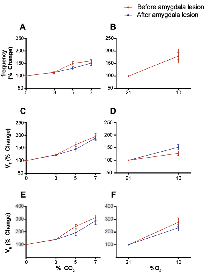
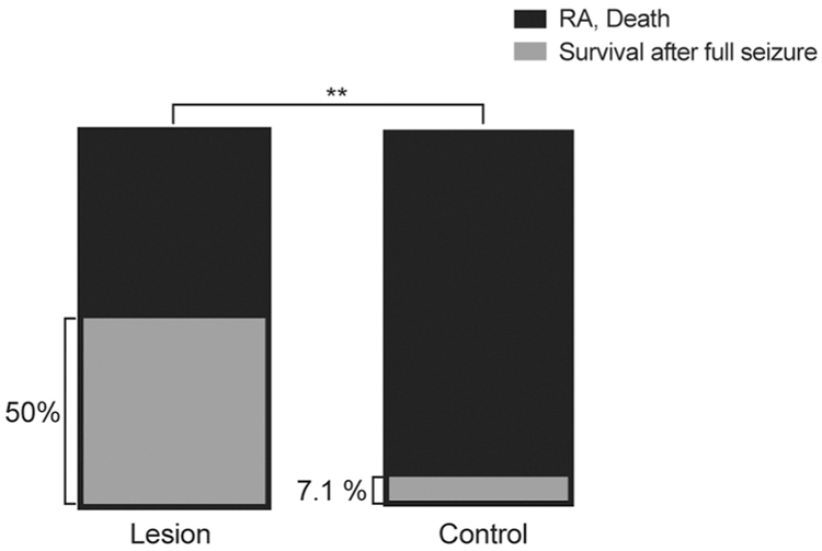
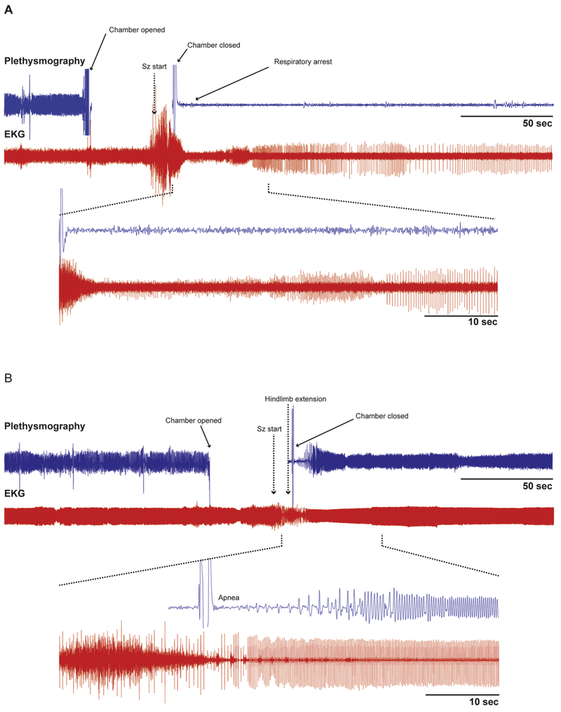
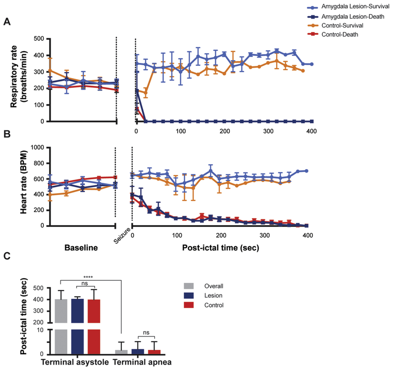
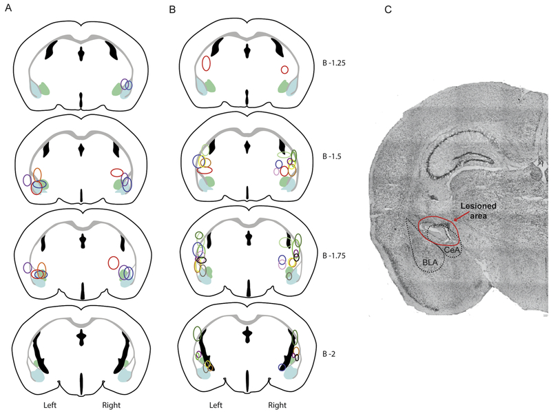
Sudden unexpected death in epilepsy (SUDEP) is the most common cause of death in patients with refractory epilepsy. Human studies and animal models suggest that respiratory arrest is the initiating event leading to death in many cases of SUDEP. It has previously been reported that the onset of apnea can coincide with the spread of seizures to the amygdala, and apnea can be reproduced by electrical stimulation of the amygdala. The aim of the current work was to determine if the amygdala is required for seizure-induced respiratory arrest (S-IRA) in a mouse model of SUDEP. Experiments were performed on DBA/1 mice that have audiogenic seizures with a high incidence of fatal postictal respiratory arrest. Electrolytic lesions of the amygdala significantly reduced the incidence of S-IRA without altering seizures, baseline breathing, or the hypercapnic ventilatory response. These results indicate that the amygdala is a critical node in a pathway to the lower brainstem that is needed for seizures to cause respiratory arrest. SIGNIFICANCE STATEMENT: Sudden unexpected death in epilepsy is the most common cause of mortality in patients with refractory epilepsy, and S-IRA is thought to be important in the pathophysiology in many cases. In a patient with epilepsy, the onset of apnea has been shown to coincide with spread of seizures to the amygdala, and in multiple patients, apnea was induced by stimulation of the amygdala. Here, we show that lesions of the amygdala reduced the incidence of S-IRA and death in a mouse model of SUDEP. These results provide evidence that the amygdala may be a critical node in the pathway by which seizures influence the brainstem respiratory network to cause apnea. This article is part of the Special Issue NEWroscience 2018.

Keywords: Amygdala; Respiration; SUDEP; Seizures.


Figures

Figure 1-
Figure 1-. Amygdala lesions did not affect baseline breathing or chemoreception in DBA/1 mice.
A, C, E- CO2 chemoreception as measured by the HCVR was not significantly different in DBA/1 mice pre- and post-amygdala lesion. Plotted are the percentage increase in frequency (p=0.179), VT (p=0.419) and VE (p=0.228) in response to hypercapnia. (2 way ANOVA, Bonferroni post Hoc test) B, D, F- O2 chemoreception as measured by the HVR was also not affected by amygdala lesions. Plotted are the percentage increase in frequency (p=0.990), VT(P=0.096), and VE (P=0.318) in response to hypoxia (2 way ANOVA, Bonferroni post Hoc test). HCVR= Hypercapnic ventilatory response HVR= Hypoxic ventilatory response VT= tidal volume VE= Minute ventilation
Figure 2 -
Figure 2 -. Amygdala lesions significantly reduced S-IRA and death in DBA/1 mice.
Lesioned animals had a significantly higher rate of survival after full seizures compared to Controls. 50% (n=10) compared to 7.14% (n=28), respectively, p=.0080, Fischer’s exact test).
Figure 3 -
Figure 3 -. A DBA/1 mouse with an amygdala lesion survived a seizure while a control mouse died of seizure-induced respiratory arrest.
A - Raw data traces of plethysmography and EKG from a control DBA/1 mouse in which terminal apnea occurred during a seizure. The heart rate (EKG) was disrupted during and shortly after the seizure, but continued for a prolonged period before it eventually slowed to terminal asystole (not shown). Inset below shows expanded traces from the time indicated by the dashed lines. B- Raw data traces of plethysmography and EKG in a DBA/1 mouse with a unilateral R sided amygdala lesion that survived an audiogenic seizure. Note the brief apnea followed by slow increase in amplitude and frequency of breathing which eventually returned to baseline. There was also brief bradycardia that recovered more rapidly. Inset below shows expanded traces from the time indicated by the dashed lines.
Figure 4 -
Figure 4 -. Terminal apnea preceded terminal asystole during seizure-induced death in DBA/1 mice with or without amygdala lesions.
A - Mean values of respiratory rate (breaths/min) in DBA/1 mice before and after seizure induction. There was no difference in the changes in respiratory rate between animals that died with or without lesions, or between animals that survived with or without lesions. B - Mean values of heart rate (beats/min) in mice before and after seizure induction. Similar to part A, there was no independent effect of amygdala lesions. The changes in heart rate were the same for animals that died, and for animals that lived, regardless of whether they had amygdala lesions. *Lesion-survival (n=5), lesion-death (n=5), control-survival (n=2), control-death (n=26). C - Terminal apnea occurred significantly earlier than terminal asystole for all animals that died (3.19±1.78 secs and 400.5 ± 77.48 secs; n=29; p<0.001, paired T-test). There was also no significant difference in time to terminal asystole or terminal apnea between the lesion and control groups (p=0.4571, paired T-test).
Figure 5-
Figure 5-. Lesion locations in mice based on seizure induction trial outcome.
Lesion locations in mice that had full generalized tonic-clonic seizures. All mice had at least unilateral lesions of the amygdala. A- Lesion locations in mice that survived audiogenic seizure induction trial after having full generalized tonic-clonic seizures. B- Lesion locations in mice that succumbed to seizure induced respiratory arrest during the audiogenic seizure induction trial. C- Example of lesion appearance on histologic examination of a slice stained w/ cresyl violet. *Note that each color corresponds to the lesion locations on an individual mouse. For each drawing lesions are plotted on the correct side of the brain (left or right). Light blue represents the area of the basolateral amygdala (BLA) and light green represents the area of the central amygdala (CeA).

References

    1. Thurman DJ, Hesdorffer DC, French JA. Sudden unexpected death in epilepsy: assessing the public health burden. Epilepsia 2014;55: 1479–85. - PubMed
    1. Nashef L Sudden unexpected death in epilepsy: terminology and definitions. Epilepsia 1997;38: S6–8. - PubMed
    1. Nelson DA, Ray CD. Respiratory arrest from seizure discharges in limbic system. Report of cases. Arch Neurol 1968;19: 199–207. - PubMed
    1. Hughlings-Jackson J On asphyxia in slight epileptic paroxysms: on the symptomatology of slight epileptic fits supposed to depend on discharge-lesions in the uncinate gyrus. Lancet 1899;1: 79.
    1. Ryvlin P, Nashef L, Lhatoo SD, Bateman LM, Bird J, Bleasel A, Boon P, Crespel A, Dworetzky BA, Hogenhaven H, Lerche H, Maillard L, Malter MP, Marchal C, Murthy JM, Nitsche M, Pataraia E, Rabben T, Rheims S, Sadzot B, Schulze-Bonhage A, Seyal M, So EL, Spitz M, Szucs A, Tan M, Tao JX, Tomson T. Incidence and mechanisms of cardiorespiratory arrests in epilepsy monitoring units (MORTEMUS): a retrospective study. Lancet Neurol 2013;12: 966–77. - PubMed

Publication types