. 2019 Oct;96(4):475-484.
doi: 10.1124/mol.119.117259. Epub 2019 Aug 9.

Intercalating TOP2 Poisons Attenuate Topoisomerase Action at Higher Concentrations

Affiliations

Intercalating TOP2 Poisons Attenuate Topoisomerase Action at Higher Concentrations

Mandeep Atwal et al. Mol Pharmacol. 2019 Oct.

Abstract

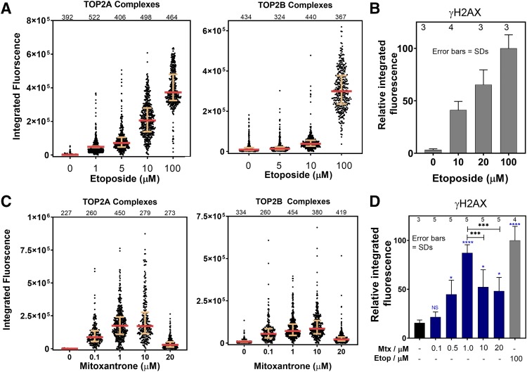
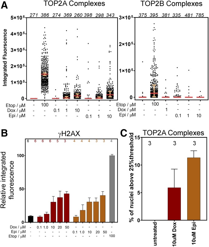
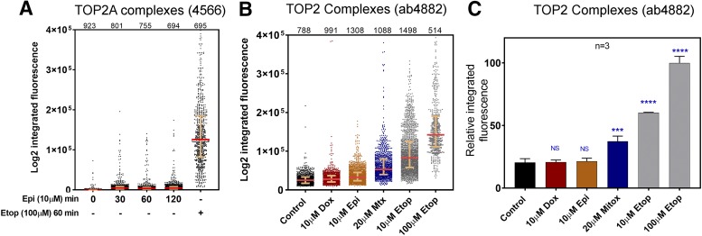
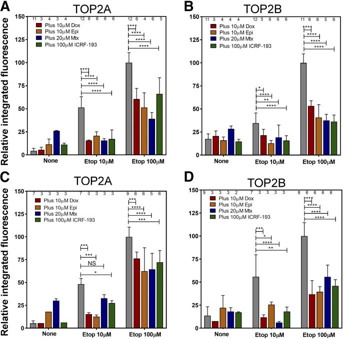
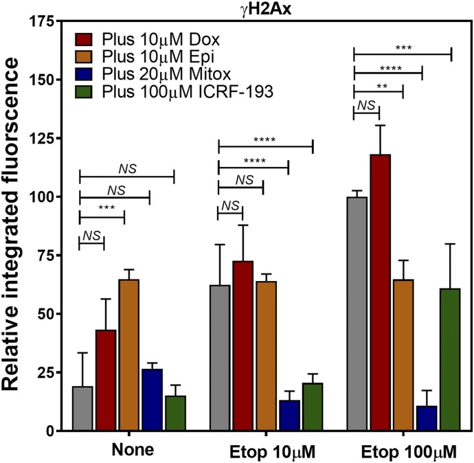
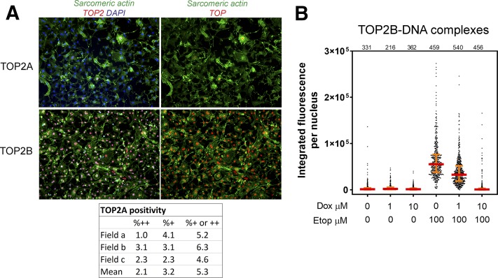
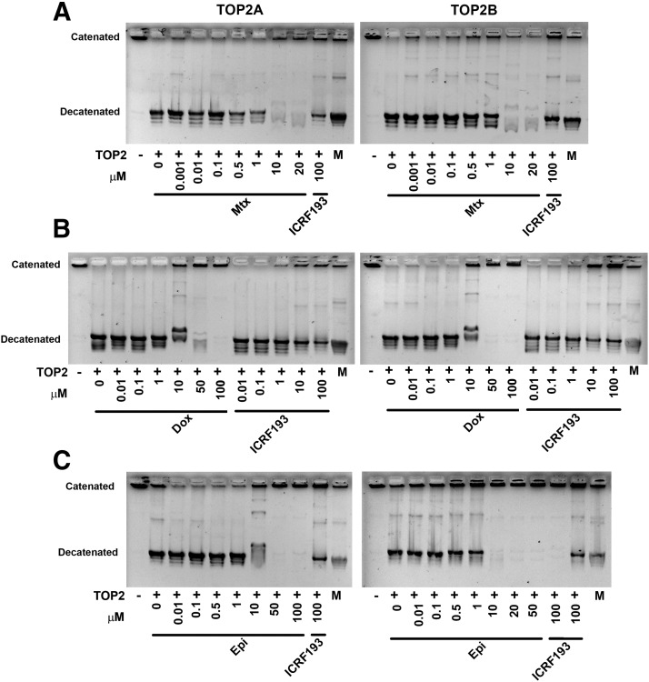
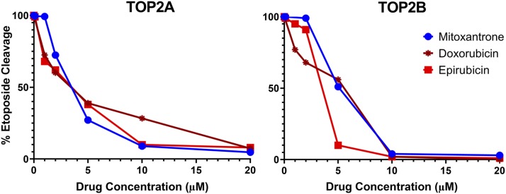
Topoisomerase II (TOP2) poisons are effective cytotoxic anticancer agents that stabilize the normally transient TOP2-DNA covalent complexes formed during the enzyme reaction cycle. These drugs include etoposide, mitoxantrone, and the anthracyclines doxorubicin and epirubicin. Anthracyclines also exert cell-killing activity via TOP2-independent mechanisms, including DNA adduct formation, redox activity, and lipid peroxidation. Here, we show that anthracyclines and another intercalating TOP2 poison, mitoxantrone, stabilize TOP2-DNA covalent complexes less efficiently than etoposide, and at higher concentrations they suppress the formation of TOP2-DNA covalent complexes, thus behaving as TOP2 poisons at low concentration and inhibitors at high concentration. We used induced pluripotent stem cell (iPSC)-derived human cardiomyocytes as a model to study anthracycline-induced damage in cardiac cells. Using immunofluorescence, our study is the first to demonstrate the presence of topoisomerase IIβ (TOP2B) as the only TOP2 isoform in iPSC-derived cardiomyocytes. In these cells, etoposide robustly induced TOP2B covalent complexes, but we could not detect doxorubicin-induced TOP2-DNA complexes, and doxorubicin suppressed etoposide-induced TOP2-DNA complexes. In vitro, etoposide-stabilized DNA cleavage was attenuated by doxorubicin, epirubicin, or mitoxantrone. Clinical use of anthracyclines is associated with cardiotoxicity. The observations in this study have potentially important clinical consequences regarding the effectiveness of anticancer treatment regimens when TOP2-targeting drugs are used in combination. These observations suggest that inhibition of TOP2B activity, rather than DNA damage resulting from TOP2 poisoning, may play a role in doxorubicin cardiotoxicity. SIGNIFICANCE STATEMENT: We show that anthracyclines and mitoxantrone act as topoisomerase II (TOP2) poisons at low concentration but attenuate TOP2 activity at higher concentration, both in cells and in in vitro cleavage experiments. Inhibition of type II topoisomerases suppresses the action of other drugs that poison TOP2. Thus, combinations containing anthracyclines or mitoxantrone and etoposide may reduce the activity of etoposide as a TOP2 poison and thus reduce the efficacy of drug combinations.


Figures

Fig. 1.
Fig. 1.
Dose response for formation of TOP2-DNA complexes and histone H2AX phosphorylation induced by etoposide (Etop) and mitoxantrone (Mtx). (A) NB4 cells were treated with the indicated concentrations of etoposide. TOP2A and TOP2B complexes were quantified on a cell-by-cell basis by the TARDIS assay using anti-TOP2A (4566) or anti-TOP2B (4555), respectively. Data are displayed as scatterplots, each dot representing the integrated immunofluorescent signal from a single nucleus. Medians and interquartile ranges are indicated. The number of nuclei quantified for each condition are indicated above each plot. For TOP2A, fluorescent intensities were significantly above those of untreated cells for 1 µM etoposide and above, whereas for TOP2B, significant increase was reached by 10 µM. (B) Cells treated as in (A) were analyzed by immunofluorescence for phospho-S139 histone H2AX (γH2AX). The results are displayed as the means of the medians of replicate experiments (number of replicas indicated above each bar) normalized to 100 μM etoposide. Error bars represent the S.D. (C and D) NB4 cells were treated with the indicated concentrations of mitoxantrone and were analyzed for TOP2-DNA complexes and γH2AX immunofluorescence as in (A) and (B), respectively. For both TOP2A and TOP2B, fluorescent intensities significantly increased compared with untreated cells at all concentrations of mitoxantrone from 0.1 to 20 µM. (D) Significance values were determined using one-way ANOVA with Tukey correction for multiple comparisons. Significance values shown in blue refer to comparison with untreated cells. Error bars indicate S.D. values.
Fig. 2.
Fig. 2.
Dose response for formation of TOP2-DNA complexes and histone H2AX phosphorylation induced by doxorubicin and epirubicin. (A) NB4 cells were treated with the indicated concentrations of anthracycline or etoposide (Etop). TOP2A- and TOP2B-DNA complexes were quantified as in Fig. 1. The number of cells analyzed for each treatment is indicated in italics at the top of each graph. (B) Cells were treated as in (A) and were analyzed by immunofluorescence for γH2AX. The results are displayed as the means of the medians of replicate experiments ±S.D, normalized to 100 μM etoposide. The number of replicates for each condition is indicated above each graph. (C) Proportion of cells treated with 10 μM dox or epi that exhibit TOP2A TARDIS fluorescence above a threshold of 25% of the median value obtained with 100 μM etoposide. Error bars indicate S.D. values.
Fig. 3.
Fig. 3.
Low anthracycline-induced TOP2-DNA complex TARDIS signal is not due to short drug incubation time or specific loss or masking of TOP2 C-terminal domain epitopes. (A) Cells were treated with 10 μM epi for the times indicated or with etoposide (Etop; 100 μM) for 60 minutes, and TOP2A complexes were quantified using antibody 4556 as in Fig. 1. (B and C) Cells were treated with the indicated TOP2 poisons for 60 minutes, and TOP2 complexes were analyzed using anti-TOP2 antibody 4882 (raised to N-terminal 140 kDa of calf thymus TOP2). Data are shown as scatterplots for one replica experiment (B) and as the means of the median values obtained from three replica experiments ±S.D. for (C). Significance values refer to comparison with untreated (control) cells by one-way ANOVA with Dunnett’s correction for multiple comparisons. Error bars indicated S.D. values. Mtx, mitoxantrone.
Fig. 4.
Fig. 4.
Anthracyclines and mitoxantrone inhibit etoposide-induced TOP2A- and TOP2B-DNA complex stabilization. NB4 cells (A and B) or K562 cells (C and D) were preincubated for 1 hour with doxorubicin (Dox), epirubicin (Epi), mitoxantrone (Mtx), or ICRF-193 at the concentration shown in the top-left corner of each graph. Cells were then treated with 10 or 100 μM etoposide (Etop; or vehicle control) for a further hour. TOP2A (A and C) and TOP2B (B and D) complexes were quantified as in Fig. 1. The first bar in each group (gray) corresponds to treatment with etoposide alone. Data in each graph are the means of the medians from replica experiments ±S.D. The number of replicates for each condition is shown at the top of each graph. Significance tests were performed by two-way ANOVA using Dunnett’s correction for multiple comparisons (comparisons were made to etoposide treatment alone).
Fig. 5.
Fig. 5.
Anthracyclines and mitoxantrone (Mitox) inhibit etoposide (Etop)-induced H2AX phosphorylation. NB4 cells were pretreated with anthracyclines, mitoxantrone, or ICRF-193 before treatment with etoposide, as in Fig 4. Histone H2AX phosphorylation was assessed by quantitative immunofluorescence. Data in each graph are the means of the medians from replica experiments ±S.D. Significance tests were performed as for Fig. 4.
Fig. 6.
Fig. 6.
TOP2 expression and induction of stable TOP2B-DNA complexes in human cardiomyocytes. (A) Induced human iPSC cardiomyocytes were stained with anti-sarcomeric actin (green) and TOP2A or TOP2B antibodies (red) and counterstained with DAPI (blue). All cells stained positive for TOP2B. Most cells were negative for TOP2A; the proportion of cells weakly (+) or strongly (++) positive for TOP2A are indicated in the table below (A). (B) Cardiomyocytes were pretreated with doxorubicin as indicated and then incubated with 100 μM etoposide (Etop). TOP2B-DNA stabilized complexes were then quantified using the TARDIS assay as in Fig. 1. Significance values refer to comparison with control (untreated) cells (Kruskal-Wallace test with Dunn’s correction for multiple comparisons).
Fig. 7.
Fig. 7.
Mitoxantrone, doxorubicin, and epirubicin inhibit TOP2A- and TOP2B-mediated in vitro decatenation activity. kDNA decatenation assays using recombinant TOP2A (left panels) or recombinant TOP2B (right panels) were performed in the presence of mitoxantrone (Mtx) (A), doxorubicin (B), or epirubicin (C). The gel positions of catenated and decatenated kDNA are indicated. The last lane in each gel (M) contains a marker consisting of catenated and decatenated kDNA.
Fig. 8.
Fig. 8.
Increasing doses of mitoxantrone, epirubicin, or doxorubicin attenuate etoposide-induced in vitro DNA cleavage activity. Plasmid (pTCS1) cleavage assays were performed using recombinant TOP2A or TOP2B in the presence of 1 mM etoposide combined with a range of concentrations of mitoxantrone, doxorubicin, or epirubicin. Gels were quantified, and the degree of cleavage was normalized to that obtained with etoposide alone.

Similar articles

Cited by

References

    1. Atwal M, Lishman EL, Austin CA, Cowell IG. (2017) Myeloperoxidase enhances etoposide and mitoxantrone-mediated DNA damage: a target for myeloprotection in cancer chemotherapy. Mol Pharmacol 91:49–57. - PMC - PubMed
    1. Austin CA, Barot HA, Margerrison EE, Turcatti G, Wingfield P, Hayes MV, Fisher LM. (1990) Structure and partial amino acid sequence of calf thymus DNA topoisomerase II: comparison with other type II enzymes. Biochem Biophys Res Commun 170:763–768. - PubMed
    1. Binaschi M, Farinosi R, Austin CA, Fisher LM, Zunino F, Capranico G. (1998) Human DNA topoisomerase IIalpha-dependent DNA cleavage and yeast cell killing by anthracycline analogues. Cancer Res 58:1886–1892. - PubMed
    1. Bodley A, Liu LF, Israel M, Seshadri R, Koseki Y, Giuliani FC, Kirschenbaum S, Silber R, Potmesil M. (1989) DNA topoisomerase II-mediated interaction of doxorubicin and daunorubicin congeners with DNA. Cancer Res 49:5969–5978. - PubMed
    1. Capranico G, De Isabella P, Tinelli S, Bigioni M, Zunino F. (1993) Similar sequence specificity of mitoxantrone and VM-26 stimulation of in vitro DNA cleavage by mammalian DNA topoisomerase II. Biochemistry 32:3038–3046. - PubMed

Publication types

MeSH terms

LinkOut - more resources