. 2019 Aug 27;116(35):17450-17459.
doi: 10.1073/pnas.1901765116. Epub 2019 Aug 9.

ARF6 and AMAP1 are major targets of KRAS and TP53 mutations to promote invasion, PD-L1 dynamics, and immune evasion of pancreatic cancer

Affiliations

ARF6 and AMAP1 are major targets of KRAS and TP53 mutations to promote invasion, PD-L1 dynamics, and immune evasion of pancreatic cancer

Shigeru Hashimoto et al. Proc Natl Acad Sci U S A. .

Abstract

Although KRAS and TP53 mutations are major drivers of pancreatic ductal adenocarcinoma (PDAC), the incurable nature of this cancer still remains largely elusive. ARF6 and its effector AMAP1 are often overexpressed in different cancers and regulate the intracellular dynamics of integrins and E-cadherin, thus promoting tumor invasion and metastasis when ARF6 is activated. Here we show that the ARF6-AMAP1 pathway is a major target by which KRAS and TP53 cooperatively promote malignancy. KRAS was identified to promote eIF4A-dependent ARF6 mRNA translation, which contains a quadruplex structure at its 5'-untranslated region, by inducing TEAD3 and ETV4 to suppress PDCD4; and also eIF4E-dependent AMAP1 mRNA translation, which contains a 5'-terminal oligopyrimidine-like sequence, via up-regulating mTORC1. TP53 facilitated ARF6 activation by platelet-derived growth factor (PDGF), via its known function to promote the expression of PDGF receptor β (PDGFRβ) and enzymes of the mevalonate pathway (MVP). The ARF6-AMAP1 pathway was moreover essential for PDGF-driven recycling of PD-L1, in which KRAS, TP53, eIF4A/4E-dependent translation, mTOR, and MVP were all integral. We moreover demonstrated that the mouse PDAC model KPC cells, bearing KRAS/TP53 mutations, express ARF6 and AMAP1 at high levels and that the ARF6-based pathway is closely associated with immune evasion of KPC cells. Expression of ARF6 pathway components statistically correlated with poor patient outcomes. Thus, the cooperation among eIF4A/4E-dependent mRNA translation and MVP has emerged as a link by which pancreatic driver mutations may promote tumor cell motility, PD-L1 dynamics, and immune evasion, via empowering the ARF6-based pathway and its activation by external ligands.

Keywords: ARF6; PD-L1; mRNA translation; mevalonate pathway; pancreatic driver oncogenes.


Conflict of interest statement

Conflict of interest statement: H.S., S. Hashimoto, and A.H. are inventors on the patent application PCT/JP2019/10925.

Figures

Fig. 1.
Fig. 1.
The ARF6–AMAP1 pathway is central to PDAC malignancy. (A) Expression of ARF6 pathway components in PDAC cells, analyzed by Western blotting. β-Actin was used as a loading control. (BE) PDGF activates ARF6 via GEP100 (B and D) and promotes cell invasion via ARF6, GEP100, AMAP1, and EPB41L5 (C and E). Two different siRNAs (#1 and #2) were used. Irr, a control siRNA with an irrelevant sequence. In C and E, results are shown as ratios by normalizing values obtained for Irr-treated cells as 1 (n = 3). Error bars, mean ± SEM; *P < 0.001. (F and G) Lung metastases of KPC cells, expressing a luciferase reporter gene and transfected with an EPB41L5 shRNA plasmid (shEPB41L5, sequence #2) or a control empty vector (Irr), in nude mice. In F, bioluminescence intensities from the chests were measured on days 0 and 9. Results are shown as the mean ± SEM; *P < 0.05 (n = 5 for each group); NS, not significant. In G, representative images of the lungs are shown. (H) Representative IHC images of PDGFRβ, AMAP1, and EPB41L5 in human primary PDACs. (I) Kaplan–Meier plots with regard to the different levels of PDGFRβ, AMAP1, EPB41L5, and their combinations (high, score of 1 or 2; low, score 0). P values were obtained by ANOVA (C, E, and F) and by the log-rank test (I).
Fig. 2.
Fig. 2.
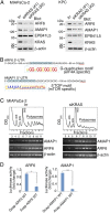
KRAS promotes ARF6 and AMAP1 expression via their mRNA translation. (A) Representative immunoblots of ARF6, AMAP1, and EPB41L5 in cells treated with KRAS siRNAs or with an Irr control. Two different siRNAs (#1 and #2) were used. β-Actin was used as a control. Irr, an irrelevant siRNA. (B) 5′-UTRs of ARF6 mRNA (the G-quadruplex motif) and AMAP1 mRNA (the TOP motif). (C) Polysome profiles, as detected by absorbance at 254 nm after 15% to 60% sucrose gradient ultracentrifugation of RNAs from MIAPaCa-2 cells treated with siKRAS or an irrelevant siRNA (Irr). Ribosomal peaks (40S, 60S, and 80S) and polysomal peaks are shown. B, Bottom shows detection of AMAP1, ARF6, and β-actin mRNAs by PCR. A representative result from 3 independent experiments is shown. Actin was used as a control. (D) Translational activity of the 5′-UTRs of ARF6 and AMAP1 mRNAs, bearing either a 5′-Gcap or 5′-Acap and constructed into the polyadenylated firefly luciferase reporter, assessed in vitro using micrococcal nuclease-treated MIAPaCa-2 extracts. Results are shown as ratios by normalizing values obtained from each of the Gcap constructs as 1 (n = 3). Error bars, mean ± SEM; **P < 0.01, ***P < 0.001 by ANOVA.
Fig. 3.
Fig. 3.
(A–J) Mechanisms by which KRAS up-regulates mRNA translation of ARF6 (AH) and AMAP1 (I and J) in PDACs. (A) Suppression of ARF6 levels by silvestrol. (B) Induction of PDCD4, coupled with reduction of ARF6 levels, upon KRAS silencing. (C) Suppression of ARF6 levels by the forced expression of PDCD4. (D) Box-and-whisker plots of PDCD4 mRNA levels in wild-type KRAS-expressing (WT) and mutant KRAS-expressing (MT) PDACs of the TCGA RNASeq dataset (n = 151). P < 0.01 by the Welch t test. (E) Schematic drawing of the 5′-upstream fragments of the PDCD4 TSS (#4 to #6) and their transcriptional activities, as assessed by the luciferase reporter assay, in response to siKRAS in MIAPaCa-2 cells (for regions #1 to #3; SI Appendix, Fig. S3A). **P < 0.01 by ANOVA. (F and G) Luciferase reporter assay results of the PDCD4 promoter region (F) and protein levels of ARF6 and PDCD4 (G) in MIAPaCa-2 cells, infected with lentiviruses bearing shRNAs, as indicated. (H) Binding of TEAD3 and ETV4 to the PDCD4 promoter region in MIAPaCa-2 cells. Input, 5% of total lysate used for the immunoprecipitation. IgG, an irrelevant IgG. For positions af, see SI Appendix, Fig. S3F. (I and J) AMAP1 levels in MIAPaCa-2 cells, infected with lentiviruses bearing the indicated shRNAs (I) or treated with vehicle (DMSO) or mTOR inhibitors, as indicated (J). Levels of ARF6, 4EBP1, and phosphorylated 4EBP1 are also shown. In B, F, G, and I, 2 different shRNAs (#1 and #2) were used, and levels of the target proteins are shown. Irr, an irrelevant shRNA. Representative results are shown in each immunoblot from at least 3 independent experiments. β-Actin was used as a control. Error bars indicate the mean ± SEM; **P < 0.01, ***P < 0.001 by ANOVA.
Fig. 4.
Fig. 4.
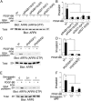
Requirement for TP53 and MVP in PDGF-induced ARF6 activation and cell invasion in MIAPaCa-2 cells. (A–F) Blockade of PDGF-induced ARF6 activation (A, C, and E) and cell invasion (B, D, and F) by silencing of RAB11b or GGT-II (A and B), by silencing of TP53 (C and D), and by simvastatin (E and F). Error bars indicate the mean ± SEM; *P < 0.05, **P < 0.01, ***P < 0.001 by ANOVA (B, D, and F).
Fig. 5.
Fig. 5.
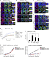
ARF6 and AMAP1, as well as KRAS and TP53, are pivotal to PD-L1 dynamics and cell surface expression. (AD) Representative images of immunofluorescence staining for PD-L1 (red) and F-actin (green) in IFNγ-treated MIAPaCa-2 cells, pretreated with siRNAs/shRNAs (A, B, and D) and simvastatin or silvestrol (C), as indicated. Nuclei were visualized by DAPI (blue). (Scale bars, 10 μm.) (E) Recycling of PD-L1 to the cell surface in the presence or absence of PDGF in IFNγ-treated MIAPaCa-2 cells, pretreated with siRNAs, as indicated. (F) PD-L1 cell surface expression in IFNγ-treated or nontreated MIAPaCa-2 cells, pretreated with siRNAs. MFI, median fluorescence intensity. (G) s.c. tumor growth of AMAP1-silenced KPCs (shAMAP1 #1 and #2) or control KPCs (Irr) in immunodeficient BALB/c nude mice and immunocompetent C57BL/6 mice. Tumors were measured every 2 to 4 d starting on day 5. Data are representative of 3 independent experiments with at least 6 mice per group. In A, D, E, and F, sequence #1 was used for each siRNA/shRNA. In A, B, and DG Irr indicates an irrelevant siRNA/shRNA. Error bars indicate the mean ± SEM; *P < 0.05, **P < 0.01, ***P < 0.001 by ANOVA (F and G).
Fig. 6.
Fig. 6.
Our proposed model of KRAS and TP53 oncogenes driving PDAC malignancy via the ARF6–AMAP1 pathway. KRAS promotes the 5′-cap–dependent translation of ARF6 and AMAP1 mRNAs, primarily via enhancing the activities of eIF4A and eIF4E, respectively. TP53 facilitates ARF6 activation by RTKs, via enhancing the expression of PDGFR (14) and MVP (15), in which MVP activity is essential to geranylgeranylate RAB11b to transport ARF6 to the plasma membrane for its activation by RTKs (21). EPB41L5 is induced during EMT by ZEB1 (23). Although TP53 mutations can induce ZEB1 and hence EPB41L5, the molecular basis of this link appears to be complicated in PDACs and is not simply mediated by miRNAs. The ARF6–AMAP1 pathway drives tumor cell motility, in which the interaction of AMAP1 with EPB41L5, PRKD2, and other proteins is necessary to promote intracellular dynamics of β1 integrins and E-cadherin, as well as the cortical actin remodeling (main text). The ARF6–AMAP1 pathway also promotes PD-L1 dynamics and is closely associated with the immune evasion of PDACs, whereas factors linking AMAP1 with PD-L1 and immune evasion are unknown.

References

    1. Siegel R. L., Miller K. D., Jemal A., Cancer statistics, 2017. CA Cancer J. Clin. 67, 7–30 (2017). - PubMed
    1. Vincent A., Herman J., Schulick R., Hruban R. H., Goggins M., Pancreatic cancer. Lancet 378, 607–620 (2011). - PMC - PubMed
    1. Yachida S., et al. , Distant metastasis occurs late during the genetic evolution of pancreatic cancer. Nature 467, 1114–1117 (2010). - PMC - PubMed
    1. Notta F., et al. , Erratum: A renewed model of pancreatic cancer evolution based on genomic rearrangement patterns. Nature 542, 124 (2017). - PubMed
    1. Jones S., et al. , Core signaling pathways in human pancreatic cancers revealed by global genomic analyses. Science 321, 1801–1806 (2008). - PMC - PubMed

Publication types

MeSH terms