

Exosomal PD-L1 promotes tumor growth through immune escape in non-small cell lung cancer
- PMID: 31399559
- PMCID: PMC6802663
- DOI: 10.1038/s12276-019-0295-2
Exosomal PD-L1 promotes tumor growth through immune escape in non-small cell lung cancer
Abstract
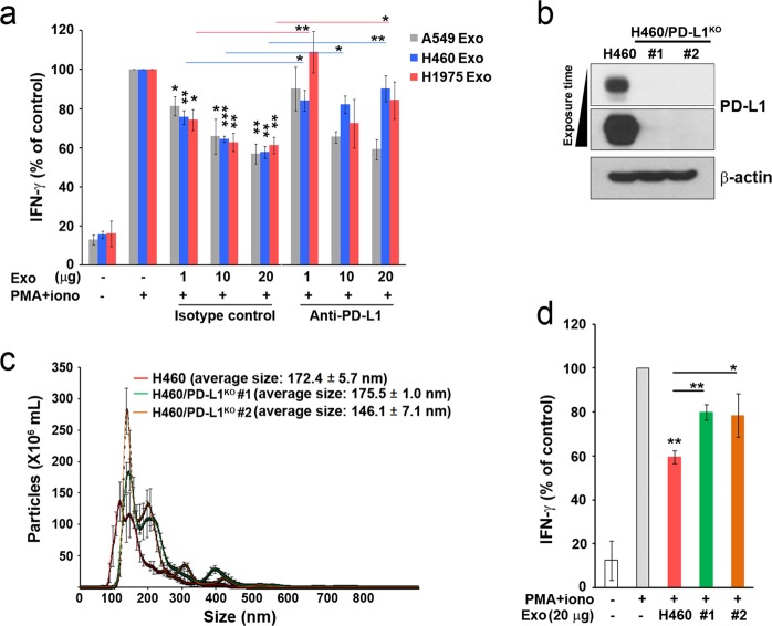
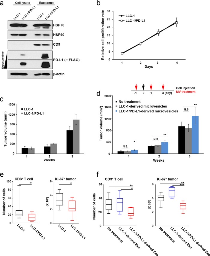
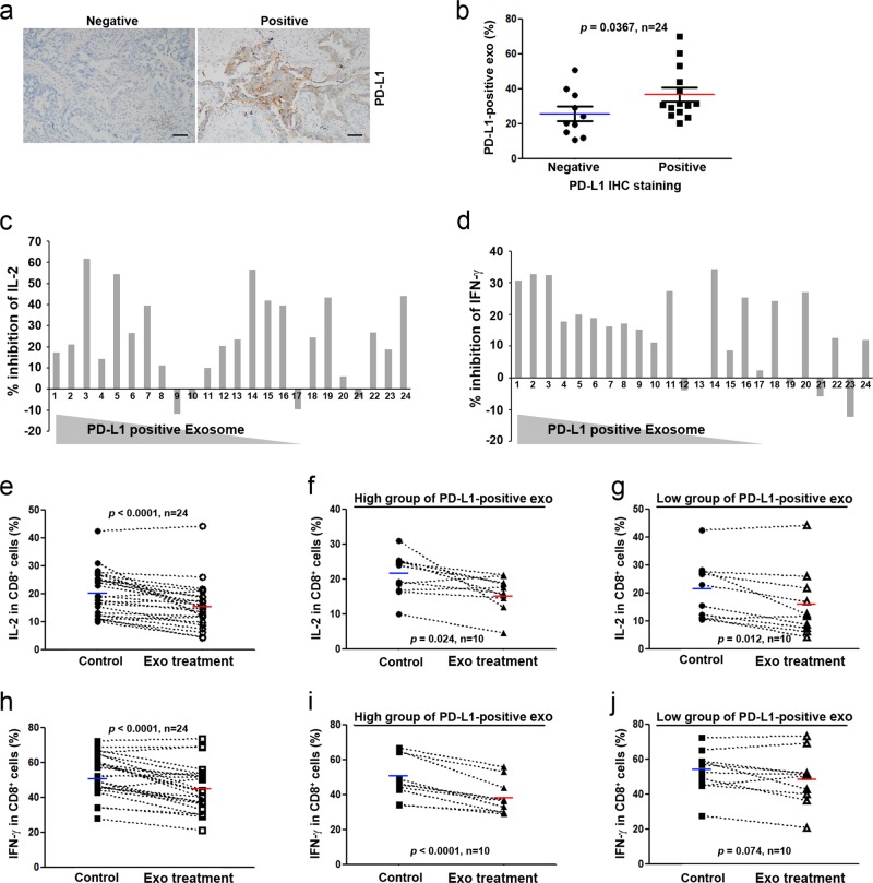
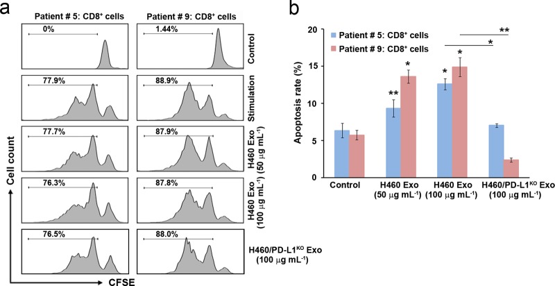
Programmed cell death protein-1/programmed cell death ligand-1 (PD-1/PD-L1) pathway blockade is a promising new cancer therapy. Although PD-1/PD-L1 treatment has yielded clinical benefits in several types of cancer, further studies are required to clarify predictive biomarkers for drug efficacy and to understand the fundamental mechanism of PD-1/PD-L1 interaction between host and tumor cells. Here, we show that exosomes derived from lung cancer cells express PD-L1 and play a role in immune escape by reducing T-cell activity and promoting tumor growth. The abundance of PD-L1 on exosomes represented the quantity of PD-L1 expression on cell surfaces. Exosomes containing PD-L1 inhibited interferon-gamma (IFN-γ) secretion by Jurkat T cells. IFN-γ secretion was restored by PD-L1 knockout or masking on the exosomes. Both forced expression of PD-L1 on cells without PD-L1 and treatment with exosomes containing PD-L1 enhanced tumor growth in vivo. PD-L1 was present on exosomes isolated from the plasma of patients with non-small cell lung cancer, and its abundance in exosomes was correlated with PD-L1 positivity in tumor tissues. Exosomes can impair immune functions by reducing cytokine production and inducing apoptosis in CD8+ T cells. Our findings indicate that tumor-derived exosomes expressing PD-L1 may be an important mediator of tumor immune escape.
Conflict of interest statement
The authors declare that they have no conflict of interest.
Figures


Similar articles
-
Integrin α2 promotes immune escape in non-small-cell lung cancer by enhancing PD-L1 expression in exosomes to inhibit CD8 + T-cell activity.J Investig Med. 2024 Jan;72(1):57-66. doi: 10.1177/10815589231207801. Epub 2023 Dec 12. J Investig Med. 2024. PMID: 37804164
-
Transforming growth factor beta orchestrates PD-L1 enrichment in tumor-derived exosomes and mediates CD8 T-cell dysfunction regulating early phosphorylation of TCR signalome in breast cancer.Carcinogenesis. 2021 Feb 11;42(1):38-47. doi: 10.1093/carcin/bgaa092. Carcinogenesis. 2021. PMID: 32832992
-
LAMTOR1 decreased exosomal PD-L1 to enhance immunotherapy efficacy in non-small cell lung cancer.Mol Cancer. 2024 Sep 2;23(1):184. doi: 10.1186/s12943-024-02099-4. Mol Cancer. 2024. PMID: 39223601 Free PMC article.
-
Exosomal PD-L1 in non-small cell lung Cancer: Implications for immune evasion and resistance to immunotherapy.Int Immunopharmacol. 2025 May 16;155:114519. doi: 10.1016/j.intimp.2025.114519. Epub 2025 Apr 8. Int Immunopharmacol. 2025. PMID: 40199140 Review.
-
Tumor-derived exosomes in the PD-1/PD-L1 axis: Significant regulators as well as promising clinical targets.J Cell Physiol. 2021 Jun;236(6):4138-4151. doi: 10.1002/jcp.30197. Epub 2020 Dec 4. J Cell Physiol. 2021. PMID: 33275291 Review.
Cited by
-
Extracellular vesicles and particles impact the systemic landscape of cancer.EMBO J. 2022 Sep 15;41(18):e109288. doi: 10.15252/embj.2021109288. Epub 2022 Sep 2. EMBO J. 2022. PMID: 36052513 Free PMC article. Review.
-
Programmed Death-Ligand 1 as a Regulator of Tumor Progression and Metastasis.Int J Mol Sci. 2021 May 20;22(10):5383. doi: 10.3390/ijms22105383. Int J Mol Sci. 2021. PMID: 34065396 Free PMC article. Review.
-
The effect of the intratumoral microbiome on tumor occurrence, progression, prognosis and treatment.Front Immunol. 2022 Nov 18;13:1051987. doi: 10.3389/fimmu.2022.1051987. eCollection 2022. Front Immunol. 2022. PMID: 36466871 Free PMC article. Review.
-
Improvement of the anticancer efficacy of PD-1/PD-L1 blockade via combination therapy and PD-L1 regulation.J Hematol Oncol. 2022 Mar 12;15(1):24. doi: 10.1186/s13045-022-01242-2. J Hematol Oncol. 2022. PMID: 35279217 Free PMC article. Review.
-
Serum exosomal miR-16-5p functions as a tumor inhibitor and a new biomarker for PD-L1 inhibitor-dependent immunotherapy in lung adenocarcinoma by regulating PD-L1 expression.Cancer Med. 2022 Jul;11(13):2627-2643. doi: 10.1002/cam4.4638. Epub 2022 Mar 28. Cancer Med. 2022. PMID: 35347894 Free PMC article.
References
Publication types
MeSH terms
Substances
Grants and funding
LinkOut - more resources
Full Text Sources
Medical
Research Materials
