. 2019 Aug 9;10(1):3355.
doi: 10.1038/s41467-019-11335-1.

Diurnal rhythms in gene expression in the prefrontal cortex in schizophrenia

Affiliations

Diurnal rhythms in gene expression in the prefrontal cortex in schizophrenia

Marianne L Seney et al. Nat Commun. .

Abstract

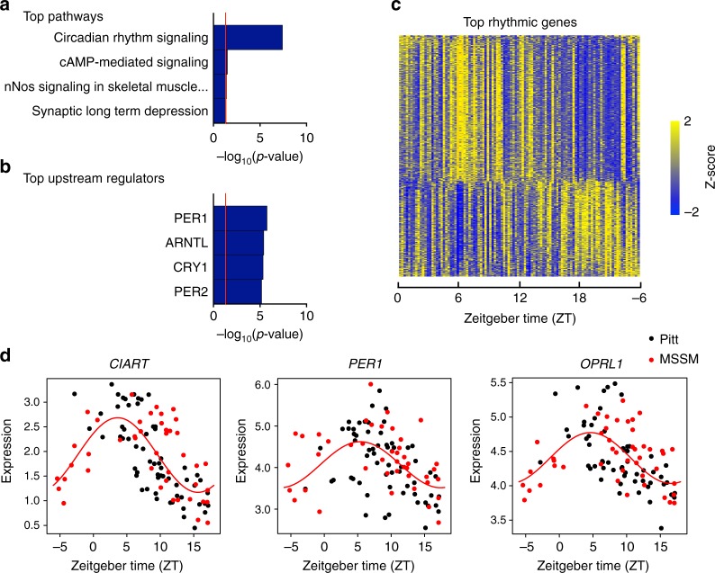
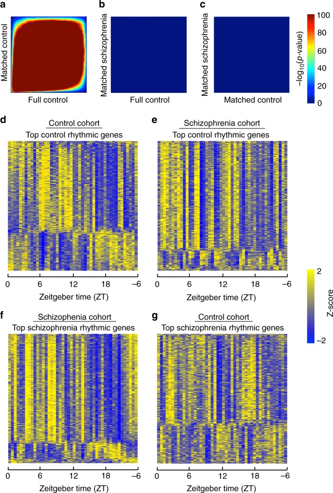
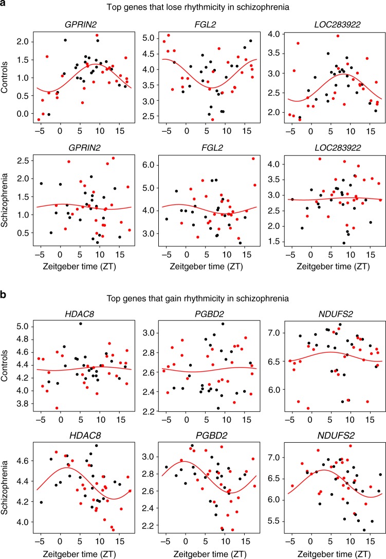
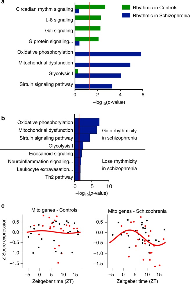
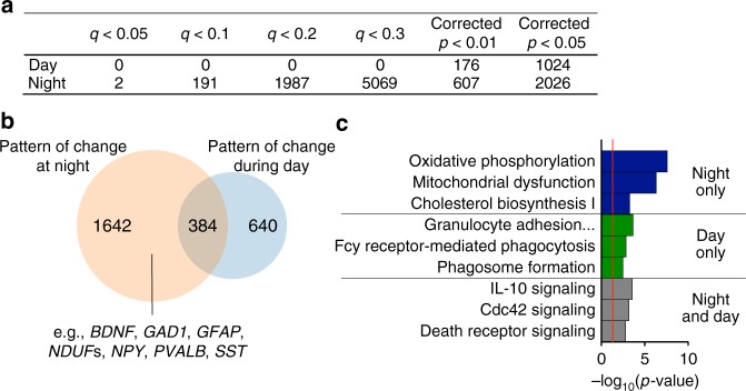
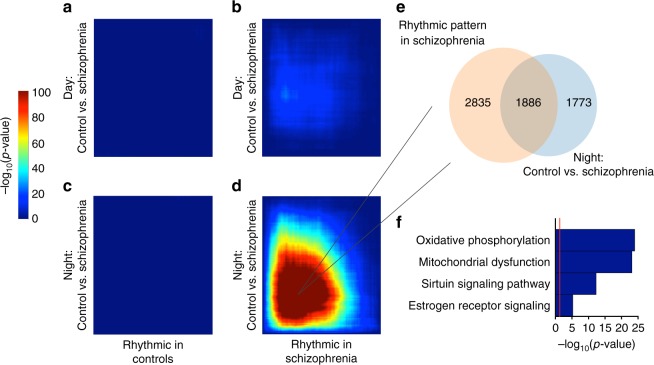
Schizophrenia is associated with disrupted cognitive control and sleep-wake cycles. Here we identify diurnal rhythms in gene expression in the human dorsolateral prefrontal cortex (dlPFC), in schizophrenia and control subjects. We find significant diurnal (24 h) rhythms in control subjects, however, most of these transcripts are not rhythmic in subjects with schizophrenia. Instead, subjects with schizophrenia have a different set of rhythmic transcripts. The top pathways identified in transcripts rhythmic only in subjects with schizophrenia are associated with mitochondrial function. Importantly, these rhythms drive differential expression patterns of these and several other genes that have long been implicated in schizophrenia (including BDNF and GABAergic-related transcripts). Indeed, differential expression of these transcripts is only seen in subjects that died during the night, with no change in subjects that died during the day. These data provide insights into a potential mechanism that underlies changes in gene expression in the dlPFC with schizophrenia.


Conflict of interest statement

The authors declare no competing interests.

Figures

Fig. 1
Fig. 1
Circadian rhythms in gene expression in human DLPFC in healthy control subjects. Nonlinear regression was used to detect circadian gene expression patterns based on individual TODs. Sinusoidal curves were fitted using the nonlinear least-squares method and the coefficient of determination (R2) was used as a proxy of goodness-of-fit. A null distribution of R2 generated from 1,000 TOD-randomized expression data sets was used to estimate the empirical p-value by comparing observed R2 and the null distribution of R2. a The top pathway represented by these rhythmic genes is Circadian rhythm signaling. b The top predicted upstream regulators are core circadian genes. c Heatmap for circadian genes for all 104 healthy subjects (p < 0.01). Expression levels are Z-transformed for each gene, and the genes are ordered by their circadian phase value (peak hour). Each column represents a subject and the subjects are ordered by time of death. d Scatterplots representing rhythms in gene expression for the top 3 circadian genes in healthy controls. Each dot indicates a subject with x-axis indicating the time of death (TOD) on ZT scale (−6 to 18 h) and y-axis indicating gene expression level. Subjects from the Pitt brain bank are in black while subjects from the MSSM brain bank are in red. The red line is the fitted sinusoidal curve
Fig. 2
Fig. 2
Rhythmic genes are largely distinct in control subjects and subjects with schizophrenia. Nonlinear regression was used to detect circadian gene expression patterns based on individual TODs. Sinusoidal curves were fitted using the nonlinear least-squares method and the coefficient of determination (R2) was used as a proxy of goodness-of-fit. A null distribution of R2 generated from 1000 TOD-randomized expression data sets was used to estimate the empirical p-value by comparing observed R2 and the null distribution of R2. a RRHO plot indicating high degree of overlap in rhythmic genes between the full healthy control cohort and the matched control cohort. b RRHO plot indicating lack of overlap in rhythmic genes between the full healthy control cohort and the matched schizophrenia cohort. c RRHO plot indicating lack of overlap in rhythmic genes between the matched healthy control cohort and the schizophrenia cohort. d Heatmap for the circadian genes in the matched control cohort (p < 0.05). Expression levels are Z-transformed for each gene, and the genes are ordered by their circadian phase value (peak hour). Each column represents a subject and the subjects are ordered by time of death. e Heatmap for the healthy control circadian genes in the schizophrenia cohort, indicating disrupted rhythmicity of normally rhythmic genes in subjects with schizophrenia. f Heatmap for the circadian genes in the matched schizophrenia cohort. g Heatmap for the schizophrenia circadian genes in the control cohort, indicating that these genes are not rhythmic in control subjects
Fig. 3
Fig. 3
Scatterplots indicating rhythmicity for genes that lose or gain rhythmicity in subjects with schizophrenia compared to controls. Each dot indicates a subject with x-axis indicating the time of death (TOD) on ZT scale (−6 to 18 h) and y-axis indicating gene expression level. Subjects from the Pitt brain bank are in black while subjects from the MSSM brain bank are in red. The red line is the fitted sinusoidal curve. a Scatterplots indicating rhythmicity of GPRIN2 (p < 0.0005), FGL2 (p < 0.002), and LOC283922 (p < 0.002) in healthy controls (top), which lose rhythmicity in subjects with schizophrenia (bottom). b Scatterplots indicating lack of rhythmicity of HDAC8, PGBD2, and NDUFS2 in control subjects, but these genes gain rhythmicity in subjects with schizophrenia (HDAC8: p < 0.0005; PGBD2: p < 0.001; NDUFS2: p < 0.006).
Fig. 4
Fig. 4
The top pathways represented by the rhythmic genes in control subjects and subjects with schizophrenia are completely distinct. While the top pathway in healthy controls relates to circadian rhythm signaling (a), the top pathways in subjects with schizophrenia relate to oxidative phosphorylation and mitochondrial dysfunction. b The top pathways for genes that gain rhythmicity in schizophrenia compared to healthy controls are oxidative phosphorylation and mitochondrial dysfunction. c Expression of mitochondrial-related genes in control (left) and subjects with schizophrenia (right). Expression for each gene was Z-transformed and averaged to create a Z-mitochondria (Z-mito) score for each subject. These Z-mito values are plotted across time of day. These mitochondrial-related genes are not rhythmic in healthy controls. In subjects with schizophrenia, mitochondrial-related genes peak between ZT 0–5
Fig. 5
Fig. 5
More genes are differentially expressed between schizophrenia and control subjects if they died during the night. a Table showing number of DE genes during day or night at various significance cutoffs. A permutation test was used to return corrected p-values and Storey’s q value was used to correct for multiple testing. b Venn diagram indicating overlap in genes that are DE at p < 0.05 in schizophrenia at night and during the day. Many genes that have previously been identified as being disrupted in schizophrenia are DE only at night (e.g., BDNF, PVALB, SST). c The pathways represented by the genes that are DE only at night are related to oxidative phosphorylation and mitochondrial dysfunction. The pathways represented by the genes that are trending towards DE only during the day relate to immune function. For the genes that are DE both during the day and night, the top pathways are related to immune function and Cdc42 signaling
Fig. 6
Fig. 6
Several genes that are rhythmic in schizophrenia are also DE in subjects that died at night. ad RRHO plots indicating that there is a high level of overlap between genes that are changed in schizophrenia during the night and rhythmic in subjects with schizophrenia. Venn diagram (e) indicating overlap in genes that are DE at night and rhythmic in schizophrenia and top pathways (f) for these genes relate to oxidative phosphorylation and mitochondrial dysfunction

References

    1. Wulff K, Joyce E. Circadian rhythms and cognition in schizophrenia. Br. J. Psychiatry. 2011;198:250–252. doi: 10.1192/bjp.bp.110.085068. - DOI - PubMed
    1. Kaskie RE, Graziano B, Ferrarelli F. Schizophrenia and sleep disorders: links, risks, and management challenges. Nat. Sci. Sleep. 2017;9:227–239. doi: 10.2147/NSS.S121076. - DOI - PMC - PubMed
    1. Wulff K, Dijk DJ, Middleton B, Foster RG, Joyce EM. Sleep and circadian rhythm disruption in schizophrenia. Br. J. Psychiatry.: J. Ment. Sci. 2012;200:308–316. doi: 10.1192/bjp.bp.111.096321. - DOI - PMC - PubMed
    1. Johansson AS, Owe-Larsson B, Hetta J, Lundkvist GB. Altered circadian clock gene expression in patients with schizophrenia. Schizophr. Res. 2016;174:17–23. doi: 10.1016/j.schres.2016.04.029. - DOI - PubMed
    1. Hjelm BE, et al. Evidence of mitochondrial dysfunction within the complex genetic etiology of schizophrenia. Mol. Neuropsychiatry. 2015;1:201–219. doi: 10.1159/000441252. - DOI - PMC - PubMed

Publication types

Substances