. 2019 Aug 9;9(1):11584.
doi: 10.1038/s41598-019-48095-3.

Extracellular vesicles isolated from porcine seminal plasma exhibit different tetraspanin expression profiles

Affiliations

Extracellular vesicles isolated from porcine seminal plasma exhibit different tetraspanin expression profiles

Isabel Barranco et al. Sci Rep. .

Abstract

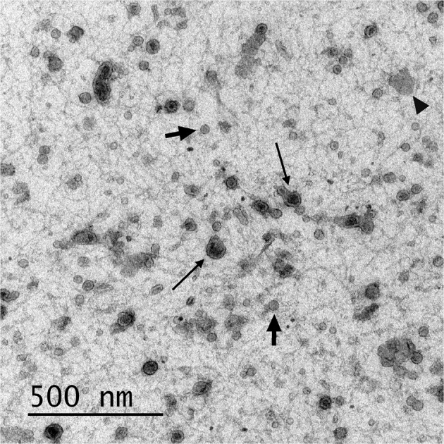
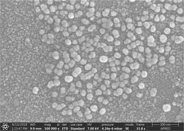
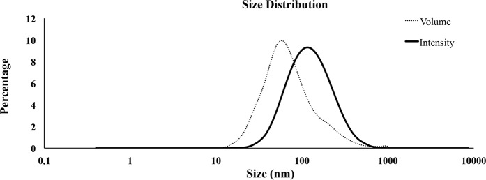
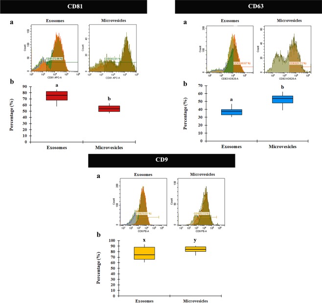
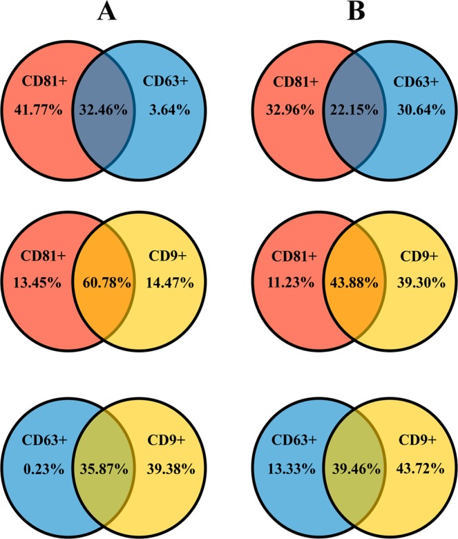
Seminal extracellular vesicles (EVs) include exosomes (ø 40-120 nm) and microvesicles (MVs, ø 120-1000 nm), which would be involved in multiple functional reproductive roles. The study aimed to establish which EV subtypes are present in pig semen, using a high-resolution flow cytometer to explore differences in their tetraspanin expression profile. The EVs were isolated from 12 pig ejaculates using serial ultracentrifugation and characterized by dynamic light scattering and electron microscopy for size and morphology as well as for tetraspanin expression using flow cytometry with Carboxyfluorescein succinimidyl ester (CFSE) and antibodies against CD9, CD63 and CD81. Pig semen contained a heterogeneous EV-population regarding size and morphology. Flow cytometric analysis demonstrated that the proportion of EVs expressing CD63 and CD9 was higher in MVs (P < 0.001 and P < 0.05, respectively) than in exosomes, while the opposite was true for CD81; higher (P < 0.001) in exosomes than in MVs. In conclusion, (1) the new generation of flow cytometers are able to accurately identify EVs and to gate them in two size-different populations named exosomes and MVs. (2) Tetraspanins CD9, CD63 and CD81 are present in both seminal EVs, albeit with exosomes and MVs differing in expression profiles, suggesting dissimilar cargo and binding affinity.


Conflict of interest statement

The authors declare no competing interests.

Figures

Figure 1
Figure 1
Transmission electron micrograph of extracellular vesicles isolated from pig seminal plasma. The extracellular vesicles were isolated by serial ultracentrifugation and contrasted in 2% uranyl-oxalate and embedded in a mixture of 4% uranyl-acetate. Larger vesicles (microvesicles) enclosed a granule electron-dense material (thick arrows) and the smaller ones (exosomes) showed a less dense inner content (short arrows). Amorphous material (arrow heads) in the background was compatible with protein aggregates.
Figure 2
Figure 2
Scanning electron micrograph of extracellular vesicles isolated from pig seminal plasma by serial ultracentrifugation.
Figure 3
Figure 3
Dynamic Light Scattering (DLS) measurements. Particle size distribution of extracellular vesicles isolated from pig seminal plasma by serial ultracentrifugation, assessed by DLS using Nano Zetasizer. Each curve represents an average of volume (dotted line) and intensity (solid line) size distributions of all semen samples (n = 12).
Figure 4
Figure 4
Discrimination of exosomes and microvesicles isolated from pig seminal plasma based on scatter parameters in CytoFLEX. Representative forward scatter (FSC-A, y-axis) vs side scatter (Violet-SSC-A, x-axis) dot plot of the two extracellular vesicles subtypes gated by size (exosomes [size: 50 to 120 nm] and microvesicles [size: 120 to 1000 nm]).
Figure 5
Figure 5
Flow cytometric analysis of tetraspanin CD81, CD63 and CD9 expression in extracellular vesicles (EVs) subtypes isolated from pig seminal plasma. (a) Histogram representative of CD81/CD63/CD9 expression in EV subtypes (exosomes and microvesicles). Fluorescence (CD81-APC-A, CD63-KO525-A and CD9-PE, x-axis), vs number of events (Count, y-axis). (b) Box-whisker plot showing variation in CD81/CD63/CD9 expression between EV subtypes of 12 ejaculates (one per boar). Boxes enclose the 25th and 75th percentiles; the line is the median; and the whiskers extend to the 5th and 95th percentiles. (a,b) and (x–y) indicate significant differences (P < 0.001 and P < 0.05, respectively) among the EV subtypes.
Figure 6
Figure 6
Venn diagrams showing the percentage of shared tetraspanins (CD9/CD63/CD81) in exosomes (A) or microvesicles (B). These extracellular vesicles, exosomes and microvesicles, were isolated from the pig seminal plasma and assessed by flow cytometry.

References

    1. Colombo M, Raposo G, Théry C. Biogenesis, secretion, and intercellular interactions of exosomes and other extracellular vesicles. Annu. Rev. Cell Dev. Biol. 2014;30:255–289. doi: 10.1146/annurev-cellbio-101512-122326. - DOI - PubMed
    1. Antimisiaris SG, Mourtas S, Marazioti A. Exosomes and Exosome-Inspired Vesicles for Targeted Drug Delivery. Pharmaceutics. 2018;10:E218. doi: 10.3390/pharmaceutics10040218. - DOI - PMC - PubMed
    1. Azoidis I, Cox SC, Davies OG. The role of extracellular vesicles in biomineralisation: current perspective and application in regenerative medicine. J. Tissue Eng. 2018;9:2041731418810130. doi: 10.1177/2041731418810130. - DOI - PMC - PubMed
    1. Gámez-Valero A, Beyer K, Borràs FE. Extracellular vesicles, new actors in the search for biomarkers of dementias. Neurobiol. Aging. 2018;74:15–20. doi: 10.1016/j.neurobiolaging.2018.10.006. - DOI - PubMed
    1. Ruhen O, Meehan K. Tumour-derived extracellular vesicles as a novel source of protein biomarkers for cancer diagnosis and monitoring. Proteomics. 2018;19:e1800155. doi: 10.1002/pmic.201800155. - DOI - PubMed

Publication types