Review
. 2020 Mar;38(3):531-545.
doi: 10.1007/s00345-019-02899-0. Epub 2019 Aug 9.

Personalizing prostate cancer diagnosis with multivariate risk prediction tools: how should prostate MRI be incorporated?

Affiliations
Review

Personalizing prostate cancer diagnosis with multivariate risk prediction tools: how should prostate MRI be incorporated?

Ivo G Schoots et al. World J Urol. 2020 Mar.

Abstract

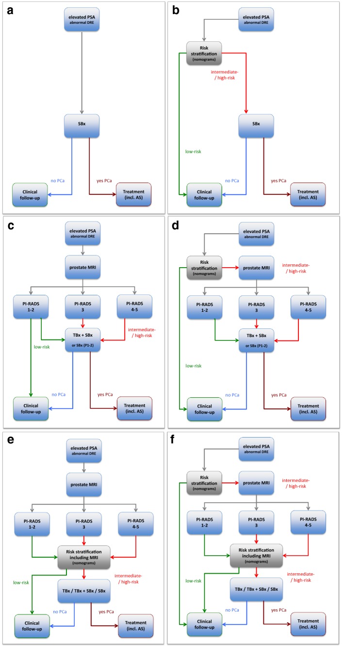
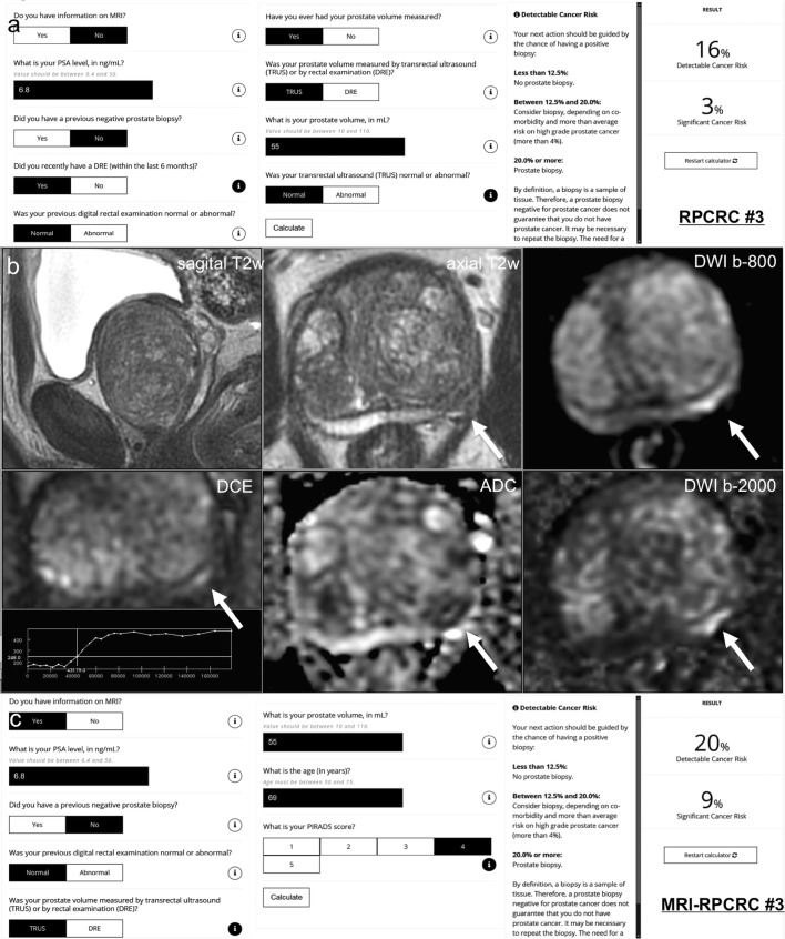
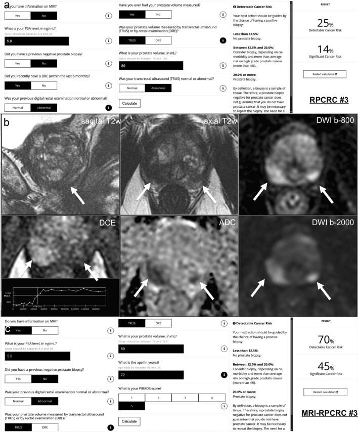
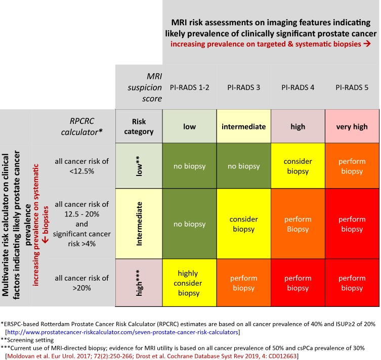
Risk-based patient selection for systematic biopsy in prostate cancer diagnosis has been adopted in daily clinical practice, either by clinical judgment and PSA testing, or using multivariate risk prediction tools. The use of multivariable risk prediction tools can significantly reduce unnecessary systematic biopsies, without compromising the detection of clinically significant disease. Increasingly multi-parametric magnetic resonance imaging (MRI) is performed, not only in men with a persistent suspicion of prostate cancer after prior negative systematic biopsy, but also at initial screening before the first biopsy. The combination of MRI and multivariate risk prediction tools could potentially enhance prostate cancer diagnosis using multivariate MRI incorporated risk-based models to decide on the need for prostate MRI, but also using MRI results to adjusted risk-based models, and to guide MRI-directed biopsies. In this review, we discuss the diagnostic work-up for clinically significant prostate cancer, where the combination of MRI and multivariate risk prediction tools is integrated, and how together they can contribute to personalized diagnosis.

Keywords: Biopsy; Magnetic resonance imaging (MRI); Multivariate risk prediction; Nomogram; Prostate cancer; Risk calculator; Risk stratification.


Conflict of interest statement

The authors have no potential conflicts of interest to declare.

Figures

Fig. 1
Fig. 1
Diagnostic flowcharts of men with elevated prostate-specific antigen (PSA) and/or abnormal digital rectal examination (DRE), with combinations of risk stratification w/o prostate MRI. Risk assessment with a PSA only, b multivariate risk prediction tools only, c prostate MRI only, and d multivariate risk prediction tools: when systematic biopsy is indicated subsequently a prostate MRI is performed to indicate a combined targeted biopsy, e multivariate risk prediction tools incorporating prostate MRI for biopsy-decision management (systematic, targeted, or both), and f multivariate risk prediction tools (step 1) may indicate systematic biopsy and subsequently a prostate MRI is performed. Based on the outcome parameters of prostate MRI incorporated in MRI-multivariate risk prediction tools, these tools may navigate into further biopsy testing or into deferring biopsy testing. PCa prostate cancer, PI-RADS MRI suspicion score, TBx MRI-targeted biopsy, SBx systematic biopsy, AS active surveillance
Fig. 2
Fig. 2
Case example: a 69-year-old man with a PSA of 6.8 ng/ml. Digital rectal examination (DRE) was normal, and transrectal ultrasound (TRUS) showed a prostate volume of 55 ml, without any hypo-echoic lesions found on ultrasound. The PSA density was 0.12 ng/ml2 (6.8/55). Does this biopsy-naive man deserve an MRI and/or biopsy? (1) In this biopsy-naïve man, the chance of finding any prostate cancer with further testing is 32%, based on the ERSPC-RC#2 (including PSA only in biopsy-naïve men) [49], suggesting a systematic biopsy is justified. (2) The chance of finding any prostate cancer and clinically significant prostate cancer with further testing is 16% and 3%, respectively, based on the ERSPC-RC#3 (including PSA, DRE, TRUS, and TRUS volume) [49] (a). Including DRE, TRUS, and TRUS volume in the risk calculation gives a lower risk estimation of having a biopsy-detectable clinically significant prostate cancer. The next action should be guided by the chance of having a positive biopsy: (1) less than 12.5% → no prostate biopsy (and no MRI); (2) between 12.5 and 20.0% → consider biopsy (and MRI), depending on co-morbidity and more than average risk on high grade prostate cancer (more than 4%); (3) 20.0% or more → prostate biopsy (and MRI). In this man, a systematic biopsy is justified, based on the ERSPC-RC#3. (3) A 3T multi-parametric MRI was performed (b), showing a large prostate gland with compression of the peripheral zone, and a suspected lesion for clinically significant prostate cancer at the left peripheral zone dorsolateral, at the mid-prostate, 12 × 4 mm, without extraprostatic extension or seminal vesicle invasion; PI-RADS assessment score 4 (4/4/+), based on low signal intensity on T2w-imaging (4), high signal intensity on DWI (b-800 and b-2000 (calculated) combined with low signal intensity on ADC (4), and focal enhancement (+). Based on high yields of clinically significant prostate cancer (> 30–40%) in an MRI lesion with a PI-RADS assessment score 4 (Sects. 3.2 and 4.2) [37], a targeted biopsy is indicated. (4) The chance of finding any prostate cancer and clinically significant prostate cancer with further testing is 20% and 9%, respectively, based on the MRI-ERSPC-RC#3 (including PSA, DRE, TRUS, TRUS volume, and MRI PI-RADS score) [49] (c). In this man, a systematic and targeted biopsy is justified. Biopsy considerations the morphology of the suspected peripheral zone lesion is long and small; targeted cores of the lesions combined with targeted cores from the penumbra (focal saturation) could be sufficient. Biopsy protocol and pathology findings Protocolled 12 systematic biopsies and 2 targeted biopsies were performed under MRI/US fusion guidance. 12 systematic biopsies showed benign prostatic tissue only. 2 targeted biopsy cores showed Gleason score 3 + 4 (ISUP group 2) with 3 mm and 4 mm maximum cancer core length, without cribriform or intraductal growth pattern
Fig. 3
Fig. 3
Case example. a 72-year-old man has a PSA of 5.8 ng/ml with lower urinary tract symptoms. Digital rectal examination (DRE) showed a right-sided abnormal prostate, classified as cT2 tumor. Transrectal ultrasound (TRUS) showed a prostate volume of 89 ml, with a right-sided hypo-echoic lesion found on ultrasound. The PSA density was 0.07 ng/ml2 (5.8/89). Does this biopsy-naive man deserve an MRI/biopsy? (1) In this biopsy-naïve man, the chance of finding any prostate cancer with further testing is 29%, based on the ERSPC-RC#2 (including PSA only in biopsy-naïve men) [49], suggesting a systematic biopsy is justified. (2) The chance of finding any prostate cancer and clinically significant prostate cancer with further testing is 25% and 14%, respectively, based on the ERSPC-RC#3 (including PSA, DRE, TRUS, and TRUS volume) [49] (a). In this man, a systematic biopsy is strongly recommended, based on the ERSPC-RC#3. (3) A 3T multi-parametric MRI was performed (b), showing a large prostate with compression of the peripheral zone, and a suspected lesion for clinically significant prostate cancer at the right peripheral zone dorsolateral, at the apex, 17 × 15 mm, without extraprostatic extension or seminal vesicle invasion; PI-RADS assessment score 5 (5/5/+), based on low signal intensity on T2w-imaging (5), high signal intensity on DWI (b-800 and b-2000 (synthetic/calculated) combined with low signal intensity on ADC (5), and focal enhancement (+). Based on high yields of clinically significant prostate cancer (> 70%) in an MRI lesion with a PI-RADS assessment score 5 (Sect. 3.2 and 4.2) [37], a targeted biopsy is strongly recommended in this biopsy-naïve man. A separate suspected lesion in the dorsal left peripheral zone in the apex categorized as PI-RADS score 4 (4/4/−) suggests multifocal prostate cancer. (4) The chance of finding any prostate cancer and clinically significant prostate cancer with further testing is 70% and 45%, respectively, based on the MRI-ERSPC-RC#3, including PSA, DRE, TRUS, TRUS volume, and MRI PI-RADS score [49] (c). In this man, a systematic and targeted biopsy is advised. Biopsy considerations The morphology of the suspected peripheral zone lesion in the right apex is large; targeted cores of the lesion will most likely hit the lesion. To limit undersampling, up to five cores might be sufficient [50]. The morphology of the suspected peripheral zone lesion in the left apex is small. Although targeted cores of the lesion most likely will hit the lesion, still, there is reasonable chance of undersampling or missing the lesion. A focal saturation biopsy might be most accurate [39]. Biopsy protocol and pathology findings Protocolled 12 systematic biopsies and 2 targeted biopsies in each suspected lesion were performed under MRI/US fusion guidance. Right-sided 6 systematic biopsies showed in 3 cores Gleason score 3 + 4 (ISUP group 2) with 1 mm, 10 mm, and 3 mm maximum cancer core length. Right-sided 2 targeted biopsies showed in 2 cores Gleason score 3 + 4 (ISUP group 2) with 14 mm and 7 mm maximum cancer core length, without cribriform or intraductal growth pattern. Left-sided 6 systematic biopsies showed benign prostatic tissue. Left-sided 2 targeted biopsies showed in 2 cores Gleason score 3 + 3 (ISUP group 1) with 6 mm and 1 mm maximum cancer core length, without cribriform or intraductal growth pattern
Fig. 4
Fig. 4
Matrix table based on the validated ERSPC-based Rotterdam prostate cancer risk calculator (RPCRC) and on MRI risk assessments. The continuum of estimated risks of having a biopsy-detectable prostate cancer in the RPCRC is categorized into low (no elevated risk; less then 12.5%), intermediate (elevated risk; between 12.5 and 20%), and high (elevated risk; 20% or more). Men with high risk of having a biopsy-detectable prostate cancer require biopsy. Also men with intermediate risk combined with a more than average risk on clinically significant prostate cancer (more than 4%) require biopsy [25]. Clinically significant prostate cancer in RPCRC is defined as any Gleason ≥ 4 grade, or primary and secondary Gleason ≤ 3 with ≥ 50% positive cores or total cancer core length of ≥ 20 mm [25]. The MRI risk assessment is categorized into low (PI-RADS 1 or 2), intermediate (PI-RADS 3), high (PI-RADS 4), and very high risk (PI-RADS 5) of having a biopsy-detectable clinically significant prostate cancer. Each cell ascribes a different biopsy action. This matrix table bridges the gap to new thresholds of developing and not-yet validated MRI prediction tools and may guide biopsy-decision management on individual patients in the increasingly complex, multivariate approach of prostate cancer diagnosis

References

    1. European Association of Urology (EAU) (2019) Guidelines on prostate cancer. http://uroweb.org/guideline/prostate-cancer/. Accessed Apr 2019
    1. Prostate cancer: diagnosis and management (update) (CG175) (2019) National institute for health and care excellence (NICE) Web site. https://www.nice.org.uk/guidance/cg175. Accessed April 2019
    1. National Comprehensive Cancer Network (NCCN) (2018) Guidelines on prostate cancer: 2018 update. https://www.nccn.org/professionals/physician_gls/default.aspx. Accessed Apr 2019
    1. Kasivisvanathan V, Rannikko AS, Borghi M, Panebianco V, Mynderse LA, Vaarala MH, Briganti A, Budäus L, Hellawell G, Hindley RG, Roobol MJ, Eggener S, Ghei M, Villers A, Bladou F, Villeirs GM, Virdi J, Boxler S, Robert G, Singh PB, Venderink W, Hadaschik BA, Ruffion A, Hu JC, Margolis D, Crouzet S, Klotz L, Taneja SS, Pinto P, Gill I, Allen C, Giganti F, Freeman A, Morris S, Punwani S, Williams NR, Brew-Graves C, Deeks J, Takwoingi Y, Emberton M, Moore CM. MRI-targeted or standard biopsy for prostate-cancer diagnosis. N Engl J Med. 2018;378(19):1767–1777. doi: 10.1056/NEJMoa1801993. - DOI - PMC - PubMed
    1. Porpiglia F, Manfredi M, Mele F, Cossu M, Bollito E, Veltri A, Cirillo S, Regge D, Faletti R, Passera R, Fiori C, De Luca S. Diagnostic pathway with multiparametric magnetic resonance imaging versus standard pathway: results from a randomized prospective study in biopsy-naive patients with suspected prostate cancer. Eur Urol. 2017;72(2):282–288. doi: 10.1016/j.eururo.2016.08.041. - DOI - PubMed