. 2019 Oct 10;47(18):9696-9707.
doi: 10.1093/nar/gkz696.

Dynamic architecture of the Escherichia coli structural maintenance of chromosomes (SMC) complex, MukBEF

Affiliations

Dynamic architecture of the Escherichia coli structural maintenance of chromosomes (SMC) complex, MukBEF

Karthik V Rajasekar et al. Nucleic Acids Res. .

Abstract

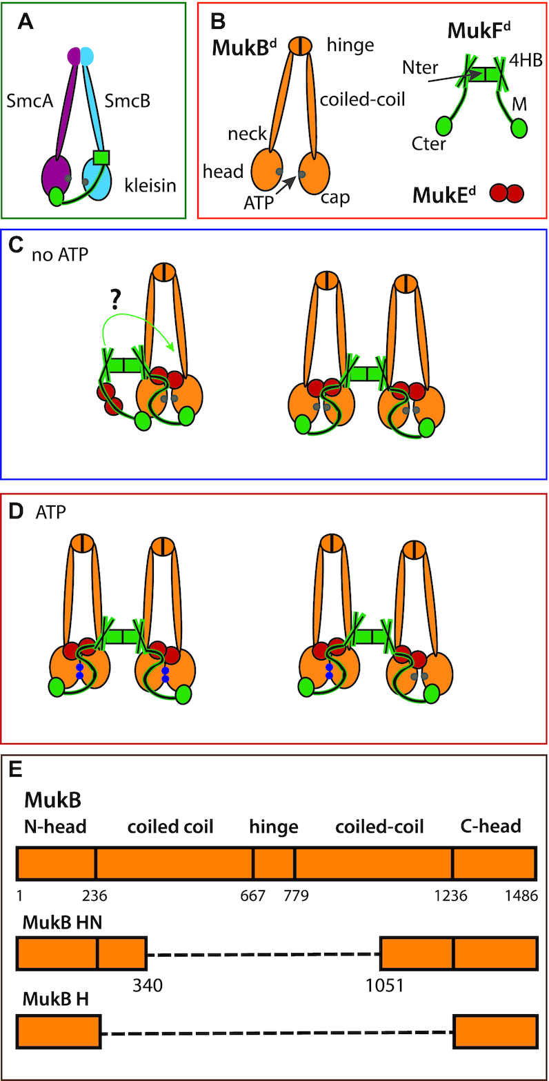
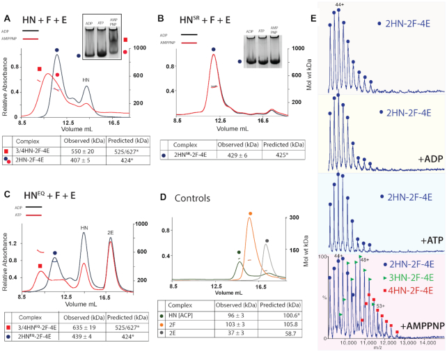
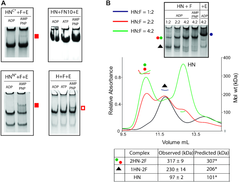
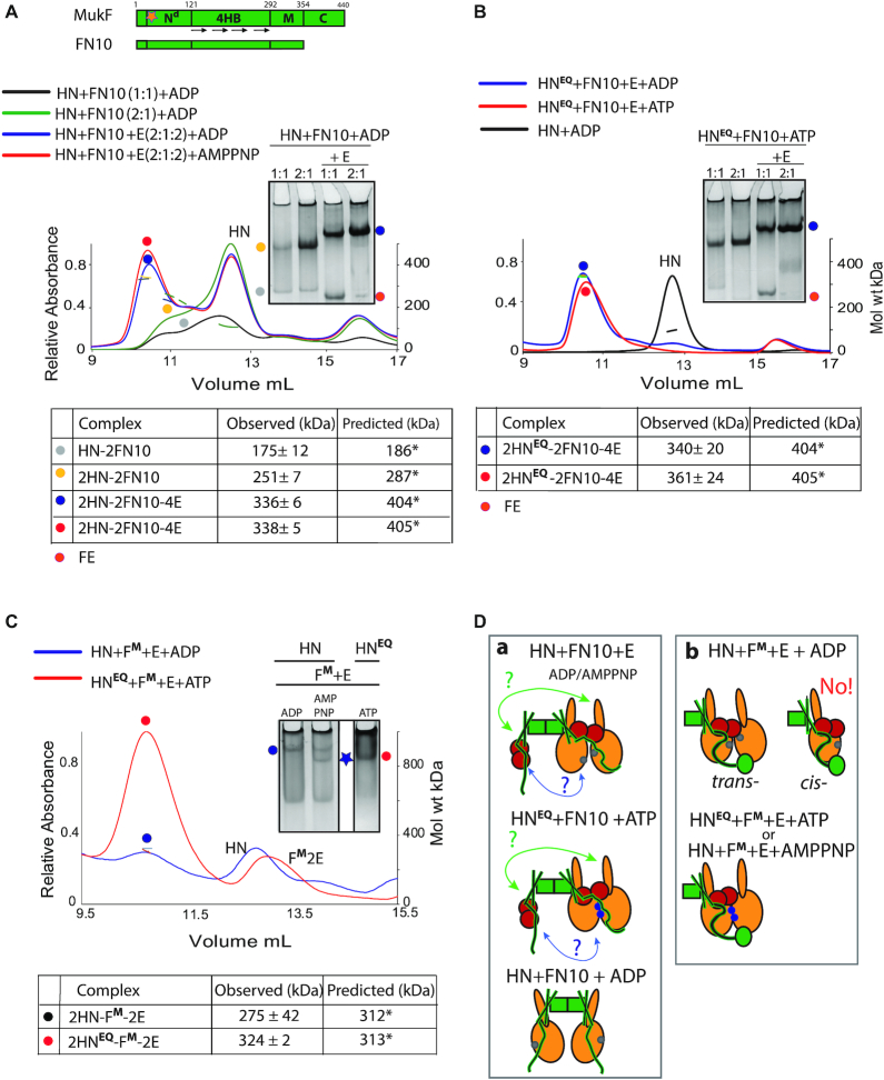
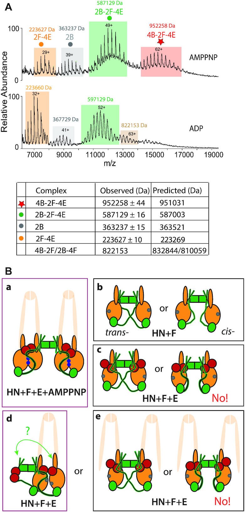
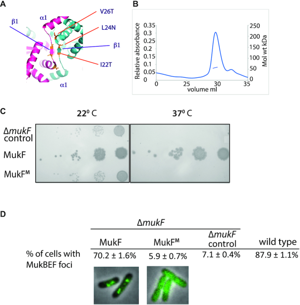
Ubiquitous Structural Maintenance of Chromosomes (SMC) complexes use a proteinaceous ring-shaped architecture to organize and individualize chromosomes, thereby facilitating chromosome segregation. They utilize cycles of adenosine triphosphate (ATP) binding and hydrolysis to transport themselves rapidly with respect to DNA, a process requiring protein conformational changes and multiple DNA contact sites. By analysing changes in the architecture and stoichiometry of the Escherichia coli SMC complex, MukBEF, as a function of nucleotide binding to MukB and subsequent ATP hydrolysis, we demonstrate directly the formation of dimer of MukBEF dimer complexes, dependent on dimeric MukF kleisin. Using truncated and full length MukB, in combination with MukEF, we show that engagement of the MukB ATPase heads on nucleotide binding directs the formation of dimers of heads-engaged dimer complexes. Complex formation requires functional interactions between the C- and N-terminal domains of MukF with the MukB head and neck, respectively, and MukE, which organizes the complexes by stabilizing binding of MukB heads to MukF. In the absence of head engagement, a MukF dimer bound by MukE forms complexes containing only a dimer of MukB. Finally, we demonstrate that cells expressing MukBEF complexes in which MukF is monomeric are Muk-, with the complexes failing to associate with chromosomes.


Figures

Figure 1.
Figure 1.
Schematics showing conserved SMC architectures and possible MukBEF stoichiometries and architectures. (A) Generic SMC complex architecture showing the tripartite proteinaceous ring formed by the kleisin and SMC proteins. (B) The components of the MukBEF complex. (C and D) Possible MukBEF architectures, without (C) and with (D) ATP. In D, the right panel shows a possible complex after ATP is hydrolysed in one of the dimers of a dimer of dimer complex. When ATP-dependent heads engagement occurs, the MukF middle region blocks the binding of a second MukF C-terminal domain to the second MukB head of a MukB dimer (27). When heads are unengaged, each MukB head can bind a MukF C-terminal domain, potentially leading to ‘daisy chain’ forms of a higher complexity than dimers and dimers of dimers (not shown). ATP-bound ATPase active sites denoted as blue dots on the heads and ADP-bound or nucleotide-unbound as grey dots. (E) Schematic showing MukB and its truncated variants head neck (HN) and head (H); a dashed line indicates a linker connecting N-and C-terminal head domains.
Figure 2.
Figure 2.
MukB head engagement is required for the formation of dimer of dimer MukBEF complexes. (AC) Native PAGE (A and B) and SEC-MALS (A–C) analyses of the stoichiometry of HN/HNSR/HNEQ complexes with MukFE in the presence and absence of ATP/AMPPNP. A total of 10 μM HN, 5 μM F and 10 μM E were incubated for 3 h at room temperature with ADP or AMPPNP/ATP (1 mM) prior to loading onto a 6% native gel, or a Superose 6 column. (D) SEC-MALS analysis of individual Muk proteins. The observed MukE mass of 37 kDa is consistent with previously published studies (30,31) and likely reflects MukE in an equilibrium mixture of monomers and dimers in solution. Predicted and observed masses of the complexes are tabulated below with the values and their uncertainties derived from a single representative SEC-MALS experiment. Predicted masses (kDa) of complexes containing ACP-4’phosphopantetheine bound to HN at stoichiometric levels (see below, Supplementary Figure S2) and bound AMPPNP when appropriate. Differences between the predicted and observed masses of the complexes are within 5%. (E) Native mass spectra of MukB HN complexes formed with MukFE in the absence of nucleotide or in the presence of ADP, ATP or AMPPNP. The complexes are indicated as follows: 2HN-2F-4E (blue dots), 3HN-2F-4E (green triangles) and 4HN-2F-4E (red squares). The observed and predicted masses (Da) of these complexes are tabulated in Supplementary Table S2.
Figure 3.
Figure 3.
Dimer of dimer formation requires MukE, and HN interactions with both the MukF C-terminal domain and the MukF 4-helix bundle. (A) Native PAGE of complexes generated with HN and F variants deficient in binding across one (HNC*, FN10), or the other (HNN*and H), HN-F interface. The position of low levels of dimer of dimer complexes is indicated with filled red squares (HN), or an unfilled red square (H). HNC* carries the following aa residues alterations: F1453S, H1458A, R1465A. (B) SEC-MALS analyses of HN-F complexes at different HN:F ratios; 2.5/5/10 μM HN was mixed with 5 μM F prior to separation through a Superdex 200 column. Predicted and observed masses of the complexes are tabulated below. An 11% difference between the predicted and observed mass of 1HN-2F reflects a presence of some 2HN-2F in the peak (black trace). The same mixtures incubated with ADP along with samples containing 10 μM HN+5 μM F and either AMPPNP or 10 μM E were analysed on 6% native gels.
Figure 4.
Figure 4.
Architecture of MukBEF complexes. (A) Top, schematic of MukF and its truncated derivative FN10; Nd, N-terminal dimerisation domain; 4HB, 4-helix bundle; M, middle region; and C, C-terminal domain, which is deleted in FN10. Orange star indicates position of dimerisation interface, which has been altered in MukFM; also see (Figure 6). (A andB) SEC-MALS and native PAGE analyses of HN and HNEQ complexes generated with FN10 dimers. For SEC-MALS, samples at the indicated protein ratios were incubated with ADP, AMPPNP or ATP before separation through a Superose 200 column. FN10 was at 5 μM and E, when present, was 10 μM. Predicted and observed masses of complexes are tabulated below traces. Native gel samples were incubated with ADP (A) or ATP (B) prior to loading onto a gel. (C) SEC-MALS and native PAGE analyses of HN and HNEQ complexes generated with FM-E in the presence of ADP, AMPPNP or ATP as indicated. The proteins were at concentrations of 10 μM HN/HNEQ, 5 μM FM and 10 μM E. (A–C) Significant (<16%) differences in observed to predicted masses of the complexes in some experiments are due to incomplete resolution of the complexes from unbound HN. (D) Schematics of the proposed architectures with FN10 (panel a) and MukFM (panel b). The green arrows (a) indicate a possible interaction between the FN10 4HB and the neck of the distal HN molecule. A second potential interaction between the ‘free’ FN10 middle region and its bound MukE to the proximal HN molecule is indicated by blue arrows. The bottom cartoon in (a) shows two HN molecules binding a FN10 dimer through interactions with the 4HBs. ATP-bound ATPase active sites denoted as blue dots on the heads and ADP-bound or nucleotide-unbound as grey dots.
Figure 5.
Figure 5.
Dimers of dimers are formed with full length MukB dimers, MukEF and AMPPNP. (A) Native mass spectra of complexes formed with MukBEF in the presence of AMPPNP (top), or ADP (bottom). The predicted and observed masses are tabulated below the graphs. The proteins were at concentrations 5 μM B, 2.5 μM F and 5 μM E. The small population of complexes (mass 822153 Da, beige) observed in the ADP sample may reflect a presence of 4B-2F or 4B-2F-E2 complexes (predicted mass 832843.68 Da and 891578 Da, respectively). (B) Schematics of the architectures demonstrated or inferred from the biochemical analyses. The extrapolation from HN to full length MukB dimers is cartooned by showing the remainder of MukB as semi-transparent. Panel (a), MukBEF-AMPPNP- and head engagement-dependent dimer of dimers. (b) Alternative possible architectures of HN + MukF. (c) As (b) in the presence of MukE. The data provide evidence for the trans-configuration shown on the left. Panels d and e, show possible configuration of 2HN-2F-4E dimers in the presence of ADP/absence of head engagement. Note that the architectures in d and e-left are topologically identical if both necks engage with a 4-helix bundle (green arrow; see Figure 4D), although if part of a full-length MukB dimer as indicated, would be a dimeric MukBEF complex (d) or dimer of dimer complex (e). ATP-bound ATPase active sites denoted as blue dots on the heads and ADP-bound or nucleotide-unbound as grey dots.
Figure 6.
Figure 6.
Escherichia coli cells expressing monomeric MukF have a Muk phenotype and fail to form MukBEF clusters on the chromosome. (A) Dimerisation interface of MukF showing the three mutated residues, I22T, L24N, V26T. (B) SEC-MALS of MukF monomers. The observed mass of 57.3 kDa corresponded to theoretical mass, of 52.9 kDa, of the monomeric variant. A small shoulder in the elution profile reflects the presence of the protein proteolytic cleavage product. (C) Temperature-sensitivity of growth in rich medium assay. 102-fold serial dilutions of ΔmukF cells containing plasmid pET21 expressing basal levels of wild-type MukF or the MukF monomer (MukFM) are shown, alongside a control in which cells contain the plasmid vector alone. (D) ΔmukF cells expressing MukF monomers (MukFM) fail to form MukBEF foci. The analysis of foci formation was performed in ΔmukF strain carrying C-terminal mukB-gfp fusion; MukF and MukFM were expressed from pET21 as in (C). For comparison, foci formation in a strain carrying the intact endogenous chromosomal copy of mukF was monitored using MukB-mYpet expression (SN192), (15).

References

    1. Bürmann F., Shin H.C., Basquin J., Soh Y.M., Gimenez-Oya V., Kim Y.G., Oh B.H., Gruber S.. An asymmetric SMC-kleisin bridge in prokaryotic condensin. Nat. Struct. Mol. Biol. 2013; 20:371–379. - PubMed
    1. Gligoris T.G., Scheinost J.C., Burmann F., Petela N., Chan K.L., Uluocak P., Beckouet F., Gruber S., Nasmyth K., Lowe J.. Closing the cohesin ring: structure and function of its Smc3-kleisin interface. Science. 2014; 346:963–967. - PMC - PubMed
    1. Huis in ’t Veld P.J., Herzog F., Ladurner R., Davidson I.F., Piric S., Kreidl E., Bhaskara V., Aebersold R., Peters J.M.. Characterization of a DNA exit gate in the human cohesin ring. Science. 2014; 346:968–972. - PubMed
    1. Zawadzka K., Zawadzki P., Baker R., Rajasekar K.V., Wagner F., Sherratt D.J., Arciszewska L.K.. MukB ATPases are regulated independently by the N- and C-terminal domains of MukF kleisin. Elife. 2018; 7:e31522. - PMC - PubMed
    1. Ganji M., Shaltiel I.A., Bisht S., Kim E., Kalichava A., Haering C.H., Dekker C.. Real-time imaging of DNA loop extrusion by condensin. Science. 2018; 360:102–105. - PMC - PubMed

Publication types

MeSH terms