Review
. 2019 Apr:144:57-77.
doi: 10.1016/j.addr.2019.07.010. Epub 2019 Aug 7.

Nanocarrier-based systems for targeted and site specific therapeutic delivery

Affiliations
Review

Nanocarrier-based systems for targeted and site specific therapeutic delivery

Joydeb Majumder et al. Adv Drug Deliv Rev. 2019 Apr.

Abstract

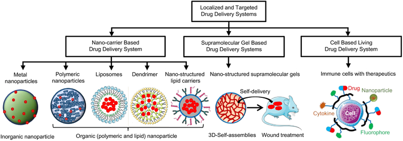
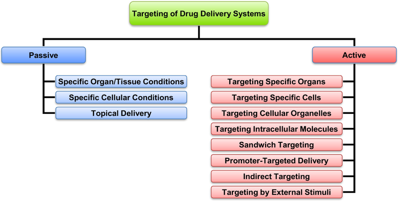
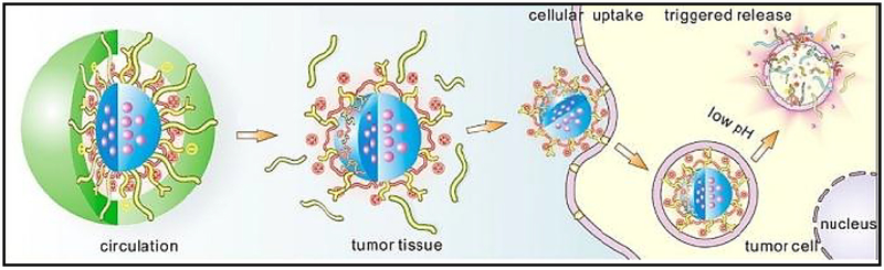
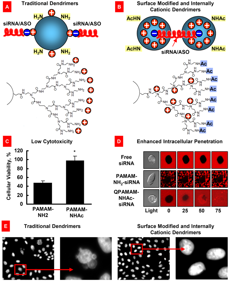
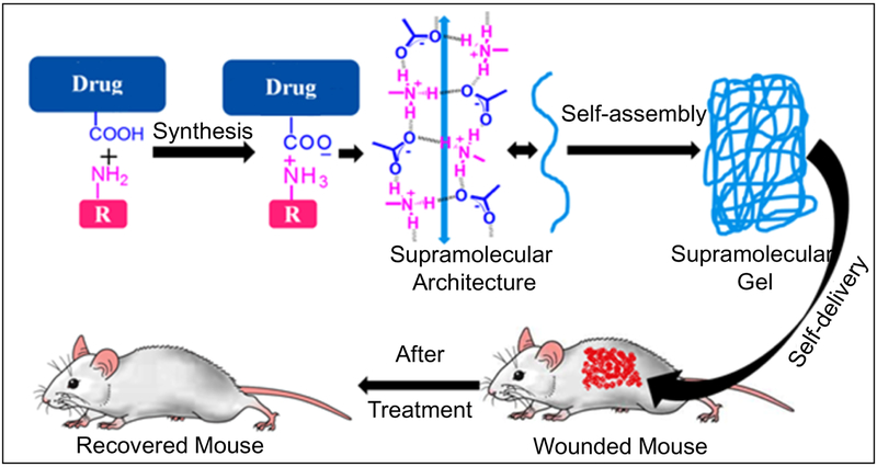
Systemic drug delivery methods such as oral or parenteral administration of free drugs possess relatively low treatment efficiency and marked adverse side effects. The use of nanoparticles for drug delivery in most cases substantially enhances drug efficacy, improves pharmacokinetics and drug release and limits their side effects. However, further enhancement in drug efficacy and significant limitation of adverse side effects can be achieved by specific targeting of nanocarrier-based delivery systems especially in combination with local administration. The present review describes major advantages and limitations of organic and inorganic nanocarriers or living cell-based drug and nucleic acid delivery systems. Among these, different nanoparticles, supramolecular gels, therapeutic cells as living drug carriers etc. have emerged as a new frontier in modern medicine.

Keywords: Cell-based carriers; Drug; Drug targeting; Organic and inorganic nanoparticles; Supramolecular gel; siRNA and nucleotide delivery.


Conflict of interest statement

Conflict of interest

The authors declare no conflict of interest in the publication of this work.

Figures

Fig. 1.
Fig. 1.
Various carrier-based systems for localized and targeted delivery of drugs and other therapeutics. Nanocarrier-based systems such as nanoparticles, liposomes, dendrimers, nanostructured lipid carriers are shown with drug/therapeutic (marked as red dot) encapsulated for their site-specific controlled delivery applications. Formation of nanostructured supramolecular topical gels and their in vivo application are shown as self-delivery system. Other drug carriers such as therapeutic living cells decorated with drug (red color), nanoparticle (olive color), cytokine (orange color), fluorophore (green color) are shown as living delivery systems for disease specific drug targeting. Part of this figure was created using Servier Medical Art images, which are licensed under a Creative Commons Attribution 3.0 Unported License (https://smart.servier.com).
Fig. 2.
Fig. 2.
Different types of targeted delivery systems. Modified from [40].
Fig. 3.
Fig. 3.
Schematic illustration of nanoparticles used in drug delivery application Reproduced with permission from [95].
Fig. 4.
Fig. 4.
Surface modified and internally cationic quaternized PAMAM dendrimers for efficient nucleic acid intracellular delivery. (A) Traditional dendrimers with non-modified surface and external positive charges for siRNA or antisense oligonucleotides (ASO) binding. (B) Modified dendrimers with acetylated surface and internal positive charges for siRNA binding. (C) Cytotoxicity of a traditional (PAMAM-NH2) and surface-modified quaternized (QPAMAM-NHAc) dendrimers. Means ± SD are shown. *P < 0.05 when compared with PAMAM-NH2. (D) Cellular internalization of free siRNA, and dendrimer–siRNA complexes with a traditional (PAMAM-NH2) and internally cationic and surface-modified (QPAMAM-NHAc) dendrimers. siRNA were labeled with a red fluorescence dye, conjugated with different dendrimers, and added to the incubation medium of living cancer cells. Real-time fluorescence was registered using a fluorescent microscope. Free siRNA and traditional dendrimer–siRNA complexes are poorly internalized by cancer cells, while modified dendrimers provided for an efficient intracellular delivery of siRNA. (E) Genotoxicity (formation of micronuclei) of traditional and modified PAMAM dendrimers. Representative fluorescence microscopy images of CHO-K1 cells incubated within 24 hours with non-modified and modified dendrimers. Modified from [6, 23, 104].
Fig. 5.
Fig. 5.
Schematic illustration of the hierarchically targetable solid lipid nanoparticles for the local chemo/thermo combination therapy against colon cancer by oral administration. Reproduced with permission from [111].
Fig. 6.
Fig. 6.
Local inhalation codelivery of anticancer drug and suppressors of pump and nonpump cellular resistance enhances apoptosis induction in the lung tumor, decreases tumor size, and prevents adverse side effects of treatment on healthy organs. An orthotopic mouse model of lung cancer was created by intratracheal instillation of human A549 lung cancer cells into nude mice. Untreated mice (1), mice treated by i.v injection of DOX (2), by i.v. injection of liposomal DOX (3), by inhalation with liposomal DOX (4), by inhalation with a mixture of the two systems (liposomal DOX + ASO targeted to MRP1 mixed with liposomal DOX + ASO targeted to BCL2 mRNA) (5), and by inhalation with one complex liposomal delivery system containing DOX, ASO targeted to MRP1, and BCL2 mRNA (6). (A) Apoptosis induction in the lungs with tumor and other organs 4 weeks after beginning of treatment. Enrichment of histone-associated DNA fragments (mono- and oligonucleosomes) per gram tissue in the tumor and organs of control animals was set to unit 1, and the degree of apoptosis was expressed in relative units. Apoptosis measurements were performed 24 h after last treatment. (B) Typical fluorescent microscopy images of tumor tissue slides labeled by TUNEL 24 h after last treatment. (C) Changes in tumor volume after beginning of treatment. Mice were treated on days 0, 3, 7, 11, 14, 17, 21, and 24. Mean ± SD are shown. *P < 0.05 when compared with untreated animals. Reproduced with permission from [120].
Fig. 7.
Fig. 7.
Changes in lung tumor volume after beginning of treatment. An orthotopic mouse model of lung cancer was created by intratracheal instillation of human A549 lung cancer cells into nude mice. Lung tumor was evaluated by bioluminescence optical imaging (IVIS), magnetic resonance imaging (MRI) and computed tomography (CT). Mice were treated on days 0, 3, 7, 11, 14, 17, 21, and 24. 1 – Untreated mice; 2 – LHRH-NLC (inhalation); 3 – TAX (i.v.); 4 – LHRH-NLC-TAX (inhalation); 5 – LHRH-NLC-TAX-siRNAs targeted to MRP1 and BCL2 mRNAs (inhalation). Means ± SD are shown. Modified from [21, 135].
Fig. 8.
Fig. 8.
Possible mechanisms of skin permeation by nanostructured lipid carriers (NLCs). Part of this figure was created using Servier Medical Art images, which are licensed under a Creative Commons Attribution 3.0 Unported License (https://smart.servier.com).
Fig. 9.
Fig. 9.
Treatment of mice with Idiopathic Pulmonary Fibrosis (IPF). IPF was induced by intratracheal instillation of 1.5 U/kg of bleomycin. Mice were treated by inhalation with substances indicated twice a week for three weeks starting next day after the bleomycin administration. (A) Expression of targeted genes in lung tissues of mice with IPF. At the end of treatment, lungs were harvested and homogenized. The expression of MMP3 (A), CCL12 (B) and HIF1A (C) genes was measured by the QPCR and presented as fold change relative to the healthy animals. Means ± SD are shown. *P < 0.05 when compared with untreated mice with pulmonary fibrosis. (B) Survival of mice (Kaplan-Meier survival plot) with IPF. Modified from [115].
Fig. 10.
Fig. 10.
Distribution of NLCs in the lungs. (A) Distribution of fluorescently labeled (Cy5.5) non-targeted and LHRH-tumor targeted NLCs in mouse lungs bearing human lung cancer. (B) Distribution of fluorescently labeled (Cy5.5) LHRH-tumor - targeted (NLC-LHRH) in mouse lungs bearing human lung tumor cells (tumor and non-tumor tissues; bright field and fluorescence microscope images; red color represents distribution of NLC-LHRH in tumor and non-tumor lung tissues). Modified from [21].
Fig. 11.
Fig. 11.
Schematic illustration of DOX-loaded MSNs@PDA–PEG–FA. Reproduced with permission from [152].
Fig. 12.
Fig. 12.
Sequential targeting drug delivery based on RGD and TAT peptides co-conjugated MSNs for effective cancer therapy. Reproduced with permission from [153].
Fig. 13.
Fig. 13.
Preparation and testing of supramolecular gels for topical drug delivery application Low molecular weight gelator molecule (derived from salt formation between the carboxylic acid functionalized drug and primary amine) first self-assemble to form 1D network which further self-assemble to form 3D cross-linked network - under which the solvent molecules are trapped to produce supramolecular gels. Drug based supramolecular topical gel is shown as self-delivery systems for treating skin diseases in mouse.
Fig. 14.
Fig. 14.
Toxicity and drug release profile of different taxol-loaded gels. (A) Structures of the gelators 5a and 5b. (B) Cytotoxicity of taxol (1) and gelators 5a, 5b, and 4 after incubated with HeLa cells for 48 h. (C) Accumulative drug release profile of two types of taxol gels in 100 mM PBS buffers. Reproduced with permission from [184].
Fig. 15.
Fig. 15.
Structure of the gelator molecule 11. Mice were treated with DOX-local administration and with DOX-gel (made with gelator 11). (A) Tumor volume. Means ± SD are shown. *P < 0.005. (B) representative pictures of excised tumors. Reproduced with permission from [187].
Fig. 16.
Fig. 16.
Modifications of nonsteroidal anti-inflammatory drug naproxen by decorating the parent drug with β-amino acid/amino alcohols/L-amino acids results in potent hydrogelators namely 2, 3, 10 and 11. (A) Structures of NSAID-naproxen derived various supramolecular gelators. (B) Opaque hydrogels and the corresponding gel-network morphologies. (C) Sustained release of hydrogelator 2 and 11 in PBS (pH 7.4). Reproduced with permission from [190].
Fig. 17.
Fig. 17.
Topical cetirizine gel application for treatment of dinitrochlorobenzene (DNCB)-induced allergic conditions. (A) Chemical structures of gelator salt 3; (B) Methylsalycylate/menthol topical gel and gel beads of salt 3; (C) Treatment of DNCB-induced allergic ears redness of BALB/c male mice with the anti-allergic gelator salt 3. (D) Histological features of the dorsal skin tissues of mice. (Group X) no skin allergic induction by DNCB as the normal mice; (Group Y1) no treatment after skin allergy induction by DNCB; (Group Y2) menthol solution application after skin allergy induction by DNCB; (Group Y3) mother drug cetirizine solution application after skin allergy induction by DNCB; and (Group Y4) methylsalycylate/menthol topical gel of salt 3 application after skin allergic induction by DNCB. Reproduced with permission from [203].
Fig. 18.
Fig. 18.
Synthesis and characterization of T cell receptor (TCR) signaling-responsive protein nanogels. (A) Scheme for protein nanogel (NG) synthesis and release of protein in response to reducing activity in the local microenvironment. (B) Representative transmission electron microscopy image of NGs prepared from a human IL-15 superagonist (IL-15Sa). Scale bar, 50 nm. (C) Hydrodynamic sizes of different NGs, as determined by dynamic light scattering. Means ± SD are shown. (D) Release kinetics of cytokines from redox-responsive or nondegradable IL-15Sa-NGs in PBS with or without added glutathione (GSH) as a reducing agent. Means ± SD are shown. (E) Representative MALDI mass spectrometric analysis of released and native cytokines. Reproduced with permission from [253].
Fig. 19.
Fig. 19.
Targeted release of cell membrane-permeable, immunomodulatory compounds from T-cell-linked nanoparticles. (A) Schematic view of strategy to modulate T-cell responses via nanoparticle conjugation to membrane proteins: Surface-conjugated drug-loaded nanoparticles slowly release their cargo compounds, which locally permeate the plasma membrane and block molecules in the cytosol that dampen T-cell activation. (B) 3D reconstruction of confocal microscopy images showing CD8+ effector T-cells (CFSE stain shown in blue) immediately after conjugation with fluorescent multilamellar lipid vesicles (yellow). Reproduced with permission from [251].

References

    1. Mainardes RM, Silva LP, Drug delivery systems: past, present, and future, Curr Drug Targets, 5 (2004) 449–455. - PubMed
    1. Robinson DH, Mauger JW, Drug delivery systems, Am J Hosp Pharm, 48 (1991) S14–23. - PubMed
    1. Viswanathan P, Muralidaran Y, Ragavan G, Chapter 7 - Challenges in oral drug delivery: a nano-based strategy to overcome, in: Andronescu E, Grumezescu AM (Eds.) Nanostructures for Oral Medicine, Elsevier, 2017, pp. 173–201.
    1. Bardal SK, Waechter JE, Martin DS, Chapter 2 - Pharmacokinetics, in: Bardal SK, Waechter JE, Martin DS (Eds.) Applied Pharmacology, Philadelphia, 2011, pp. 17–34.
    1. Koushik OS, Rao YV, Kumar P, Karthikeyan R, Nano Drug Delivery Systems to Overcome Cancer Drug Resistance - A Review, J Nanomed Nanotechnol, 7 (2016) 378–387.

Publication types