Review
. 2019 Oct:58:214-223.
doi: 10.1016/j.sbi.2019.04.006. Epub 2019 Aug 7.

CryoEM maps are full of potential

Affiliations
Review

CryoEM maps are full of potential

Mayra A Marques et al. Curr Opin Struct Biol. 2019 Oct.

Abstract

Electron microscopy is based on elastic scattering due to Coulomb forces between the incident electrons and the sample; thus, electron scattering is dependent on the charge distribution in the sample. Unlike atomic scattering factors for X-rays, electron scattering factors for some atoms are strongly dependent on scattering angle, and the scattering factor for ionic oxygen is negative at low scattering angle. This phenomenon can result in a significant negative contribution to Coulomb potential maps by oxygen and can result in deviations in the positions of positive map features from atomic centers. An important factor that can also complicate the interpretation of cryoEM maps is the exquisite sensitivity of macromolecules to damage from electron irradiation, especially the carboxylates of acidic amino acids. Ideally, when compared with electron density maps derived by X-ray crystallography, Coulomb potential maps can provide additional details about the electrostatic environment and charge state of atoms. Enhancements in model building, refinement and computational simulation will be required to realize the full potential of EM-derived maps to reveal deeper insight into the electronic structure and functional properties of macromolecular complexes and their interactions with binding partners, ligands, cofactors, and drugs.


Conflict of interest statement

Author disclosures

The authors declare no conflicts of interest.

No conflict of interest exists.

We wish to confirm that there are no known conflicts of interest associated with this publication and there has been no significant financial support for this work that could have influenced its outcome.

Figures

Figure 1
Figure 1. Electron (a, c, e, g) and X-ray (b, d, f, h) scattering amplitudes of charged and neutral atoms.
Electron scattering factors of charged atoms vary considerably over a range of spatial frequency, depending on the charged state. Electron scattering factors of negatively charged atoms can even become negative (a) and those of positively charged atoms show high amplitudes at low scattering angles [39]. The same is not observed for X-ray data where the scattering amplitudes are always positive and proportional to atomic number, Z (e.g., compare the amplitudes for the light atoms in (b) and (d) versus the heavy atoms in (f) and (h). Electron and X-ray scattering factors of neutral and ionized atoms are based on values at given scattering angles from the International Tables for Crystallography [40] (a and c adapted from [39]).
Figure 2
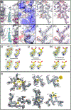
Figure 2. Coulomb potential maps of macromolecules can reveal protein drug interactions, partial charged states of carboxylates, and bound water molecules.
(a,b) MicroED map at 2.9-Å resolution showing a cross-section view of the 6-helix bundle formed by the CTD (slate blue) and SP1 (magenta) domains of HIV-1 Gag. (a) en face and (b) profile views of a single molecule of the maturation inhibitor bevirimat (BVM, cyan) stabilizes the six-helix bundle via both electrostatic interactions between the K359 residues and the dimethylsuccinyl moiety and hydrophobic interactions with the pentacyclic triterpenoid ring. HIV protease cleaves the 6 CTD-SP1 domains at L363, which reside on the interior of the 6-helix bundle. (c,d) The CTD-SP1-BVM Coulomb potential surface [−8 kcal/mol e (red) to 8 kcal/mol e (blue)] shows significant negative charge for solvent-exposed acidic residues (D295, E307 and E319) and neutralization in the case of an acidic residue involved in a salt bridge (E291). (e-h) CTD-SP1-BVM MicroED maps showing acidic and basic residues in a variety of environments. Electron scattering factors are strongly dependent on charge at low resolution, and excluding these reflections results in additional side chain density for residues containing O; e.g., note the reduced density of D295, E307, and E319 (red ellipses) in e and f for the map calculated over the resolution range from 20–2.9 Å (gray mesh), compared with g and h for the map calculated from 8–2.9 Å resolution (pink mesh) [10]. Scattering factors for neutral oxygen are much less dependent on resolution and are positive at all scattering angles; therefore, the maps are nearly identical for acidic side chains involved in salt bridges (e.g., E291 in e and g) or neutral oxygen-containing side chains (e.g., N315 and T318 in f and h), whether the full or high-resolution range is used in map calculations [10]. Simulated Coulomb potential maps (i-k) demonstrate the ability to identify charged states of acidic residue carboxylates (top and bottom, 2.0-Å and 3.0-Å resolution, respectively): aspartic acid (i) fully protonated, (j) fully deprotonated with the negative charge distributed equally on the two oxygen atoms, and (k) fully deprotonated with the charge localized on one of the two O atoms (adapted from [19]). (l) Coulomb potential map of β-galactosidase with bound phenylethyl β-D-thiogalactopyranoside (PETG) at 2.2 Å resolution reveals tightly bound water molecules (yellow) (from [6]).
Figure 3
Figure 3. Biological macromolecules are exquisitely sensitive to damage from electron irradiation.
(a) Coulomb potential map of β-galactosidase at 3.2 Å resolution, showing that aspartic and glutamic acid are more sensitive to radiation damage (total electron dose 10, 20, or 30 e2) than cationic and neutral amino acids (adapted from [7]). (b) Coulomb potential map of proteinase K displays initiation of disulfide bond breakage and decarboxylation of amino acidic side chains at an electron dose of ~1 e2 (adapted from [21]). The program EMringer is a sidechain-directed approach to study model-to-map agreement in cryoEM. Using the cryoEM map of the proteasome, the Cγ atom of a particular side chain was rotated around the X(note to copy editor: Greek letter chi)1 angle, and the peak value between the model and map is interpreted as the correct position of the side chain. In this analysis lower scores were observed for acidic amino acid side chains, consistent with greater radiation damage (adapted from [23]).
Figure 4
Figure 4. Charge density maps are an alternate way to represent experimental cryoEM maps.
(a) Relative amplitudes for phosphate groups of a G:C base pair in the large ribosomal subunit increases from the unsharpened experimental electrostatic potential (ESP) map (EMD2847) [5]. (b) ESP maps sharpened by the original authors with B factor of −120 Å2. (c) Experimental charge density (CD) map contoured at +6.0σ. Appearance for all recognizable side chains (EMD 2984) can be improved from the sharpened experimental ESP map by the original authors (d, +2.2σ, salmon), and the experimental CD map (e, +2.2σ, cyan) viewed at the same contour level (adapted from [5]). (f) A published figure of the sharpened 1.9-Å Coloumb potential map of β-galactosidase (EMD-7770) [8], (g) the deposited map after Phenix Autosharpen [26] and (h) the deposited map following conversion to a charge density map in Chimera [13,25]. Figures g and h were generated using PyMOL 1.8 [41].
Figure 5
Figure 5. Analysis of cryoEM images of P22 bacteriophage capsid demonstrates the local electrostatic environment around negatively charged (a) and positively charge (b) amino acids.
Acidic residues are surrounded by strongly negative electrostatic potential (red), whereas basic residues are surrounded by strongly positive electrostatic potential (green). (c) Fit of atomic model to the experimental cryoEM density map. (d) Map calculated from the atomic model in (c) in which all atoms have equal weight, showing model bias. (e) Calculated map derived from the atomic model in (c) compensating for the electrostatic potential of each atom using atomic displacement (ADP) factors. The map in (e) demonstrates that the properly weighted model is capable of faithfully recapitulating the experimental Coulomb potential map (from [24]).

References

    1. Henderson R: The potential and limitations of neutrons, electrons and X-rays for atomic resolution microscopy of unstained biological molecules. Q Rev Biophys 1995, 28:171193.

      • It is impossible to overstate Richard Henderson’s contributions to the field of structural biology and to the larger scientific community. This review provides an excellent background to imaging of macromolecules with different radiation sources and a view from the past into the present.

    1. Vinothkumar KR, Henderson R: Single particle electron cryomicroscopy: trends, issues and future perspective. Q Rev Biophys 2016, 49:e13.

      • An excellent overview of the current state of cryoEM and future challenges and opportunities.

    1. Wang J, Moore PB: On the interpretation of electron microscopic maps of biological macromolecules. Protein Sci 2017, 26:122–129.

      •• This paper and two others by Jimin Wang in the same issue of Protein Science present in depth discussions and analysis of the fundamental nature of Coulomb potential maps and current issues and shortcomings in the use of these maps for structure modeling.

    1. Merk A, Bartesaghi A, Banerjee S, Falconieri V, Rao P, Davis MI, Pragani R, Boxer MB, Earl LA, Milne JLS, et al.: Breaking Cryo-EM Resolution Barriers to Facilitate Drug Discovery. Cell 2016, 165:1698–1707. - PMC - PubMed
    1. Fischer N, Neumann P, Konevega AL, Bock LV, Ficner R, Rodnina MV, Stark H: Structure of the E. coli ribosome-EF-Tu complex at <3 A resolution by Cs-corrected cryo-EM. Nature 2015, 520:567–570. - PubMed

Publication types

MeSH terms

LinkOut - more resources