. 2019 Oct 3;76(1):11-26.e7.
doi: 10.1016/j.molcel.2019.06.043. Epub 2019 Aug 7.

RAD51AP1 Is an Essential Mediator of Alternative Lengthening of Telomeres

Affiliations

RAD51AP1 Is an Essential Mediator of Alternative Lengthening of Telomeres

Jonathan Barroso-González et al. Mol Cell. .

Erratum in

  • RAD51AP1 Is an Essential Mediator of Alternative Lengthening of Telomeres.
    Barroso-González J, García-Expósito L, Hoang SM, Lynskey ML, Roncaioli JL, Ghosh A, Wallace CT, Modesti M, Bernstein KA, Sarkar SN, Watkins SC, O'Sullivan RJ. Barroso-González J, et al. Mol Cell. 2019 Oct 3;76(1):217. doi: 10.1016/j.molcel.2019.08.009. Mol Cell. 2019. PMID: 31585101 Free PMC article. No abstract available.
  • RAD51AP1 Is an Essential Mediator of Alternative Lengthening of Telomeres.
    Barroso-González J, García-Expósito L, Hoang SM, Lynskey ML, Roncaioli JL, Ghosh A, Wallace CT, de Vitis M, Modesti M, Bernstein KA, Sarkar SN, Watkins SC, O'Sullivan RJ. Barroso-González J, et al. Mol Cell. 2020 Jul 16;79(2):359. doi: 10.1016/j.molcel.2020.06.026. Mol Cell. 2020. PMID: 32679078 Free PMC article. No abstract available.

Abstract

Alternative lengthening of telomeres (ALT) is a homology-directed repair (HDR) mechanism of telomere elongation that controls proliferation in aggressive cancers. We show that the disruption of RAD51-associated protein 1 (RAD51AP1) in ALT+ cancer cells leads to generational telomere shortening. This is due to RAD51AP1's involvement in RAD51-dependent homologous recombination (HR) and RAD52-POLD3-dependent break induced DNA synthesis. RAD51AP1 KO ALT+ cells exhibit telomere dysfunction and cytosolic telomeric DNA fragments that are sensed by cGAS. Intriguingly, they activate ULK1-ATG7-dependent autophagy as a survival mechanism to mitigate DNA damage and apoptosis. Importantly, RAD51AP1 protein levels are elevated in ALT+ cells due to MMS21 associated SUMOylation. Mutation of a single SUMO-targeted lysine residue perturbs telomere dynamics. These findings indicate that RAD51AP1 is an essential mediator of the ALT mechanism and is co-opted by post-translational mechanisms to maintain telomere length and ensure proliferation of ALT+ cancer cells.

Keywords: RAD51AP1; SUMOylation; autophagy; cancer; homology-directed repair; telomere.


Conflict of interest statement

DECLARATIONS OF INTERESTS

The authors declare no competing interests.

Figures

Figure 1.
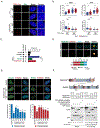
Figure 1.. RAD51AP1 is required for telomere length maintenance in ALT cells.
(A) IF-FISH showing endogenous RAD51AP1 co-localization with telomeres (TTAGGG) and PML in ALT+ (U2OS, Saos2) but not in TEL+ (HeLa LT, SJSA1) cells. (B) Analysis of ALT phenotypes indicating percentage of ALT-associated PML Bodies (APBs), C-circles, telomeric sister chromatid exchanges (t-SCEs) and fragile telomeres in U2OS with non-targeting (NT) and RAD51AP1 siRNAs. (C) Telomere length analysis of control and RAD51AP1 knockout (KO) clones at early (E, ~25) and late (L, ~50) population doubling (PD) by pulsed field gel electrophoresis (PFGE). (D) Top: Western blot of GFP-tagged WT-RAD51AP1 expression in RAD51AP1 KO clones. Below: Quantification of APBs percentages found in RAD51AP1 KOs expressing GFP-empty vector (EV) or GFP-WT-RAD51AP1. (E) Representative IF-FISH showing co-localization of GFP-WT-RAD51AP1 with telomeres and PML bodies. (F) PFGE of RAD51AP1 KO clones shown in (D) that were cultured for ~20PDs. Mean telomere length (kb) is indicated by the red dot. All data represents the mean +/− SEM. n=2 biological replicates. *p< 0.05, **p<0.01, ***p<0.001 (Student’s t test). All scale bars, 5μm.
Figure 2.
Figure 2.. RAD51AP1 is a mediator of HR and BITS at ALT telomeres.
(A) IF images of TRF1-FokI-mediated telomere clustering analysis. (B) Graphs display the number and size of mCherry-TRF1-FokI foci observed in the indicated conditions. Data represents the mean +/− SEM. n=3 biological replicates. *p<0.05, ***p<0.001 (one-way ANOVA). (C) TRF1-FokI-mediated break-induced telomere synthesis assay. POLD3 knockdown was performed as a positive control. Data represents the mean +/− SEM. n=3 biological replicates. ***p<0.001 (Student’s t test). (D) Representative structure illumination microscopy (SIM) images and graph of RAD51AP1 localization in S and G2-phase. (E) Representative images and quantification of POLD3 (left) and RAD52 (right) localization at TRF1-FokI induced telomeric DSBs. Mean +/− SEM, n= 2 biological replicates **p<0.01, ***p<0.001 (Student’s t test). (F) Top: Schematic of RAD51AP1 and RAD52 functional domains. Dotted line indicates the region in each that mediates the interaction. Below: Co-IP of GFP-RAD52 with FLAG-RAD51AP1 in U2OS cells. The average % co-IP between RAD51AP1 and RAD52 proteins is shown. n= 3 biological replicates. All scale bars, 5μm.
Figure 3.
Figure 3.. The fate of RAD51AP1 KO ALT+ cells.
(A) Images and quantification of colony formation assays with U2OS control and RAD51AP1 KO clones. Mean % colonies +/− SEM, n=3 biological replicates. ***p< 0.001 (Student’s t test). (B) Western blot of phospho-ATM (S1981), phospho-Chk2 (T68) and γH2AX. (C-D) IF-FISH images and quantification of 53BP1 positive telomere dysfunction-induced foci (TIFs) and micronuclei containing telomeres and/or cGAS. Means +/− SEM. n=2 biological replicates. *p< 0.05, **p<0.01, ***p<0.001 (Student’s t test). (E) Western blot of autophagy markers +/− DMSO or bafilomycin A1 (100nM for 12hrs). Cleavage of apoptosis associated proteins PARP1 and Caspase-3 were also examined. Etoposide (25γM, 12hrs) treated U2OS cells were used as a positive control for PARP1 and Caspase-3 cleavage. (F) IF images and quantification of mCherry-GFP-LC3B puncta in U2OS control cells and RAD51AP1 KO clones treated with DMSO and bafilomycin A1. Data represents the mean +/− SEM. n=3 biological replicates. ****p<0.0001 (one-way ANOVA). All scale bars, 5μm.
Figure 4.
Figure 4.. Activation of autophagy in RAD51AP1 KO ALT+ cells.
(A) Western blot of autophagy proteins from U2OS control and RAD51AP1 KO clones +/− DMSO or SBI-0026945 (ULK1i) (10γM, 72hrs). (B) Western blot of shRNA knockdown of cGAS, ULK1, ATG7 or ATG5 in U2OS control and RAD51AP1 KO clones. (C) Images of plates from colony formation assays with U2OS control and RAD51AP1 KO clones expressing the indicated shRNA. (D) Quantitation of mean % colonies and percentage of cleaved caspase-3 positive cells detected by flow cytometry from U2OS control and RAD51AP1 KO clones expressing the indicated shRNA. # colonies was normalized to % by comparing to the corresponding clone expressing a non-targeting (NT) shRNA. Statistical significance was derived from comparisons to the control U2OS clone for each shRNA. The % cleaved caspase-3 positive cells and statistical significance was derived from comparisons to the control U2OS clone. Dotted red line indicates the mean level (%) of basal cleaved caspase-3 positive cells detected in control cells. Data represents the mean +/− SEM. n=3 biological replicates. *p< 0.05, **p<0.01, ***p<0.001, ****p<0.0001 (Student’s t test). (E) Quantification of mCherry-GFP-LC3B puncta in control cells and RAD51AP1 KO clones expressing control (NT) or shRNAs to deplete cGAS, ULK1, ATG7 or ATG5. Data represents the mean +/− SEM. n=3 biological replicates. *p<0.01, ****p<0.0001 (one-way ANOVA). (F) Western blot of LC3A/B, p62, γH2AX and cleaved Caspase-3 levels in the indicated conditions.
Figure 5.
Figure 5.. Stabilization of RAD51AP1 in ALT+ cells by MMS21 mediated SUMOylation
(A) Western blot of RAD51AP1, RAD51, POLD3 and RAD52 protein expression in primary, TEL+ and ALT+ cell lines. ATRX is used as a control to validate the ALT+ cell lines. (B) Western blots and quantification of RAD51AP1 protein levels after cycloheximide (CHX)-western analysis in ALT+ versus TEL+ cell lines. p53 was used as a positive control for CHX efficiency. Black trend line indicates RAD51AP1 turnover calculated as a function of one-phase decay. Mean RAD51AP1 protein half-life is also indicated. *p< 0.05, **p<0.01, ***p<0.001 (Student’s t test). (C) IP-western of RAD51AP1 SUMOylation and ubiquitination with WT or C215A GFP-MMS21 in HA-SUMO3 or HA-Ubiquitin (both WT/ΔGG)-U2OS cells. (D) IP-western of RAD51AP1 SUMOylation and ubiquitination after depletion of MMS21, SENP6 and UBC9. (E) Schematic of functional domains and consensus SUMO sites in RAD51AP1 protein and IP-western of RAD51AP1 SUMOylation and ubiquitination after expression of myc-tagged WT, K269R and ΔSIM RAD51AP1. (F) Western blots and quantification of RAD51AP1 protein after CHX-western in the indicated conditions. Data represents the mean +/− SEM. n=2 biological replicates. *p< 0.05 (Student’s t-test).
Figure 6.
Figure 6.. SUMOylation of RAD51AP1 is necessary to sustain ALT activity and telomere length.
(A) Western blots showing complementation of RAD51AP1 knockdown U2OS cells with exogenous GFP-tagged WT, K269R and ΔSIM RAD51AP1. (B-C) Quantification of the percentage (%) of APB positive cells and mCherry-TRF1-FokI foci size in control (NT) and RAD51AP1 depleted U2OS cells complemented with GFP-tagged WT, K269R and ΔSIM RAD51AP1. Data represents the mean +/− SEM. n=3 biological replicates. *p< 0.05, **p<0.01, ***p<0.001 (one-way ANOVA). (D) Top: Western blot of GFP-tagged WT, K269R and ΔSIM RAD51AP1 expression in RAD51AP1 KO clones. Below: Images and quantification of colony formation assays with U2OS control and RAD51AP1 KO clones expressing GFP-tagged WT, K269R and ΔSIM RAD51AP1. Mean % colonies +/− SEM, n=3 biological replicates. ***p< 0.001 (Student’s t test). (E) PFGE of RAD51AP1 KO clones expressing GFP-tagged WT, K269R, ΔSIM RAD51AP1 for ~20 PDs (F) Western blots and PFGE with U2OS cells stably expressing myc-tagged WT, K269R, DSIM RAD51AP1 for ~20 PDs. DNA loading is shown by section of EtBr stained DNA gel. Mean telomere length is indicated as kb and by the red dot.

References

    1. Banani SF, Rice AM, Peeples WB, Lin Y, Jain S, Parker R, Rosen MK, 2016. Compositional Control of Phase-Separated Cellular Bodies. Cell 166, 651–663. doi:10.1016/j.cell.2016.06.010 - DOI - PMC - PubMed
    1. Bhowmick R, Minocherhomji S, Hickson ID, 2016. RAD52 Facilitates Mitotic DNA Synthesis Following Replication Stress. Mol. Cell 64, 1117–1126. doi:10.1016/j.molcel.2016.10.037 - DOI - PubMed
    1. Bryan TM, Englezou A, Dalla-Pozza L, Dunham MA, Reddel RR, 1997. Evidence for an alternative mechanism for maintaining telomere length in human tumors and tumor-derived cell lines. Nat. Med. 3, 1271–1274. - PubMed
    1. Cesare AJ, Kaul Z, Cohen SB, Napier CE, Pickett HA, Neumann AA, Reddel RR, 2009. Spontaneous occurrence of telomeric DNA damage response in the absence of chromosome fusions. Nat. Struct. Mol. Biol. 16, 1244–1251. doi:10.1038/nsmb.1725 - DOI - PubMed
    1. Chandramouly G, McDevitt S, Sullivan K, Kent T, Luz A, Glickman JF, Andrake M, Skorski T, Pomerantz RT, 2015. Small-Molecule Disruption of RAD52 Rings as a Mechanism for Precision Medicine in BRCA-Deficient Cancers. Chem. Biol. 22, 1491–1504. doi:10.1016/j.chembiol.2015.10.003 - DOI - PMC - PubMed

Publication types

MeSH terms